Pyeloectasia of the kidneys in a child

There are diseases that are considered finds. That is, they can be detected only by chance, during examination for other pathologies. These "hidden" diseases include pyeloectasia of the kidneys. The accidental discovery of this pathology raises many questions - what is it, where it came from and how to treat it. All this you will learn from this article.

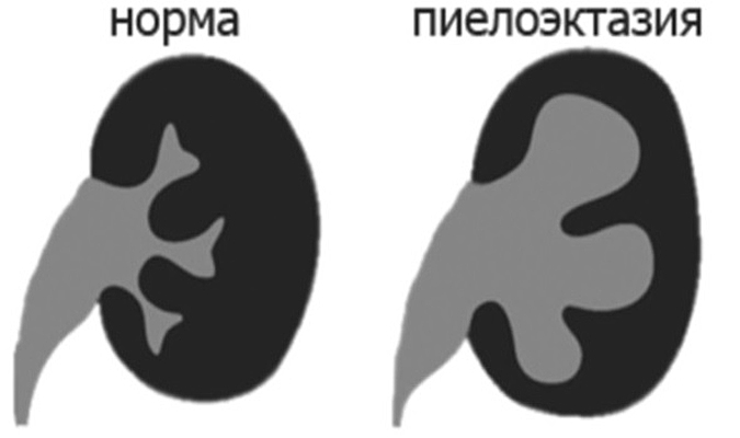
What it is
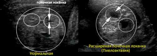
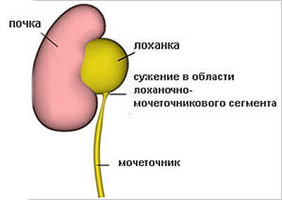
Pyeloectasia of the kidneys is a condition in which the renal pelvis and sometimes the calyx expand. In itself, this is not dangerous, but the expansion causes certain changes in the work of the urinogenital system, provoking inflammatory processes. Urine outflow is impaired, which is a prerequisite for the development of various diseases of the kidneys and urinary system.
Pathological expansion of the pelvis can not be felt, the disease is completely asymptomatic, which is why it is considered a "random finding".
The fact of detection allows us to explain why the child has other problems with the urinary system. In other words, pyeloectasia is considered as the root cause.
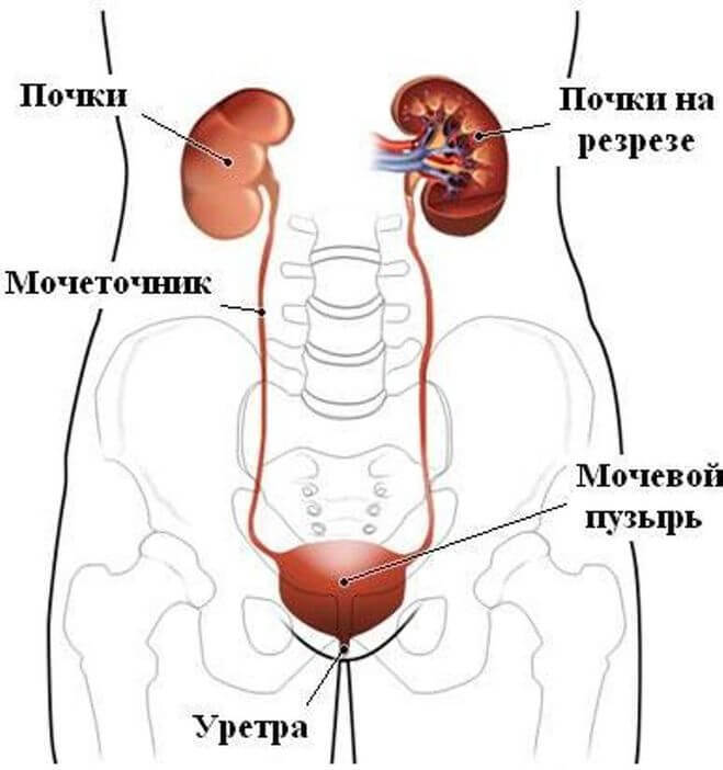
School knowledge in the field of physics is enough to understand exactly how is the expansion of the pelvis. If the urine outflow on some part of the urinary tract is disturbed, the paths are narrowed, there are obstacles, then the pelvis becomes full and stretches. From here it becomes clear why boys pathology occurs more often than girls about 4 times. The urinogenital system of the girl is designed so that stenosis is possible only in rare cases, but in the boy the narrowing of any part of the urinary tract is not uncommon, and quite often it is normal, that is, physiologically conditioned.

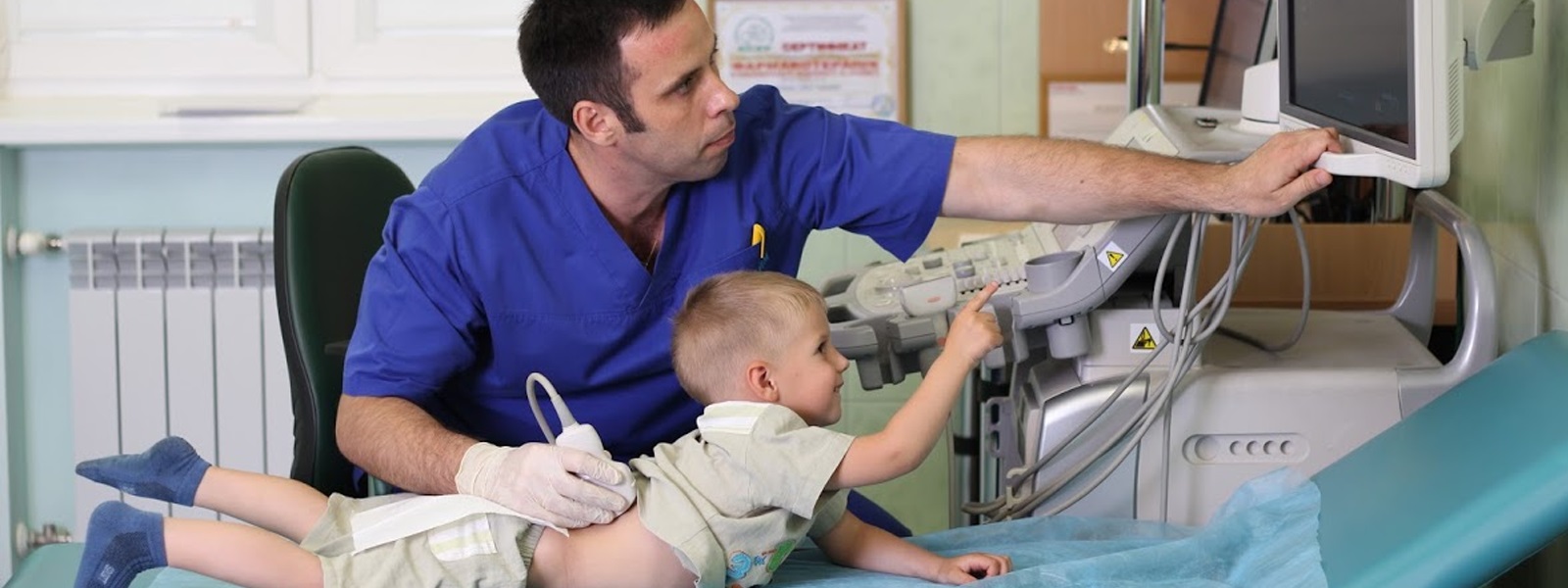
Find pyeloectasia can still have a fetus on ultrasound in the antenatal clinic. Less commonly, pathology can be found in newborns, since ultrasound diagnosis is not included in medical examinations in the first month of a baby’s life. But in infants to detect the expansion of the renal pelvis is quite simple, if at 3 months or 1 year, an ultrasound scan of the kidney is done to the child at a mandatory scheduled medical examination at the clinic.
But this type of research is not always done, and therefore it is often possible to find a pathological expansion much later, when the baby begins to disturb and a kidney ultrasound is required. Many learn about such a diagnosis only in adulthood.
The reasons
Approximately every tenth child with pyeloectasia causes are congenital. They are formed under the influence of some unfavorable factors while the child is in the womb:
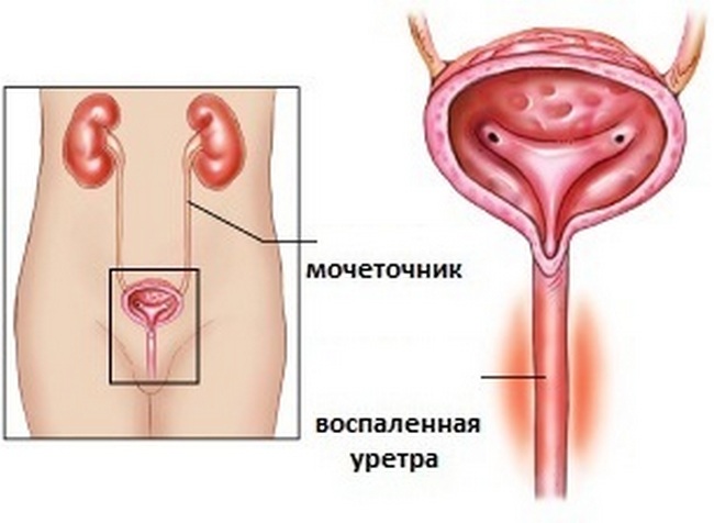
- narrowing of the lumen of the urethra;
- lesions of the central nervous system, which are reflected in dysfunction of urination;
- abnormal development of the kidneys, ureters, urethra due to the "error" during the laying of organs;
- urethral stenosis;
- disorders of the circulatory system.

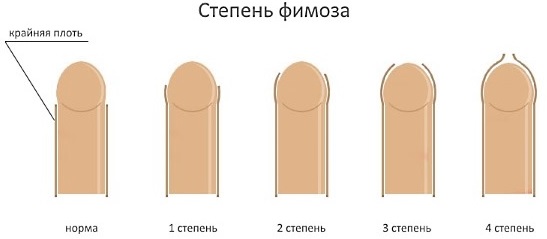
Separately, it should be said about phimosis. For newborn boys, the narrowing of the foreskin is a physiological innate rate.
Most of them have such phimosis on their own. A small percentage of children with persistent phimosis is the risk group for the development of pyeloectasia.
Most often, pyeloectasia is acquired. The pelvis and kidney cavities are able to expand under the influence of certain internal processes:
- hormonal disorders;
- inflammatory diseases of the genitourinary system (cystitis, pyelonephritis and others);
- acute infectious diseases, poisoning by chemicals and toxins that increase the burden on the kidneys;
- trauma to the pelvic organs;
- tumors;
- diabetes;
- urolithiasis and salt deposition.

Stenosis (contraction) may occur in one of five areas:
- urethra and bladder;
- external pressure on the ureter;
- bend of the ureter;
- a narrowing or other obstruction in the lumen of the ureter;
- changes in the structures of the walls of the ureter and upper divisions.
Acquired causes can be due and quite physiologically - premature babies have a weak abdominal wall, the musculature of the urinary tract is not well developed, so the pathology is often found in children born before the obstetric period. Organs in newborns grow unevenly, in some cases, the load on the kidneys, which "do not have time" for growth rates for the rest of the organs, becomes so large that the pelvis due to fluid accumulation begins to expand.
The most “dangerous” age from the point of view of the development of pyeloectasia, when the growth of the child is the most rapid, is 5-6 months, 1 year, 3 years, 5-7 years.
Types of disease and symptoms
Since the kidney is a paired organ, the disease can be unilateral or bilateral. The unilateral form is more often represented by pyeloectasia of the left kidney. Pyeloectasia of the right kidney is 45% less common. Pathological enlargement of the pelvis of both kidneys (bilateral form) is quite often peculiar to children. One-sided form is also not uncommon in childhood, but is more characteristic of adults.
There are three degrees of illnessthey are determined by the degree of damage: mild, moderate and severe. If not only the renal pelvis, but also the calyx of these organs (cavities) are enlarged, the ailment is called calicopyelectasia.
In case of unilateral disease, there may be no symptoms at all, because with pyeloectasia of the right kidney, the left takes its functions and vice versa.

Compensatory abilities of the child's body are incredibly high. Some signs that should become an “alarm bell” can be observed (but not necessarily!) Only in bilateral pathology. At the same time the probability of occurrence of complications increases. And as soon as they begin, the child is taken to the doctor’s appointment, who prescribes an ultrasound scan of the kidneys, and the fact of pyelectasis becomes obvious.
Most often the pelvis enlargement causes:
- pyelonephritis;
- urethrocele;
- prolapse of the ureter.
To prevent such and other equally serious diagnoses, at the first suspicion of a malfunction of the kidneys, you should immediately take the child to the doctor. Parents should be alerted by such signs as swelling of hands and feet, face, especially towards evening, turbid urine, blood in urine, frequent or rare urination, pain when emptying the bladder, worsening of the child’s general well-being, private headaches, pulling pain lumbar region.

Diagnostics
You can notice in a child the pathological expansion of the renal pelvis by ultrasound, starting from 18-20 weeks of pregnancy mom. An attentive diagnostician is able to see pyeloectasia in a fetal boy already from the 17th week of pregnancy. In no case should a future mother need to panic if such a conclusion was made. The fact is that in many cases the expansion of the renal pelvis may be physiological and will pass on its own.
Sometimes the problem is first detected in the fetus shortly before delivery - at 34-36 weeks of pregnancy. In this case, you should not be nervous either.
For pregnant after the establishment of the fact of possible pyeloectasia in a child, enhanced monitoring is carried out.
After the birth of a child, neonatologists must be examined with the assistance of a urologist and a nephrologist. Often, observation remains until the child is one and a half years old.It is to this age that many kids solve the problem by itself. If this does not happen, the question of treatment is resolved.
Medical diagnostic monitoring for children with a mild disease is carried out every six months - an ultrasound scan is done, dynamic indicators of urine tests are evaluated. The average degree of pathology needs to be diagnosed every three months. And only the severe form of the disease requires urgent medical measures and follow-up.

Echographic signs of pathology - the expansion of the size of the pelvis. Normal size of the pelvis in the fetus to 31-32 weeks of pregnancy should not exceed 4 mm. At 36-37 weeks, the renal pelvis normally increases to 7 mm. If the expectant mother is told that the renal pelvis of the fetus exceeds 10 mm, this is an alarming signal, which indicates the likely development of pyeloectasia.
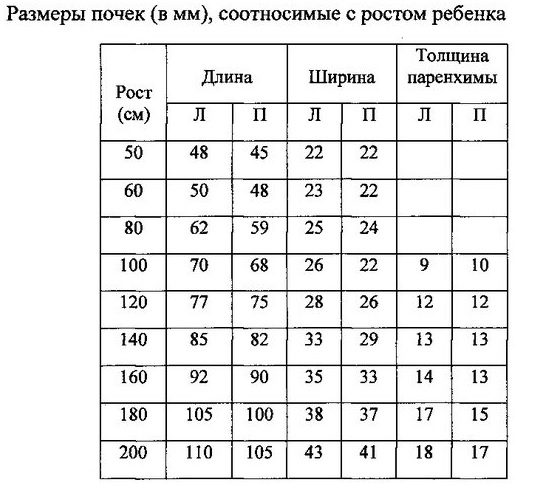
The size of the renal pelvis for children after birth - 6-7 mm, a slight excess up to 8-9 mm can be considered an individual inherited feature. In children older than 3 years, the size of the pelvis may be within 8 mm. Exceeding the threshold of 10 mm at any age is the basis for visiting a nephrologist and a urologist.
Treatment
A mild degree of pathological enlargement of the pelvis does not need special treatment, it is enough to dynamically monitor the child’s condition, he may be prescribed a urine test a little more than other children. Medium degree does not always require treatment. Quite often, doctors choose the tactics of observation, because the problem in a growing children's body may well be resolved on their own.
Severe and moderately severe forms of pyeloectasia often require surgery, even in an infant. Surgical intervention is recommended in case of moderate bilateral enlargement of the pelvis or in case of severe pyeloectasia of the right or left kidney.
The main purpose of the operation is to restore the patency of the urinary tract, so that nothing more prevents the passage of urine, so that the fluid does not accumulate in the pelvis and does not expand them.
The operation itself is not considered traumatic, it is carried out without direct incisions. To achieve the goal is quite enough endoscopic method.

Miniature instruments are inserted directly through the urethra, the surgeon conducts all the manipulations, referring to the picture on the monitor, which is “transmitted” by a microscopic camera located on the endoscope. Narrowed ways expand, obstacles (salt deposits) are removed. If the ureters are bent, they are returned to normal. After the operation, which is performed under general anesthesia, the child receives a course of anti-inflammatory drugs in order to avoid the addition of infection and the development of postoperative inflammation.
If the surgery is performed at an early age, there is a likelihood of relapsebut. During a period of rapid growth (at 5–7 years), pyeloectasia very often returns, but this usually occurs in a less complex and severe degree. therefore re-operation is not always needed.

There are no special preparations for the conservative treatment of pyeloectasia. In some cases, the doctor may prescribe a symptomatic treatment - drugs to relieve edema, diuretics, antibiotics. But usually there is no need for them in milder forms of the disease. And in severe cases, drugs are powerless, it is necessary to have surgery.
Folk remedies, herbs and homeopathic remedies can not cure this ailment. Therefore, it is not necessary to water the child with parsley decoction and give homeopathic drops, advertised as "the best remedy for all kidney problems."
Recommendations to parents
If a child has a mild or moderate pyeloectasia, do not panic. Proper observation of the condition of the baby will provide doctors. And on their own parents can only make sure that the load on the kidneys was maximally reduced. For this:
- should limit the amount of fluid consumed, the amount drunk should not exceed the age norm;
- Be sure to keep track of how much the baby is pissing - ideally, the amount allocated will be slightly less than what you have drunk or equal to;
- the child should not supercool, sit on cold surfaces;
- all infectious diseases (ARVI, flu and others) should be treated under the supervision of a physician, since the burden on the kidneys increases during the period of the disease, self-treatment is completely excluded;
- Special attention should be paid to taking medications. Many tablets and syrups for children with kidney problems are contraindicated or dosed out strictly individually.
See how the kidneys work in the next video.


