Retinopathy of prematurity
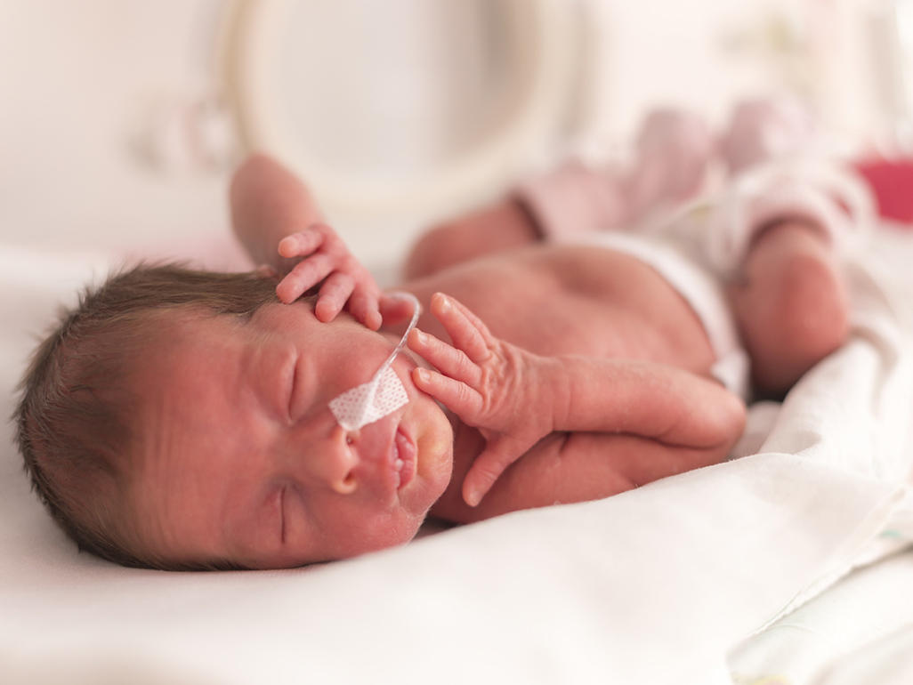
If the baby is born much earlier than the expected term of birth, it increases the risk of various health problems. One of the rather serious and common pathologies in deeply premature babies is retinopathy.
What is it?
So called the problem with the retina in babies born much earlier than expected. Its code is 10 mkb - H 35.1. The main danger of retinopathy is risk of irretrievable loss of visual function.
The reasons
The disease is caused by many factors, among which the main one is the immaturity of the infant who was born before 34 weeks of intrauterine development. However, the baby can be born in time, but still remain immature. Other factors provoking retinopathy are:
- Low weight baby.
- Multiple fertility.
- Concomitant pathologies of the fetus, for example, the development of sepsis, anemia, or acidosis.
- Chronic diseases of the genital organs of the mother.
- Gestosis.
- Bleeding during childbirth.
- Ventilation and use of oxygen after birth.
For more information about the causes of retinopathy, see the video:
Features of pathogenesis
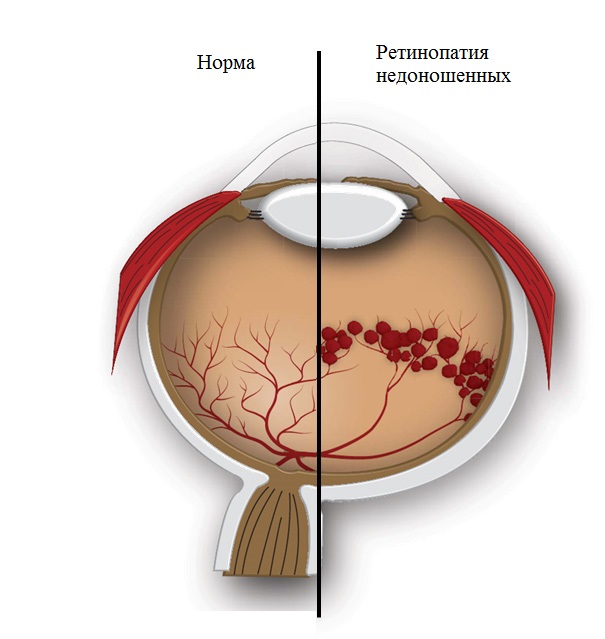
The vessels inside the retina begin to develop from the 16th week of intrauterine development of the infant. Until that time, there are no vessels in this part of the eye at all. They begin to grow from the site where the optic nerve goes, toward the periphery. Vessels are actively growing until the beginning of labor, therefore, the earlier the crumb is born, the fewer vessels in its retina have time to form. In deeply premature babies, quite extensive areas without vessels (they are called avascular) are detected.
When a child is born, various external factors (first of all, oxygen and light) begin to affect its retina, which provokes the appearance of retinopathy. The normal process of vascular formation is disrupted. They begin to germinate in the vitreous body, and the simultaneous growth of connective tissue leads to a retinal tension and its detachment.
Retinopania in preterm infants develops as follows:
- First passes active period (from birth to 6 months). In this period, the veins expand, the arteries change, the vessels become convoluted, the vitreous opacity, traction detachment of cellulose are possible, less often its rupture or tearing.
- Next comes reverse phase of development. This period starts from 6 months of age and lasts on average up to a year.
- From 1 year begins cicatricial period. Myopia can form during it, lens opacification, detachment or retinal tearing can often occur, eyeballs can decrease, and intraocular pressure may also increase. Sometimes the iris and lens are shifted forward, which causes corneal clouding and dystrophy.

Retinopathy stages
Retinopathy is a process in several stages, which can be completed both by scar formation and complete regression, in which all manifestations disappear.
Stage I
In the place of separation of normal vascular tissue and avascular sites, a whitish line appears. The identification of such a dividing line is the reason for conducting weekly inspections of the baby.
Stage II
In place of the line of separation of the avascular retina with vascular areas, a shaft appears. At 70-80% of newborns at this stage there is a spontaneous improvement, while minor changes remain on the fundus.
Stage III
In the resulting shaft, fibrous tissue appears, and the vitreous body above it is compacted, resulting in retinal vessels being drawn into the vitreous body. This causes retinal tension and a high risk of detachment. This stage is also called the threshold, because with its progression retinopathy becomes almost irreversible.
Stage IV
The retina partially exfoliates without involving the central part (transition to stage 4A) and with detachment in the central region (transition to stage 4B). This stage and the subsequent changes are called terminal, since the prognosis for the child worsens, and his vision is sharply disturbed.
Stage V
The retina exfoliates completely, which leads to a sharp deterioration in the baby's vision.
Separately isolated plus-diseasewhich has no clear stages. This form develops earlier and progresses much faster, which causes retinal detachment and the rapid onset of the terminal stages of retinopathy.

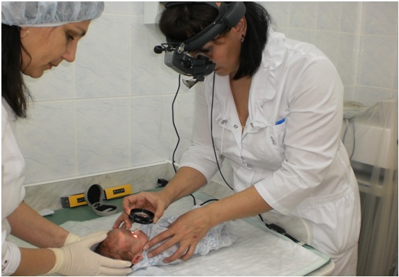
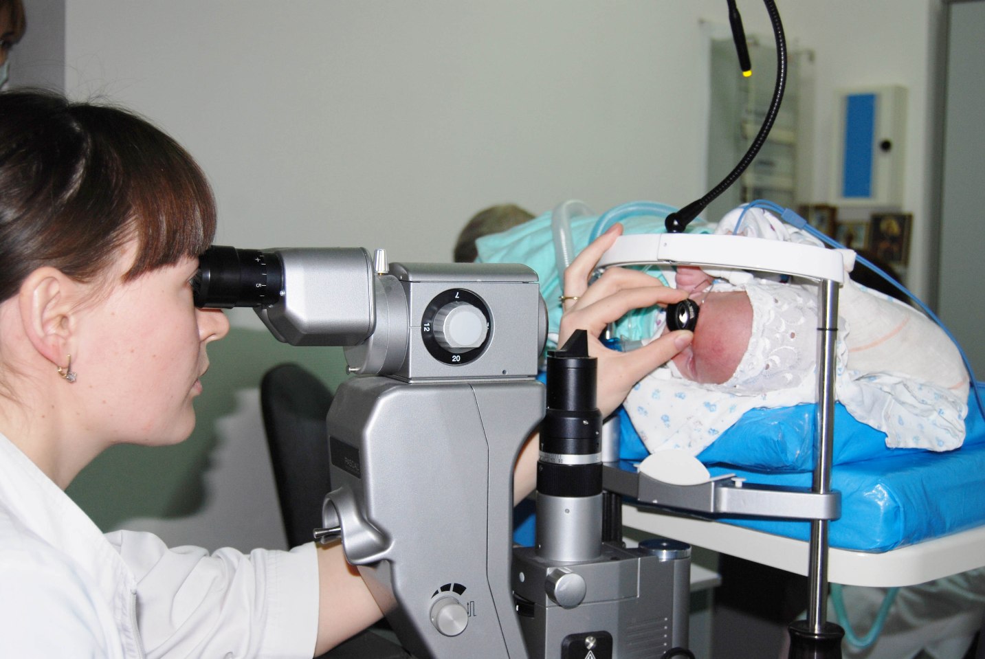
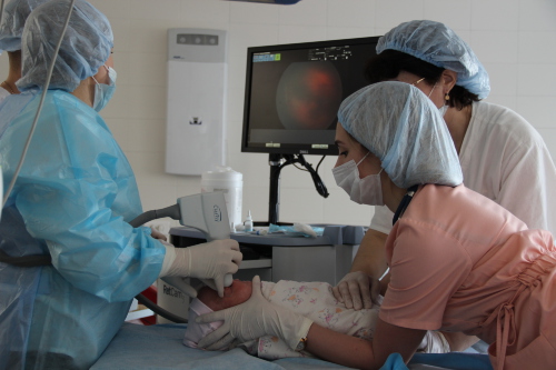
Diagnostics
All children born before 35 weeks or with low weight (less than 2 kg) should be examined by an ophthalmologist using special equipment for such an examination. If symptoms of retinopathy have been identified, the newborn is continued to be examined once a week, and in the presence of plus-disease, more often every three days.
Examinations continue until the disease fully regresses or requires surgical treatment. As soon as the disease regresses, the baby is inspected every two weeks.
During the inspection, the baby must expand the pupil, and also use special speculators for the procedure (they will eliminate pressure on the eyes from the fingers). An additional method of examination for retinopathy is ultrasound of the eye.
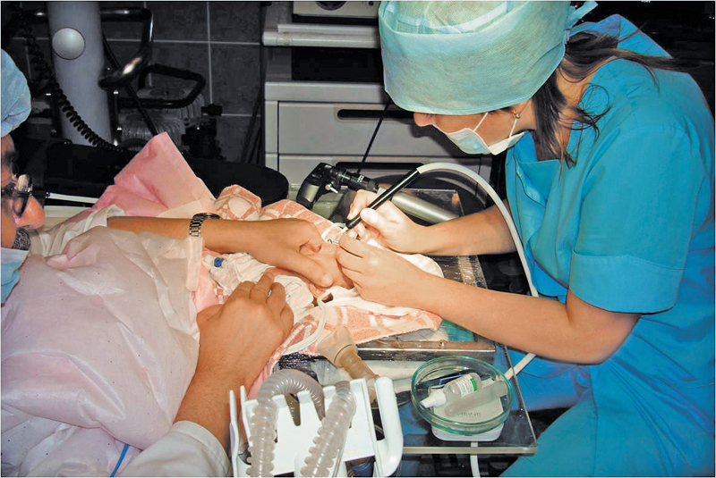
Treatment
The stage of the disease affects the treatment of retinopathy, but all methods of treatment can be divided into:
- Conservative. The child is instilled with medicines that are prescribed by an ophthalmologist. This treatment is considered ineffective.
- Surgical. The method of this treatment is chosen taking into account the course of retinopathy (usually it is carried out at stage 3-4 of the disease). Many children are prescribed cryosurgical or laser treatment. When the detachment begins, the vitreous body is removed to the child in specialized clinics.
Prognosis and prevention
The duration of active retinopathy is on average 3-6 months. The result of the development of the disease may be spontaneous cure. (often observed in the first or second stages) and scarringin which residual changes have varying severity. On their basis, cicatricial changes are divided into degrees:
- At the first degree the fundus of the eye is almost not changed, so the visual functions are not impaired.
- Second degree characterized by a shift in the center of the retina, as well as changes in peripheral areas, which increases the risk of secondary detachment in the future.
- When developing third degree, in the retina there is a deformation of the area in which the optic nerve enters into it. The central part of the retina is greatly shifted.
- Fourth degree manifested pronounced folds on the retina, causing severe visual impairment.
- At the fifth degree note total retinal detachment.
Prevention of retinopania is to prevent preterm labor. In addition, it aims to properly care for a premature child.

When a stage 1 disease is detected in a child, prophylactic administration is required. corticosteroid hormones, and with oxygen therapy, antioxidants are additionally prescribed. Defining at the crumbs stage 2, the dosage of hormonal drugs increase, and supplemental oxygen and drugs that dilate the vessels, if possible, limit it.
Cryocoagulation and laser coagulation, such as the destruction of avascular areas in the retina, are quite effective in preventing retinopathy. Their use reduces the incidence of adverse outcome by 50-80%. Manipulations are performed under general anesthesia, the result is evaluated after 7-14 days and, if necessary, repeat the procedure.
In severe stages of retinopathy, the child’s eyesight suffers greatly. Even surgical treatment can only improve light perception and provide an opportunity to navigate the room and monitor objects.
It is also worth noting that in children with mild stages of the disease further violations of the organ of vision are possible, such as amblyopia, glaucoma, myopia and late detachment. For this reason they should be monitored regularly by an ophthalmologist until the age of 18.
Retinopathy is a process in several stages, which can be completed both by scar formation and complete regression, in which all manifestations disappear.