First ultrasound after IVF

The first ultrasound after the IVF protocol is a very responsible and exciting moment. It is this examination that shows how successful the efforts of doctors and patients were. When the first ultrasound scan is done after embryo grafting and what it shows, we will explain in this article.
Why do ultrasound after replanting?
Strange as it may seem, the first after the ECO ultrasound protocol is a completely paradoxical situation - a long-awaited and suffered pregnancy turns out to be so important for a woman that she is afraid to go for an ultrasound, so as not to harm the baby if she successfully develops in her body. Sometimes no argument can force a woman who has been unsuccessfully struggling with infertility for several years to go to the ultrasound room.
You can definitely say that ultrasonic waves, on which the principle of operation of the ultrasound scanner is based, do not have any negative effect on the fetus, and therefore it is not necessary to be afraid of ultrasound scanning. The first ultrasound after the IVF protocol is of great importance. It is made not only to make sure that the pregnancy has really come, but also to find out how many embryos have taken root, if more than one embryo was planted.
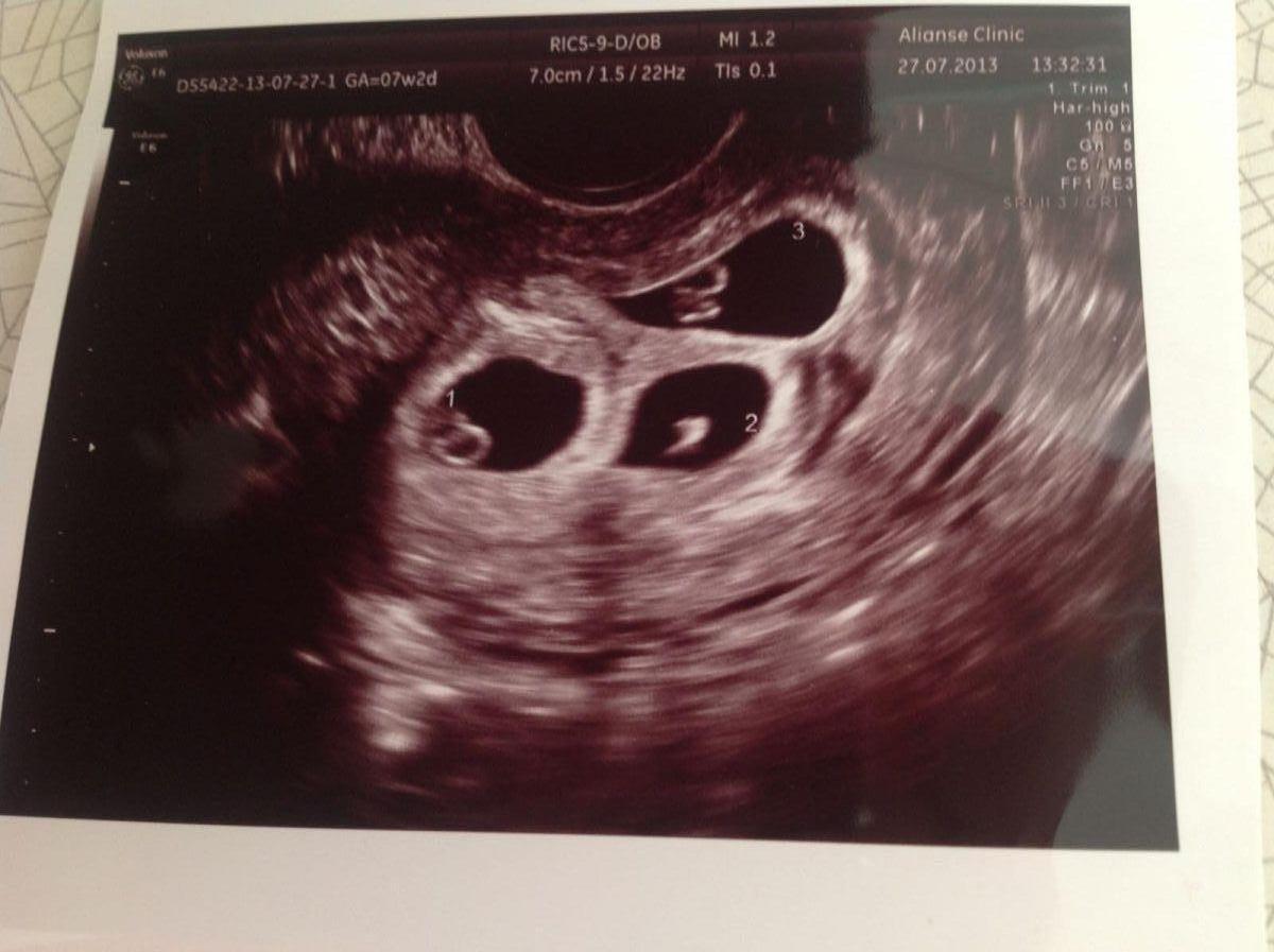
In 60% of cases when two or three embryos are planted, two of them take root. The likelihood of multiple pregnancy is high. Pregnancy of twins or triplets requires a slightly different obstetric approach, which is why it is very important to determine it at the earliest possible time.
A woman whose pregnancy has become possible as a result of in vitro fertilization should generally get used to the idea that she will have to go to the ultrasound more often than to a woman who becomes pregnant naturally. Pregnancy after IVF requires more careful observation, since it has higher risks of threatened miscarriage, missed abortion, premature birth and pathologies of placenta formation.
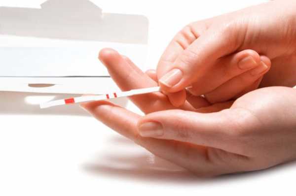
Pregnancy after IVF is determined by a blood test for hCG - this is reliable evidence of the “interesting situation” that has come. You can do it already at 12-14 days after embryo grafting. However, the growth of hCG and the dynamics of the increase in the concentration of this hormone, although they give an idea of the development of pregnancy, cannot describe all its features. This can be done only by the results of ultrasound.
The first ultrasound after IVF is recommended to be carried out a week after the start of the delay, that is, at 21 DPP (day after transfer). It is the age of three gestational weeks that is diagnostically important. It reveals the fact of pregnancy after successful IVF, determines the number of fetuses in the uterus, the viability of each of them, and also helps to assess the state of the ovaries after hormonal stimulation performed in the protocol and eliminate the possibility of a threat of early miscarriage.

In addition, an ultrasound scan, which is done on 21 DPP, allows you to find out if a woman has complications such as fading fetal development and an ectopic pregnancy, which, according to statistics, occurs in 2-3% after IVF.
After an unsuccessful IVF, at about the same time, you should also visit the ultrasound examination room, because the pelvic ultrasound makes a considerable contribution to determining the cause of failure and planning the new IVF protocol.
What shows?
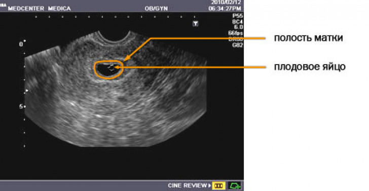
Ultrasound can theoretically be done before 21 days after embryo transfer, but the problem is that even on state-of-the-art equipment it will be virtually impossible to see microscopic embryos. Only by the end of the third gestational week the doctor will be able to assess the thickness of the endometrium of the uterus, the place of attachment of the ovum and its size, the size of the yolk sac as the main food store of the developing baby.
If you do the first ultrasound a little later - in the interval between 21 and 28 days after transplantation, then you can register the true signs of the life of the baby - at 5-6 obstetric week (this period corresponds to 3-4 weeks after transplantation) audible fetal heartbeat or two fruits.
Of great importance is the early diagnosis of multiple pregnancy.
If, due to increased risks of non-survival, 3-4 embryos were transferred to the woman, and it turns out that, contrary to forecasts, everything has taken root, then, at the wish of future parents, resection may be performed - the extra embryos will be removed. In order for such an opportunity to exist, the diagnosis must be really early.
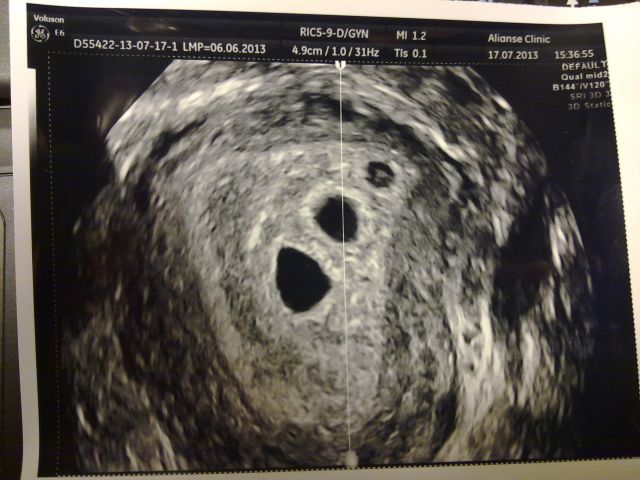
3-4 weeks after embryo transfer, regardless of the number of fetuses, healthy embryos show approximately the same size. Lagging behind the norms may indicate the inferiority of the implanted embryo, the developmental delay, or its death. The most important size is the inner diameter of the ovum, the so-called SVD. On the first ultrasound of the SVD normally ranges from 18 to 22 mm. Compliance of this parameter with the norm suggests that the growth rate is adequate, nutrition and oxygen to the fetus is enough. The shape of the ovum must be correct, with smooth contours, without deformation.
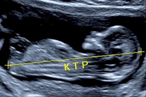
Closer to the 28th day after the transfer, another important size is defined - coccyx-parietal, or KTP. This is the distance from the coccyx to the vertex, which changes at the time of maximum extension of the embryo. Normally, it is 3-6 mm. The average diameter of the yolk sac for the first ultrasound is about 3 mm.
After IVF, sizes can be quite individual, since implantation occurs at different times. In one woman, transferred embryos are implanted in the functional layer of the uterus on the third day, and in another, only after 7 days. Therefore, these ultrasound data are necessarily compared with the growth dynamics of hCG - a hormone that is produced by the chorionic villi after implantation. If the concentration of the hormone increases, it is safe to say exactly when the implantation took place.
Naturally, the size of the embryo that was implanted earlier will be slightly larger than the size of the embryo that was implanted later. According to statistics, five-day-old embryos are implanted with a higher percentage of success than three-day embryos.
The heart of babies at the beginning of the sixth obstetric week (22-24 days after the transfer) is just starting its activity, it beats at a frequency of 80-85 beats per minute. On the 28-29th day after the transfer, the heart rate of the crumbs of the heart is already normal at 103-123 beats per minute.
On the first ultrasound, the probability of threatened abortion is determined - the doctor looks to see whether the cervical canal of the cervix is closed, whether there is an increased tone of the uterine muscles.

How is it going?
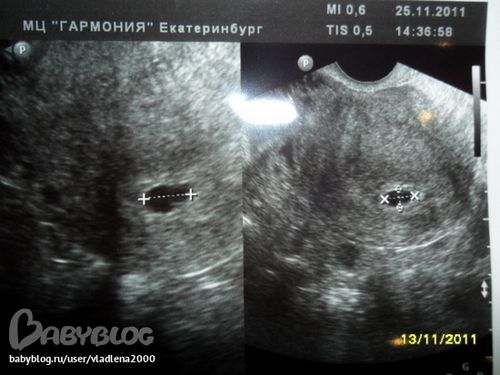
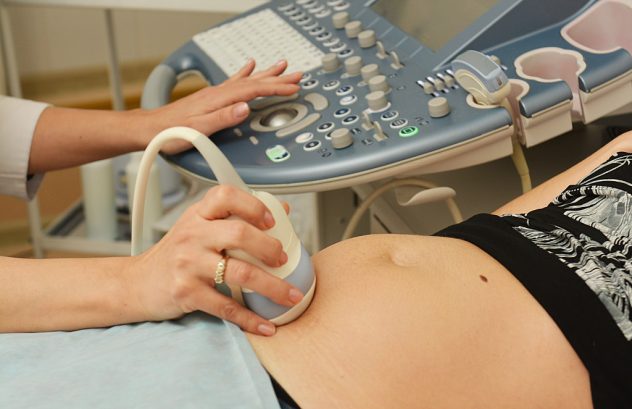
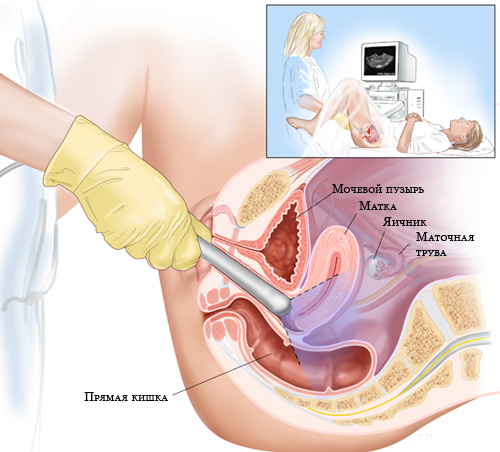
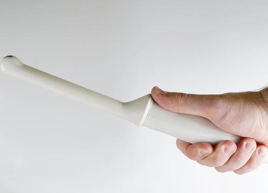
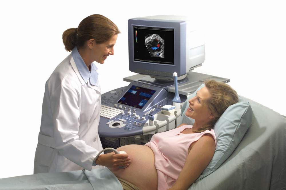
The first ultrasound scan can be done after embryo transfer in two ways: by placing the sensor along the anterior abdominal wall and using an intravaginal sensor. Which way to choose, the doctor will decide. The most preferred is considered intravaginal scanning method, which allows you to accurately assess the condition of the cervix, the size and structure of the ovaries, the size of the uterus and the presence of possible signs of the threat of spontaneous interruption.
Quite often, vaginal (internal scanning) gives more accurate results in women with overweight and large amounts of fat on the abdomen, which makes it difficult to visualize with the abdominal examination.Sometimes there is a need for a combined survey, in which the inspection will be conducted by two sensors alternately.
Preparing for an ultrasound is much easier than it seems. Recommendations from the Internet to go to the doctor on an empty stomach is completely absurd. On the informativeness of the ultrasound examination eating does not affect, as having sex on the eve of the survey.
But to refrain from soda and beans with cabbage still stands, so as not to increase gas formation in the intestine. Intestinal loops may swell with gas, put pressure on the uterus, and this may create some diagnostic difficulties. Therefore, before visiting the doctor for several hours it is recommended to take "Smektu" or "Simethicone".
Whether to drink before ultrasound fluid, the question is common. Drink about half a liter of water is needed only before transabdominal ultrasound, so that the bladder is filled and better pass ultrasonic waves. Before vaginal ultrasound drink is not necessary, on the contrary, you should empty the intestines and bladder before the examination.
Do not persuade the doctor to make you the first ultrasound in the form of 3D or 4D, as well as require an ultrasound with Doppler. You will be able to do these types of research later, since in the earliest terms they are not considered informative and the difference between the newest 4D and the usual two-dimensional ultrasound scan will not be felt by you or the doctor.
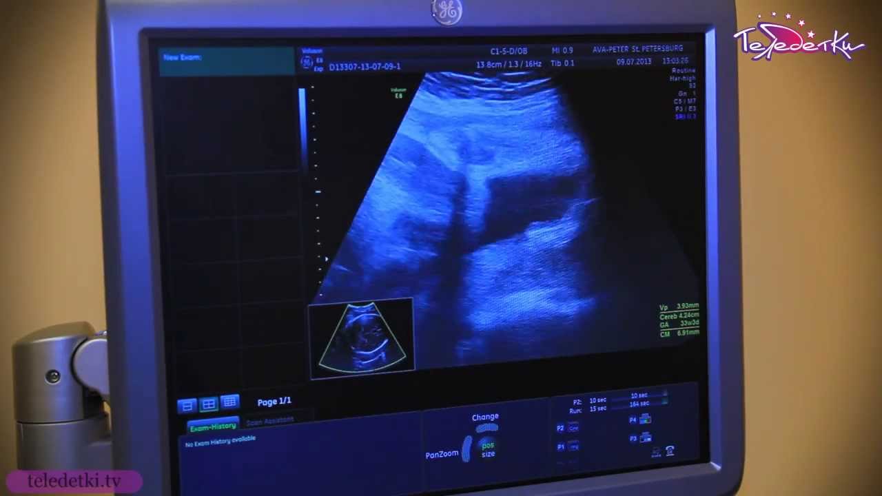
The procedure takes about a quarter of an hour. Results are given to the woman immediately after passing the examination.
Special features
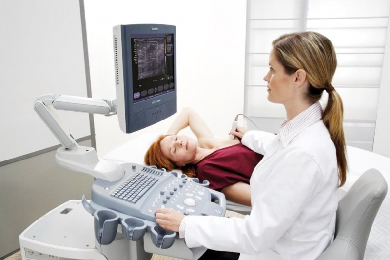
One of the distinguishing features of the first after the transfer of ultrasound is an additional ultrasound diagnosis of the mammary glands. This is done to assess how well a woman has undergone hormonal stimulation of the ovaries and if she has any problems and tumors in the breast - most breast tumors are hormone-dependent and can begin to grow under the influence of hormones.
If a woman managed to become pregnant without hormonal stimulation in the natural cycle with replanting of a fertilized egg during the implantation window, then an ultrasound of the mammary glands is not necessary.
The rest of the ultrasound scan after IVF is not much different from a similar confirmatory diagnostic procedure after the occurrence of pregnancy in a natural way.
When is the next study?
After the first ultrasound scan, carried out on days 21-28 after embryo transfer, it is often necessary to re-examine to clarify the gestation period and the rate of development of the fetus (or fetuses). In this case, women are invited to repeat the procedure in 2-3 weeks.
If the first-second ultrasound scan does not show any pathologies, the woman is recommended to do such an examination in the standard terms:
- 10-12 weeks (as part of the first prenatal screening);
- at 19-21 weeks (as part of the second prenatal screening);
- at 30-32 weeks (as part of the final screening, which summarizes the results of the first two);
- before childbirth (to choose the tactics of delivery and determine the accuracy of the proposed day of birth).
In case of multiple pregnancies, ultrasound can be prescribed not only during the designated periods, but also between them; the number of ultrasound scans is not regulated by WHO requirements. The doctor himself can determine when and how many times to do an examination for a particular pregnancy.
Reviews
According to women, the first ultrasound scan causes fear and great excitement, even if blood tests by this point already confirm that the pregnancy has begun. Not everyone can hear the baby’s heart, but there’s nothing wrong with that, after 1-2 weeks on the control ultrasound, the heartbeat will definitely be determined.
In most cases, women say, after the first or second ultrasound scan, the reproduction specialist wants a couple of happy pregnancy and childbirth and passes the patient to an ordinary gynecologist at the antenatal clinic for further observation and registration at the dispensary.
The disadvantage is, according to women, that the results of tests that were done before IVF are not accepted in the consultation, all the tests have to be done again, but by OMS, that is, completely free.

For early pregnancy after IVF, see the following video.


