Analysis of AFP during pregnancy and its rate by week

Not every future mother knows exactly what tests she gives in the direction of the doctor during the carrying of the child. Yes, it is not always required. However, the AFP test is well known to pregnant women. What kind of research this is, what it says and what are the standard values of AFP during pregnancy, we will describe in this article.

What it is?
The abbreviation AFP stands for alpha-fetoprotein. This protein in the maternal organism is formed during the development of the embryo and fetus. Initially, the substance was discovered as a marker of cancer, and only in the second half of the twentieth century, doctors and scientists noticed a pattern - protein appears in the blood of pregnant women who do not have any malignant tumors.
Alpha-fetoprotein is very similar to another protein, serum albumin.
In adults, it transports various substances with low molecular weight into tissue. In a baby that develops in the mother's womb, the AFP replaces albumin and performs its functions — it carries the substances necessary for the growth and development of the blood to all tissues.

All the functions of this amazing protein science are not yet known. Therefore, in the encyclopedias and scientific reference books there is the formulation “probable functions”, “possible and presumptive functions”.
These possible functions of the AFP include immunosuppressive effect - protein unknown while the mechanisms affects the immunity of women, suppressing its activity and possible rejection of the fetus, which is only half "native". Also, the AFP is “suspected” of suppressing fetal immunity. Otherwise, the baby could react negatively to new compounds and proteins that it receives from maternal blood.
At the very beginning of pregnancy, AFP protein is produced by the corpus luteum. However, three weeks after conception, the baby-embryo begins to produce the necessary protein compound itself. The substance enters with the urine of the crumbs in the amniotic fluid, and from there - into the bloodstream of the mother, to be brought out.

The amount of AFP gradually increases, and from 11-12 weeks it is well defined in the blood of a woman.
By week 16-17, the concentration of the substance rises to high levels.
The highest content of AFP in the blood of a woman is observed at 33-34 weeks, after which the amount of embryonic (fetal) protein begins to slowly decrease.
Protein is widely used in the treatment of many diseases, including oncological diseases. It is extracted from placental and abortion blood. During pregnancy, the level of AFP may indicate possible complications and genetic abnormalities of the fetus.

Why do the analysis?
The analysis for alpha-fetoprotein is also called the Tatarinova-Abelev test. He enters the so-called "triple test", which is assigned to all pregnant women as part of the second prenatal screening.
The best for analysis is considered 16-17-18 weeks of pregnancy, since during this period, the protein is well defined in the blood of a woman and has a reliable diagnostic value.
A fetus that has chromosomal abnormalities (Down syndrome, Turner, Patau and others, non-molar trisomies, as well as malformations of the neural tube - the brain and spinal cord) produces certain amounts of this protein.

The level of AFP can indirectly judge the possible deviation in the development of the child.
To make the picture more complete, the concentration of fetal protein is compared with the level of hCG (chorionic gonadotropic hormone), as well as the level of free estriol.
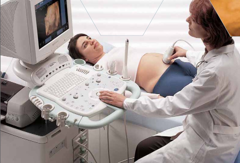
Some clinics also determine inhibin level - hormone of the placenta, and then the analysis is called "quadruple test." Complements the laboratory picture of ultrasound, which is necessarily carried out as part of a screening survey.
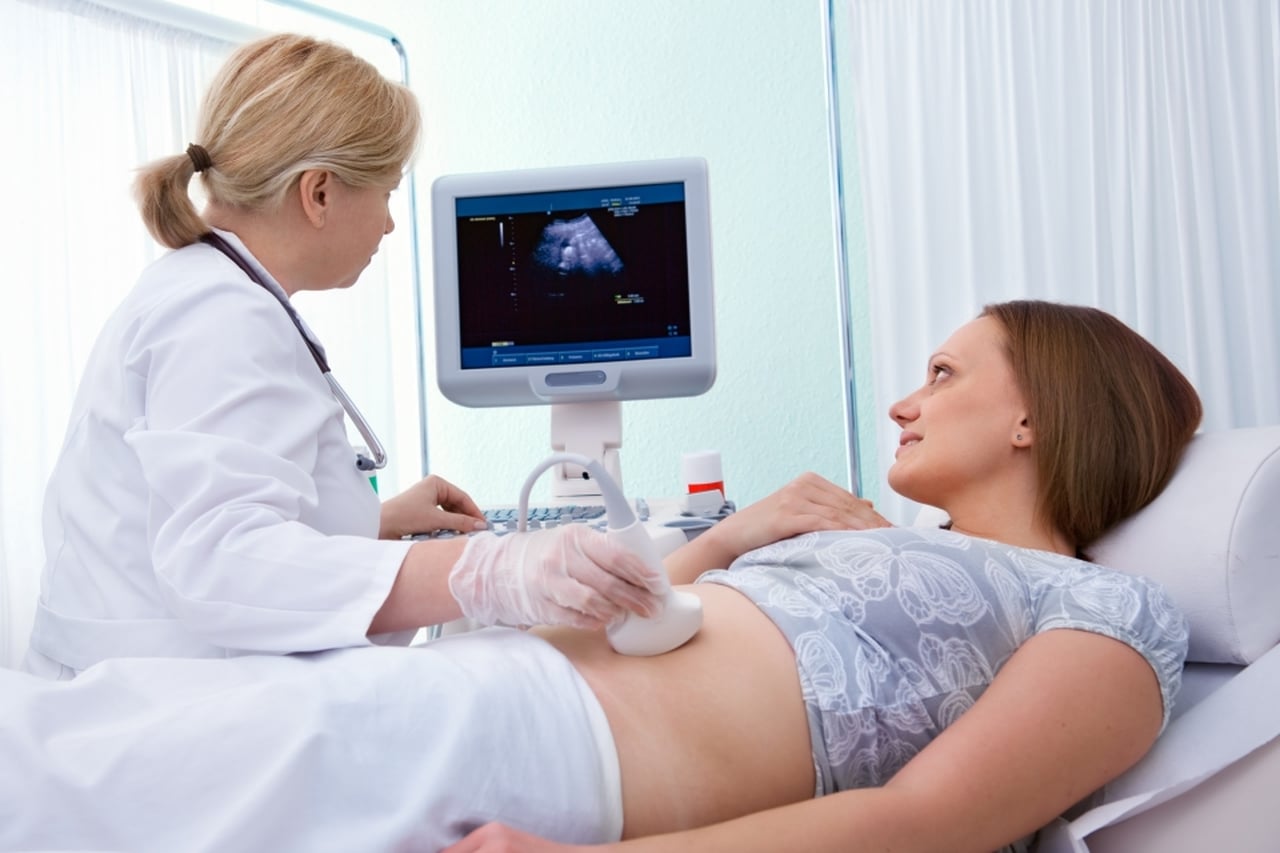
Blood for analysis taken from a vein. A woman should come to the laboratory or treatment room on an empty stomach, in the morning, after having canceled all hormones and antibiotics, if they were prescribed by a doctor.
A few days before a blood test is taken, a pregnant woman should refuse to take fatty and sweet foods, large amounts of salt, carbonated drinks, coffee. If the expectant mother smokes, despite the obvious harm of this habit, you should refrain from smoking for 3–4 hours before giving blood.
The nervous factor may also affect the results of the analysis, therefore a woman is advised not to be nervous.

Weekly rates - decoding
The level of fetal protein AFP grows with the gestational age, it is not difficult to understand the table:
Obstetric term | The concentration of AFP (in U / ml) |
First trimester up to 12 weeks | Less than 15 |
13 to 15 week | 15 to 60 |
From 15 to 19 week | 16 to 95 |
20 to 24 week | From 27 to 125 |
From 25 to 27 week | From 52 to 140 |
28 to 30 week | From 67 to 150 |
31 to 33 weeks | 100 to 250 |
From 33 weeks to birth | The tendency to a gradual decline |
Since laboratories rely on their tables, which depend on the units of measurement of the concentration of a substance, sensitivity and quality of reagents, research methods, there is a generally accepted world practice to measure the level in a multiple of the median - MoM (multiples of median).
AFP is considered normal during pregnancy. from 0.5 to 2.0 MoM. Thus, if the conclusion indicates that the level of AFP is 0.2, it is a reduced protein concentration. If the value of AFP exceeds 2 units, then we will talk about an increased level.

If the laboratory does not indicate the result in MoM, and the amount of the substance in units per milliliter appears in the conclusion, then the value of the median should be clarified in a specific laboratory in order to understand how normal the indicated indicator is.
By itself, the level of alpha-fetoprotein can not talk about the presence of pathologies in the child, in combination with other parameters of the triple or quadruple test it merely suggests any deviations:
- Chromosomal and non-chromosomal pathology of the fetus - a sharp increase in the level of AFP at the same time as the normal level of hCG.
- The threat of termination of pregnancy - a slight excess of the level of AFP and twice and more low levels of hCG.
- Down syndrome in a child - a very high level of hCG and a very low level of AFP.
- The death of a child in utero - very low hCG and moderately reduced AFP.

In fact, there are much more variants and combinations, since the somnologist can determine some markers of pathologies on ultrasound, the picture will be supplemented by the level of the hormone Estriol in the unbound state.
Analyzes the data obtained by the computer, which, given the individual risks (age of the woman, bad habits, dysfunctional heredity), determines the probability of having a child with a particular pathology in the form of 1: 400, 1: 1400, 1: 3000 and so on.

Causes of deviations
According to the existing medical statistics, deviations in the concentration of the AFP protein are observed in approximately 7% of pregnant women. This does not mean that all these expectant mothers have sick children in their womb.
The level of alpha-fetoprotein may differ from normal values for other reasons. Let's look at the most common.
Increased value
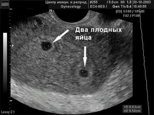
During pregnancy, fetal protein can be double or triple, as several fruits produce more AFP, respectively, the level of protein in the mother’s blood will also be 2 or 3 times higher than the average. By the time of the second screening, a woman usually already knows for sure about her multiple pregnancy, so an increased level of protein in a laboratory report will not come as a surprise.
AFP may be slightly elevated in a pregnant woman if she has a tendency toward a large fetus. The remaining reasons for the increase in AFP, unfortunately, do not have such pleasant and harmless reasons.
High levels of alpha-fetoprotein can speak about liver necrosis of the baby. An important organ of life for the baby could suffer in the process of a viral disease, from which his mother could not protect herself in the first trimester of pregnancy.
If this happens, an ultrasound diagnostic specialist will be able to see a reduced and uneven liver.
An elevated AFP is sometimes a convincing marker of irreparable pathologies in the development of a child — anencephaly (absence of the brain), microcephaly (a decrease in the brain), and crevices in the spine. Most of these pathologies make the child's life impossible, they are lethal.
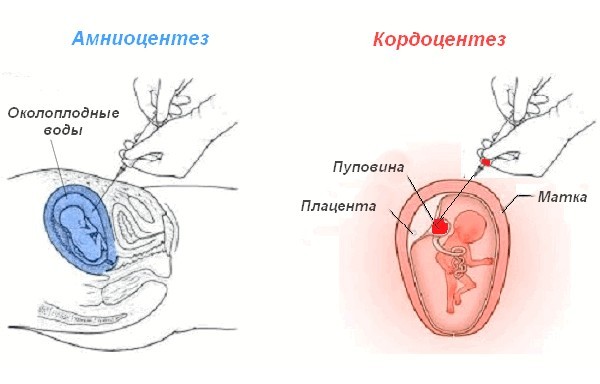
Such a suspicion can be confirmed or disproved by a good ultrasound specialist, and the final decision will be made by an expert commission, which will include genetics and gynecologists. May need amniocentesis or cordocentesis.
Congenital umbilical hernia, in which the internal organs of the baby are located outside the abdominal cavity in the hernial sac, may also be accompanied by an increased level of AFP.
Sometimes a large amount of fetal protein is observed in various malformations of the kidneys and urinary tract of the child, esophageal atresia. All these vices can be seen on ultrasound.
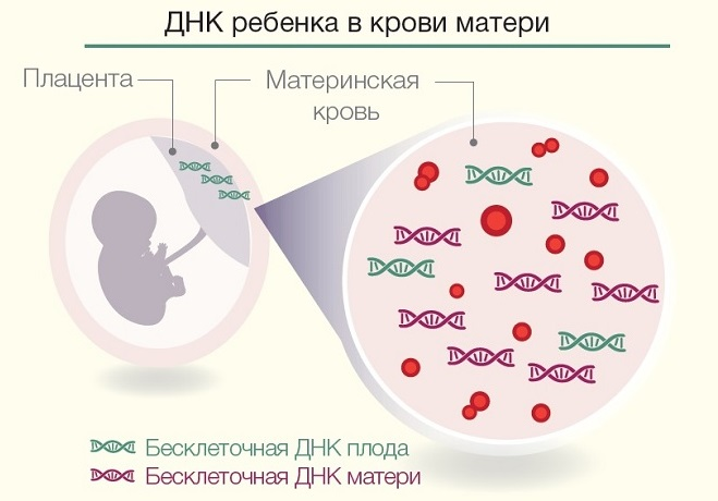
Among genetic chromosomal pathologies, an increased concentration of AFP is characteristic of Turner syndrome. It can be confirmed or refuted by invasive diagnostics (cordocentesis, amniocentesis), as well as a non-invasive DNA test for which maternal venous blood is used.
Low value
The reduced level of AFP protein detected during the second screening may indicate the presence of chromosomal abnormality in a child - Down syndrome. Also, a low concentration of alpha-fetoprotein is characteristic of trisomy 18 (Edwards syndrome).
These suspicions are checked by geneticists, who offer the expectant mother to undergo invasive diagnostic procedure. It involves a puncture in the abdominal area and a long needle of amniotic fluid (amniocentesis) or cord cord blood (cordocentesis) for genetic analysis. These procedures are associated with certain risks for the mother and the fetus. A woman may refuse such an examination.

There is a less traumatic way to find out the truth - to do a non-invasive DNA test. A woman takes regular venous blood. The fetus's red blood cells are found in it, their unique DNA is isolated from them, and they learn whether it has chromosomal abnormalities. Such an examination is very expensive - several tens of thousands of rubles.
The downside is that the conclusion of passing such a test It is not a reason to terminate a pregnancy due to medical indications.
If the sad suspicions are confirmed, and the woman wants to end the pregnancy, she will still have to go for cordocentesis or amniocentesis.

AFP levels may be lowered when fetal development is delayed. The reasons for the delay itself can be any number, and the ultrasound specialist can easily determine the lag of the baby from normal size. Also reduced AFP indicates the probability of miscarriage or fetal death of the baby.
In any case, it is important for a woman to tune in to an additional examination, which will include an ultrasound scan, other blood tests, and sometimes a visit to the medical genetic center.
In case of detection of total defects and chromosomal abnormalities, the woman is offered an abortion. In the case of confirmation of the threat of miscarriage or delayed development of the crumbs assigned supportive treatment it can be at home or in a hospital.

All about the early diagnosis of genetic diseases of the fetus, including the analysis of AFP, see the following video.


