How to measure the size of the pelvis during pregnancy, and what standards exist?

Assessment of the lengths of bone landmarks is a necessary diagnostic procedure carried out by a specialist to all women during the carrying of a baby.
Borders and structural features of the pelvic apparatus have been evaluated by future moms for centuries. Such a simple and informative study allows doctors to get a lot of diagnostic information they need.
Little about anatomy
The pelvis is a bone formation. Quite a lot of different bones and joints are involved in its formation. The pelvic bone apparatus is a complex architectural element. Every woman has its own peculiarities of its anatomy.
The pelvic bone apparatus consists of several bones: a pair of pelvic, sacral and coccyx. Each pelvic bone, in turn, consists of three more: ileal, sciatic and pubic. Between themselves, they are connected with the help of cartilage tissue.
During pregnancy, this structure is functionally beneficial. It helps the baby to move quietly through the birth canal.

The pelvis is a kind of repository for the reproductive organs. During the carrying and birth of a child, it has a very important function. It is in it that the birth canal passes, along which the baby subsequently moves during its birth into the light.
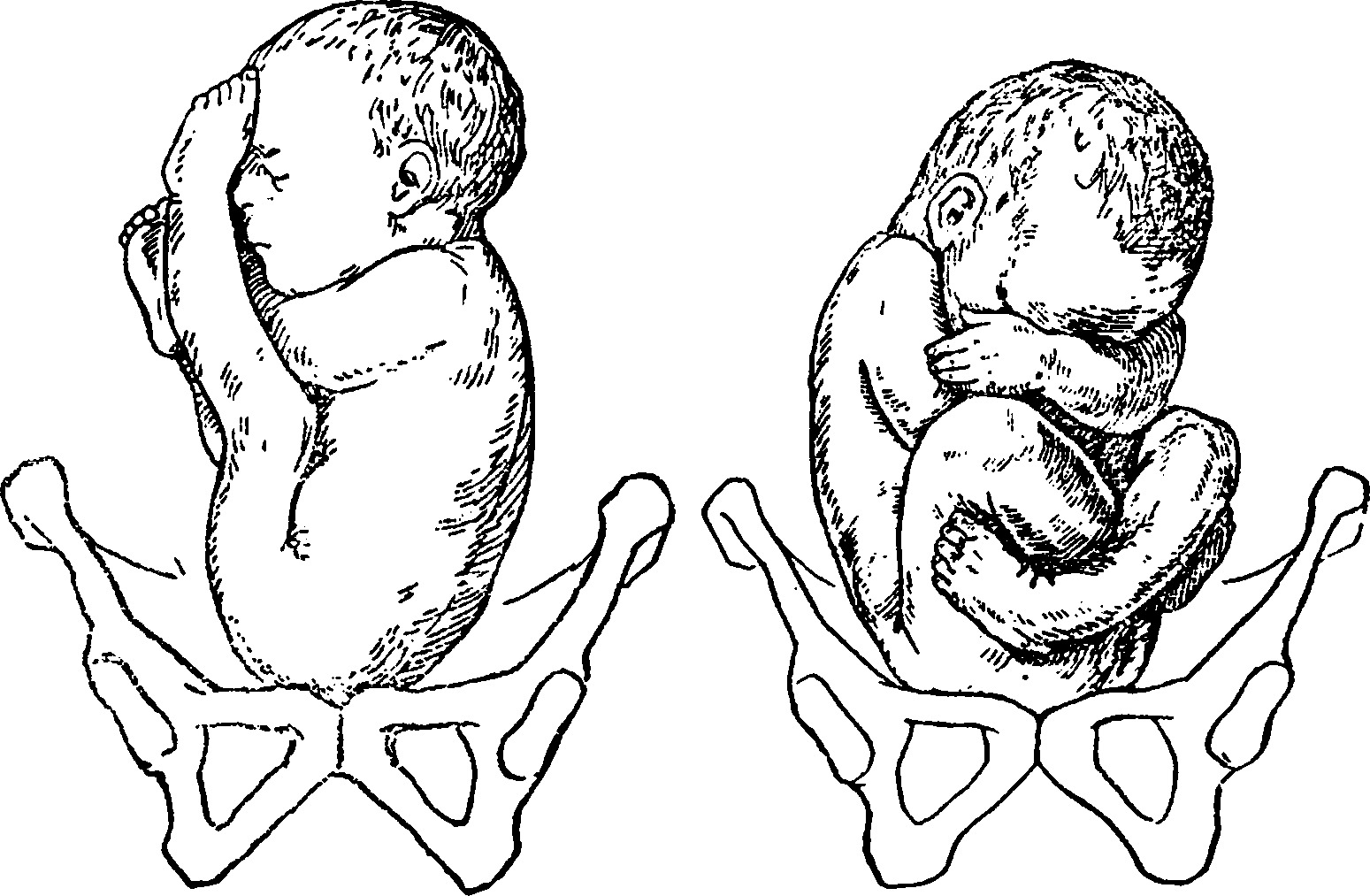
Sizing of this bone apparatus is very important. It is especially important to perform this in the event that the baby in the mother's womb is located not physiologically. Breech presentation of a child with a narrow or asymmetrical pelvis of the mother requires a more careful relationship with the woman during the course of her pregnancy.

Determination of clinical parameters
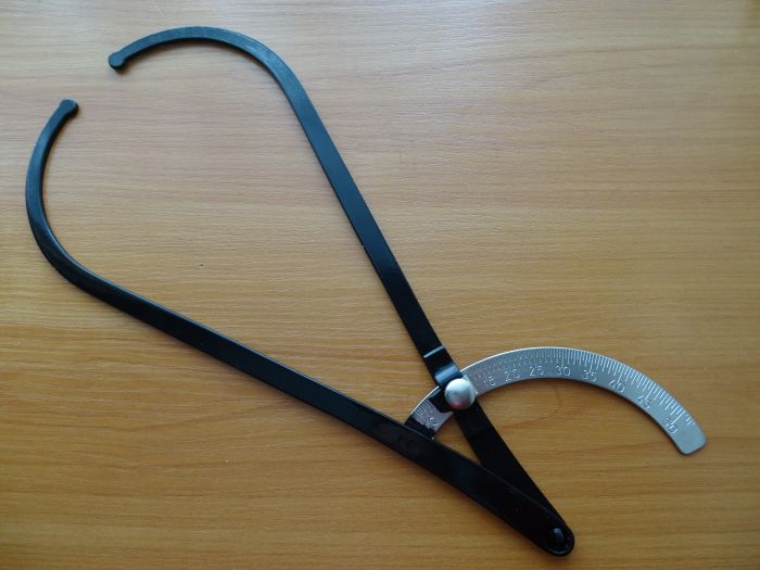
For many years, doctors have conducted an external study of the pelvis in different ways. The first of these is the determination of pelvic indices by palpation. The second method is to determine the studied lengths using a special instrument - a tasometer.
Doctors perform this diagnostic procedure when carrying a baby at least twice. For the first time, these clinical indicators are determined at the very beginning of pregnancy. The obtained values necessarily fit into the personal medical card of a pregnant woman. Usually, pelvic measurement is performed on women who are registered for pregnancy.


Also, the size of the pelvic bone apparatus in future moms doctors determine already on the term closer to childbirth. This is a very important prognostic indicator that allows you to assess how the delivery will take place. He also helps doctors to choose the optimal way of delivery to the specific patient.
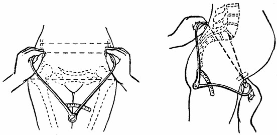
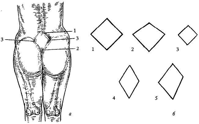
In conducting the study, the doctor will be particularly interested in a special anatomical zone - Michaelis rhombus. This site is located in the sacroiliac segment of the spine.
Its changes are very important diagnostic criteria for doctors.


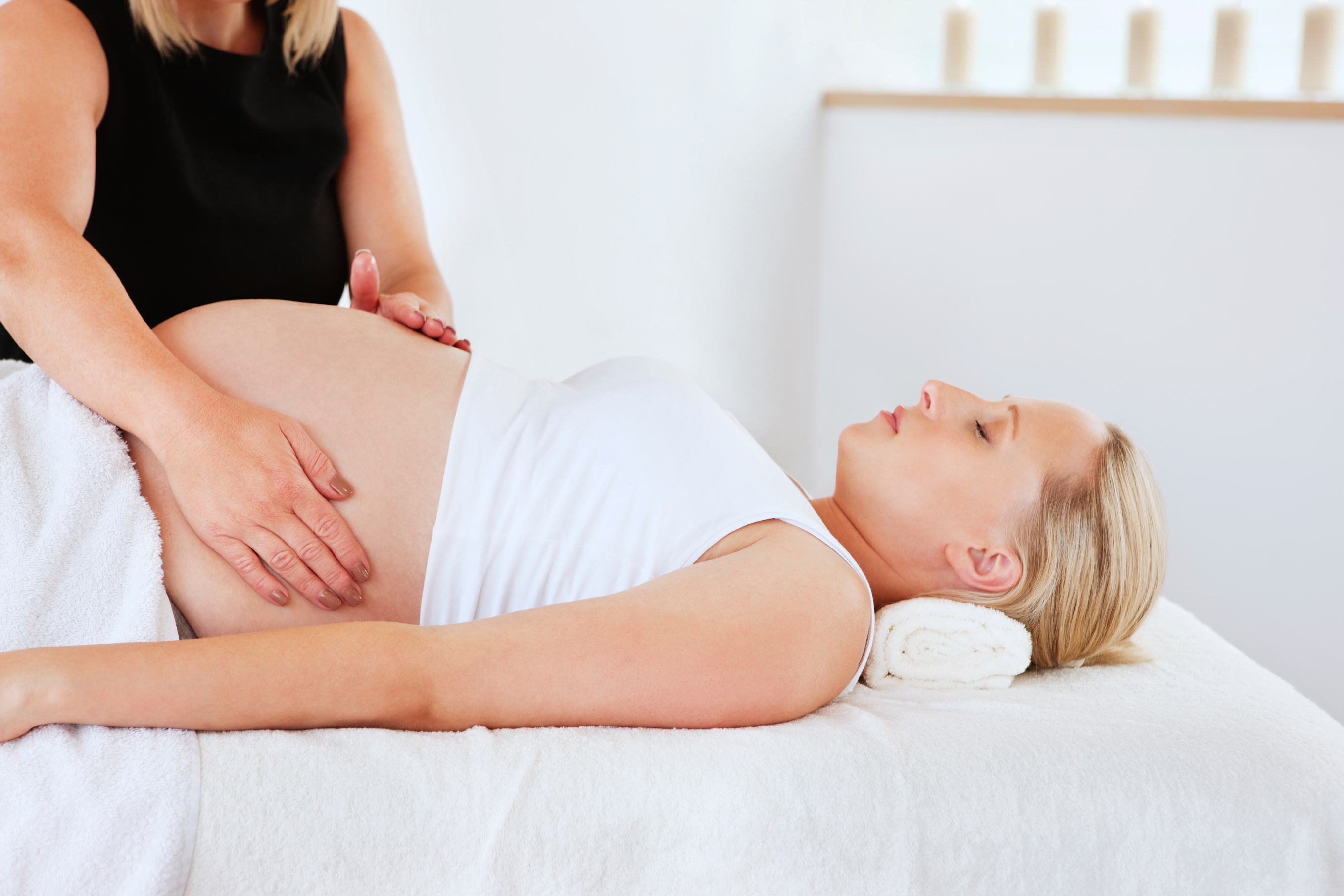
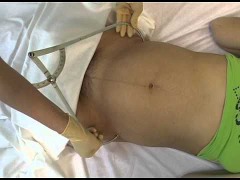
Measures the size of the pelvis obstetrician-gynecologist, who will continue to observe a woman for 9 months of carrying her baby. The study is conducted in a regular room.
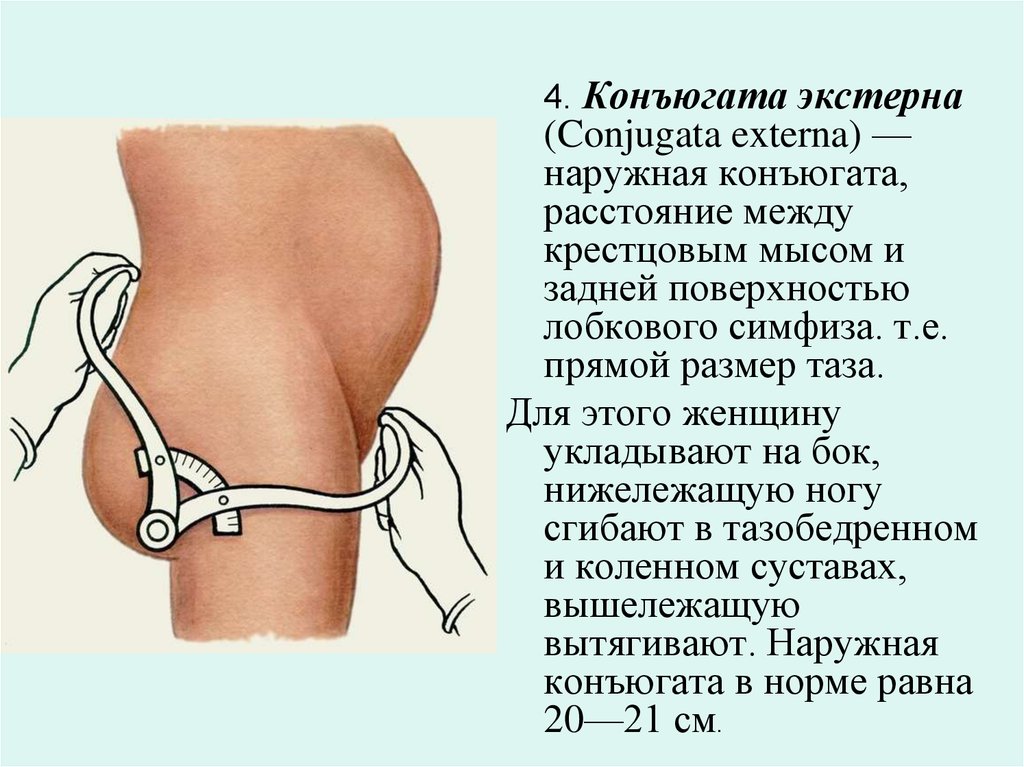
The measurement of the pelvis is carried out when the expectant mother is lying on the couch. The starting position of a pregnant woman is on the back. In order to simplify the diagnostic procedure, the expectant mother should lift the clothes from the measured area. To determine the indicators, the doctor uses a tazometer.
How is the rate determined?
An obstetrician-gynecologist measures several sizes at once. One of them is longitudinal. And the other three are transverse. For each of these values has its own criteria standards. They are used by doctors to accurately determine the type of pelvic structure in a particular patient.
Several of the parameters studied are called the special term - Distantia or abbreviated D. To determine the first of these, doctors measure the distance between the two helical zones of the hips. This parameter they call D. trochanterica. For most women, its values range from 28 to 33 cm.
To determine the next parameter to be studied, the distance between the combs of the iliac seeds is determined. It is called D. cristarum. Its normal values range from 24 to 27 cm.


Another no less important determinable indicator is the outer conjugate. To determine it, doctors measure the distance from the upper part of the womb to the edge of the end of the lower back (at the level of the fifth vertebra). Its values range from 20 to 21 cm.
After the measurement, the doctor can calculate true conjugate. This figure is less than the outside by 9 cm.
In medical practice, there is another method for determining this size. To do this, the doctor must determine the diagonal measurement. To this end, he measures the distance between the most protruding point of the sacral cape to the lower edge of the symphysis.
Most often, this clinical indicator is determined during a palpatory examination by a gynecologist on a chair. Its norm is 10-13 cm.

The physician can still measure the direct measurement of the pelvic outlet. For this, the distance from the top of the coccygeal bone to the lower corner of the womb is measured. This indicator is equal to eleven centimeters.
To refine this parameter, another refined criterion is also used - true direct measurement. Its rate is already nine and a half centimeters. The mathematical difference between these two determined sizes is, as a rule, one and a half centimeters.
Pelvic angle It is also a very important clinical indicator. Two planes horizontally and vertically participate in its formation. Tazouglomer is used to determine this clinical criterion. In the vertical position, the normal values of this defined parameter are 45-50 degrees.
During the study, the doctor may also determine several other sizes. They have additional diagnostic value. Usually they are necessary in order to identify the individual characteristics of the structure of the bone apparatus, available in a particular patient.
If the specialist has identified any asymmetry in determining the size of the pelvis, he will also measure the following parameters. They are presented in the table below:
Studied parameter | Length (cm) |
The distance between the two upper spines (on the right and left side) and the supracarpal fossa | 18 |
The distance between the rear upper bones and the central region of the upper edge of the symphysis | 17,5 |
Distance between front and rear spines | 21 |
Clinical options
The doctor takes into account the ratio of all these indicators. This allows him to assess the type of pelvis in a pregnant woman. For this, several sizes are evaluated at once: a specialist does not make a conclusion on only one clinical parameter.

The table below shows the different types of pelvic structures in women:
Pelvis shape | Distantia spinarum (cm) | Distantia cristarum (cm) | Distantia trochanterica (cm) | Conjugata externa (cm) |
Normal | 25-26 | 28-29 | 30-31 | 20 |
Cross pinned | 24-25 | 25-25 | 28-29 | 20 |
Simple flat | 26 | 29 | 30 | 18 |
Flat-ravine | 26 | 26 | 31 | 17 |
Pelvis with a decrease in the direct size of the wide part of the cavity | 26 | 29 | 30 | 20 |
General uniformly constricted | 24 | 26 | 28 | 18 |
How is the decryption of the obtained values?
If the pelvis has a normal structure, then the Michaelis rhombus looks like a square, which is turned upside down.Its diagonal is about 11 cm.
When measuring this indicator, it happens that the sides of the square begin to shift. This leads to a change in its shape: it becomes more elongated. If, when measured, the doctor determines a pair of acute and a pair of obtuse angles, then in this case it means the presence of a narrow pelvic bone apparatus.
Wide pelvis is most common in fairly tall and large women. This is influenced by the feature of the structure of the musculoskeletal system of the future mother. Also, a wide pelvis can also occur in women who have an average physique. In miniature ladies and future mothers who have a small stature, this structure is practically not found.

The wide pelvis is characterized by an increase in all detectable sizes. When measuring dimensions, it is very important to exclude the influence of a large amount of subcutaneous fat. For this exception, a gynecological examination is carried out on the chair. By determining the true conjugates, the doctor can determine how true a wide pelvis is in a particular patient.
Many expectant mothers think that the larger and wider the pelvic bones, the easier it will be for them to give birth on their own. This is not entirely true.
Indeed, the possibility of natural childbirth sizes pelvic bone apparatus are of great importance. However, in the case of a wide pelvis, the future mother may have various pathologies.

This is also no exception. for the appointment of a cesarean section. Surgical delivery can be shown with a large and deep structure of the pelvic apparatus. The choice of the method of delivery is determined by an obstetrician-gynecologist who monitors the course of pregnancy.
Symmetry - This is a very important parameter, which the doctor must fix. For this there is a certain medical algorithm. The doctor must measure the size on both halves of the body. If the obtained dimensions of the dimensions on the left side are more right-sided by 1 cm or more, then the doctor fixes the presence of asymmetry.
It is also important to evaluate the measured lateral dimensions. To do this, the doctor will measure the distance between the edge of the anterior and posterior anterior bones. These clinical parameters are determined both on the left and on the right side. Normal values of this indicator are 14 cm.
If the values obtained are substantially less than 12.5 cm or are distinctly different from each other, this also indicates the presence of asymmetry in the pelvis of the pregnant woman. In this situation, the bones are shifted in the vertical plane.
Doctors also call this variant of the structure of the pelvic apparatus asymmetric. In this situation, as a rule, a caesarean section will be required. Childbirth in a natural way can be dangerous both for a woman and for her baby. The risk of various injuries in this case increases many times.

How to measure yourself at home?
You can try to measure the size of the pelvis without the participation of a doctor. However, such measurements can only be approximate. Still, determines the type of structure of the pelvis and its main dimensions obstetrician-gynecologist, who observes the course of pregnancy in a particular woman.
The specialist has the necessary experience and knowledge to enable him to successfully carry out this important diagnostic procedure.

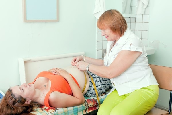
It often happens that the expectant mother wants to independently determine what her pelvis. To do this, it simply measures the circumference of the hips or the distance between the pelvic bone formations as far apart as possible.
Such a measurement has nothing to do with the clinical determination of the size of the pelvic structure. To conduct a comprehensive and comprehensive study is possible only with the participation of a doctor.

For information on how to measure the size of the pelvis during pregnancy, see the following video.


