Diagnosis and treatment of hematoma in the uterus during pregnancy
In time to recognize the various pathologies in the uterus can be quite difficult. One of these pathologies is hematoma. This article will help you understand how to diagnose and treat hematomas in the uterus during pregnancy.

What it is?
For a variety of reasons, hematomas can develop in the uterus during pregnancy. Doctors call hematoma a pathological condition in which blood accumulates in a confined space. The clinical form of the hematoma depends on where the blood has accumulated.
Doctors distinguish several variants of this pathology. Each of them has its own characteristics and in a certain way influences the forecast of the development of pregnancy. In some types of hematomas localized in certain parts of the uterus, complications that are dangerous for the future mother and her baby can develop.
It should be noted that hematomas in the uterus may appear at different stages of pregnancy. Some pathologies occur only in the first trimester, while others occur in the second half of pregnancy.
The severity of the resulting violations in this case is different. Sometimes the symptoms that occur can worsen the general condition of the future mother so much that it becomes extremely difficult for her to carry the baby before the prescribed time.
Options
In determining the diagnosis and clinical type of pathology, doctors use a variety of terms. Such specific medical terminology can confuse anyone. The difficult and long name of the diagnosis can finally confuse the future mother. It is necessary to understand what hematomas occur most often in the uterus during pregnancy.
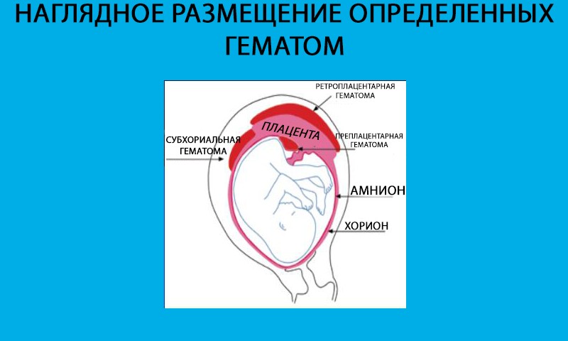
Subchorial hematoma is one of the pathology variants. At the same time, blood accumulates between the chorial membrane that covers the embryo and the uterine wall. Unfortunately, this pathology is often encountered in obstetric practice. The risk of development in this case, spontaneous abortion is quite high.
In medicine it happens that often can be used several terms to refer to the same process. Many specialists also call the subchorial hematoma retro-chorionic or subobolar. Doctors have learned to diagnose such a pathology quite well. Zabolochnaya hematoma is currently determined by ultrasound.
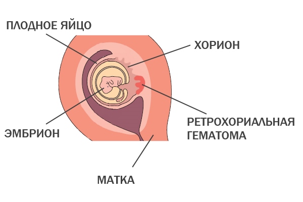

Blood can accumulate in various places of the uterus. It can spread between the shells of the chorion, as well as completely leak over them. In this case, interswitch and subhepatic hematomas develop. The accumulation of blood directly behind the shells of the chorion may also be referred to as retro-enlarged hematoma.
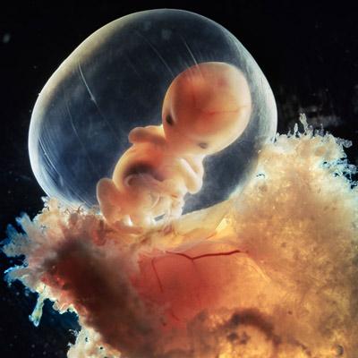
Pathologies that are associated with the chorion usually develop in the very early stages of pregnancy. It was at this time that the chorion is very important for the intrauterine development of the fetus. Then it is gradually transformed into the placenta, which already "assumes" the provision of a number of functions necessary for the full growth of the baby in the womb.
Another unique organ that appears in the female body only during pregnancy is the amnion. This is the water shell in which the baby is located.It provides a special aquatic environment, without which the prenatal development of the child is impossible.
Hemorrhages that occur for various reasons, can lead to accumulation of blood in the amniotic membrane. In this case, hematomas appear in different parts of the uterus. They may be subamniotic, retroamniotic.
Pathologies associated with the germinal membranes develop in the first half of pregnancy. Gradually, a placenta begins to form from the embryonic membranes. This feature explains the differences in the names of different hematomas.
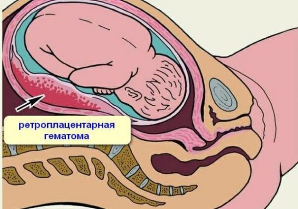
With the accumulation of blood behind placental tissues, a retro-placental hematoma appears. This condition can be extremely dangerous and even lead to the development of placental insufficiency. With this pathology, not enough oxygen and nutrients enter the children's body. Long-term placental insufficiency can even lead to the formation of various defects and developmental abnormalities in the baby.
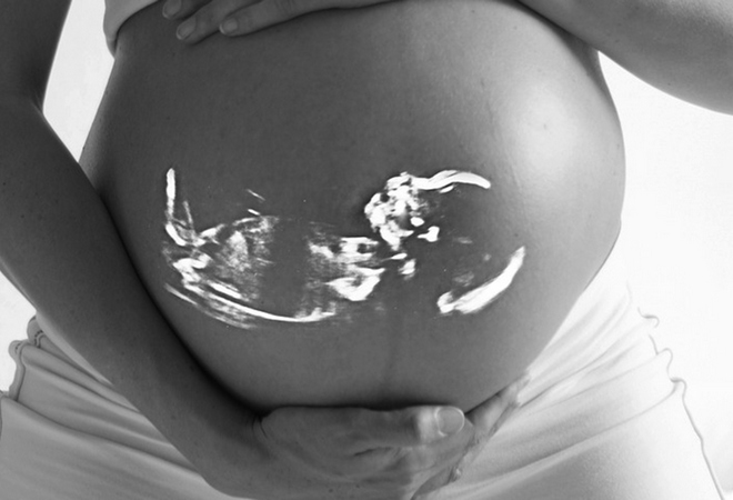
The accumulation of blood in the uterus can lead to the fact that a pregnant woman develops adverse symptoms. Bloody and dark brown discharge from the genital tract are from hematoma emptying. The accumulated blood can flow, which leads to the appearance of specific secretions from the genital tract. This symptom can really scare the expectant mother.
In such a situation, a pregnant woman should definitely seek medical help. Only a doctor can determine the exact reason why the expectant mother has a uterus.
How is the diagnosis?
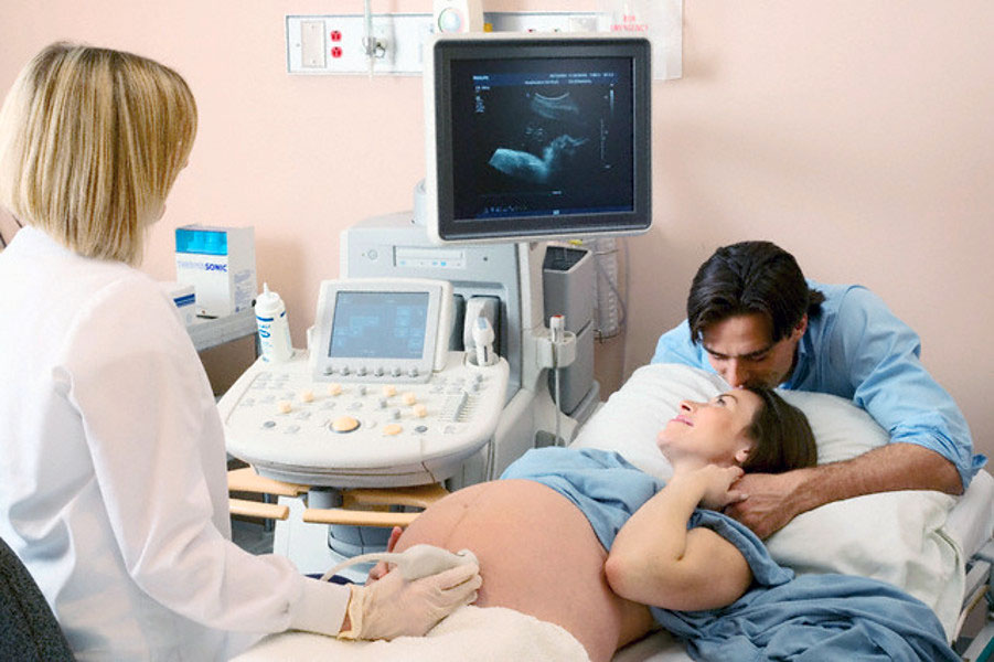
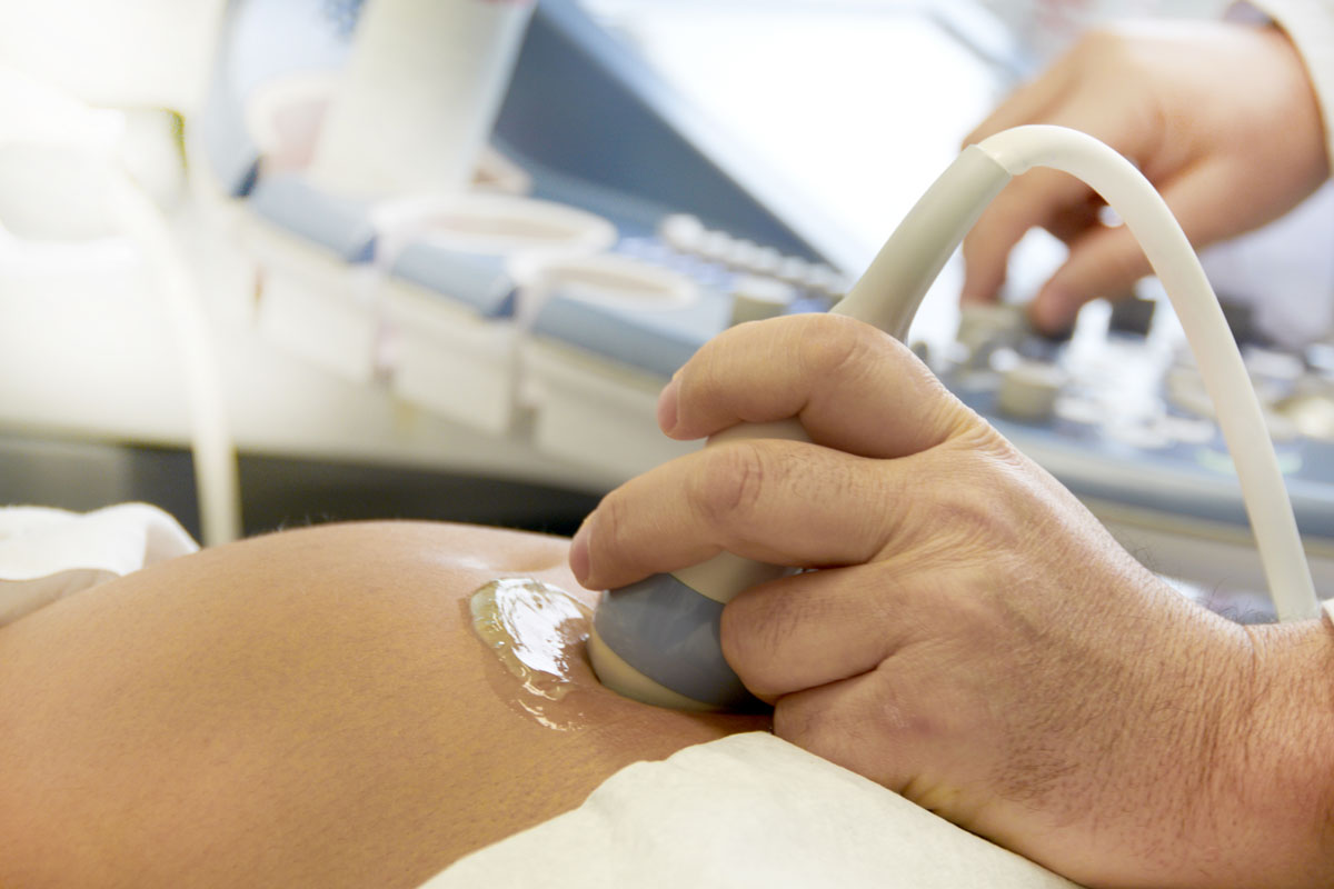
At present, thanks to the development of ultrasound examinations, doctors can make a dangerous diagnosis of a hematoma in the uterus quite accurately and simply. Modern devices have a fairly high resolution. With their help, even small accumulations of blood can be identified. Ultrasound helps doctors identify even complex pathologies that were previously impossible to identify in a timely manner.
The main method for diagnosing various hematomas localized in the uterus is ultrasound.
Main method
Reviews of many women who have already become mothers, but are faced with the appearance of hematomas in the uterus during pregnancy, indicate that the diagnosis was made after the ultrasound. In most cases, a hematoma in the uterus is determined during scheduled or screening examinations. It often happens that with this pathology a woman does not develop any discomfort symptoms. In such cases, a hematoma is detected only during screening. Quite often retroamnal hematoma is diagnosed at 11-13 weeks of pregnancy.
Modern ultrasound machines not only help to accurately determine the localization and size of the hematoma, but also reveal the development of dangerous complications. An ultrasound specialist can quite easily determine the onset of placental abruption — a dangerous pathology that can contribute to premature birth or even intrauterine fetal death.
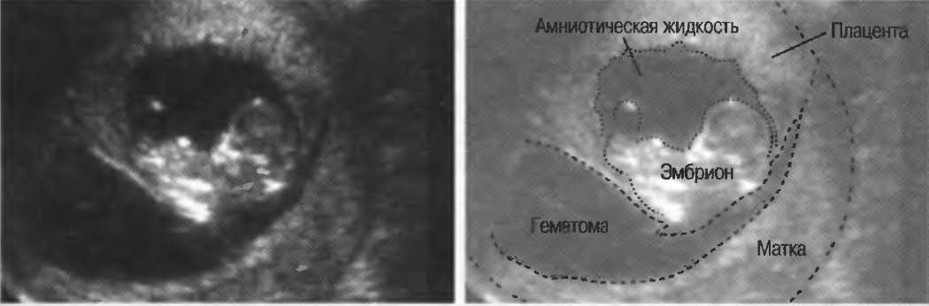
The ultrasound specialist during the analysis determines the localization of the ovum, as well as any deformation. He assesses the thickness of the uterine walls. Thickening of the uterus machines can be a sign of hematoma.
In conducting the study, the doctor necessarily assesses not only the general development of the fetus and the state of the reproductive organs of the woman. He studies the structure of the placental sheaths or placenta. Such a detailed diagnosis and allows the ultrasound doctor to identify all defects and damage, as well as accumulations of blood between the membranes.
Many doctors believe that the prognosis of pregnancy depends on the size of the hematoma. It can have a variety of sizes. The prognosis for a hematoma with a size of 14 mm is usually better than for a hematoma in 60-70 mm. The development of pregnancy largely depends on the location of the hematoma in the uterus, as well as on the state of the body of the pregnant woman.
If the expectant mother has no concomitant chronic diseases, and the pregnancy proceeded well before the appearance of a hematoma, then the probability of a favorable outcome in this case is quite high.

Auxiliary surveys
An important study that helps assess the severity of functional disorders arising from a hematoma in the uterus is doppler sonography. This diagnostic method allows you to evaluate the blood flow in the blood vessels feeding the fetus. Very often with hematoma localized in the uterus, intrauterine hypoxia develops, a condition in which a sufficient amount of oxygen does not enter the children's body. This pathological condition contributes to the fact that the formation of internal organs in a child can slow down. In such a situation, the risk of developing dangerous anomalies is quite high.
Through dopplerography, doctors can quite easily assess the indicators of uteroplacental blood flow and, if necessary, correct them (by prescribing medication). Doppler studies of women with hematoma in the uterus may be performed several times during pregnancy. This is necessary so that doctors can assess the dynamics of the development of pathology, as well as, if necessary, correct the chosen tactics of pregnancy.
Doplerography is an absolutely painless method. Expectant mother does not need to conduct any special training. All the obtained parameters of blood flow, the doctor must fix in his conclusion, which is glued to the medical card of a pregnant woman.
Hematoma in the uterus may be accompanied by the development of a number of complications. In order not to miss them, doctors resort to the appointment of some tests. Simple laboratory tests also allow you to assess the degree of violations, and therefore - to choose the right regimen of therapy.
The following tests may be given to the expectant mother, who has a hematoma in the uterus during pregnancy:
- general clinical trials (general blood and urine tests);
- biochemical test with blood clotting;
- removal of material from the vagina and genital tract (smear) for bacteriological examination;
- determination of hormone levels.

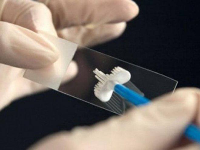
In each case, the list of tests may vary. The examination scheme is selected individually by an obstetrician-gynecologist.
If necessary, the doctor may refer the expectant mother to other doctors (for the purpose of additional consultations). If the hematoma in the uterus is complicated by the development of disorders in the blood coagulation system, the pregnant woman is referred for consultation to a hematologist. This specialist will conduct a more advanced diagnosis of the resulting pathological condition and select the necessary therapy for the correction of the developed functional disorders.

Therapy
The choice of treatment tactics for hematoma localized in the uterus depends on different conditions. Doctors necessarily evaluate the general condition of the pregnant woman and the fetus, determine the possible risk of complications, take into account the duration of pregnancy. Treat future mother can both in a hospital, and it is out-patient. It depends on how strong the disturbances are.
When hematoma in the uterus, which is almost asymptomatic and is not accompanied by the appearance of any adverse symptoms, doctors usually conduct outpatient monitoring. At the same time, the expectant mother is told that she should limit classes in the gym and visits baths or saunas. It is also strictly forbidden for a woman to lift heavy objects. In some cases, doctors prescribe and sexual rest. It is very important that the expectant mother should eat properly and consume enough protein foods, and also get enough sleep.
It often happens that women do not listen to these recommendations, considering them too banal and ineffective.However, such a frivolous attitude towards one’s own health and an unborn child can be very dangerous and even contribute to the aggravation of pathology.


If the doctor recommended the expectant mother to relax more and be more attentive to her well-being, then these recommendations should be heeded. The appearance of adverse symptoms (bloody vaginal discharge, abdominal pain) should be the reason for an emergency visit to the doctor. The future mother should pay attention to how she looks. Sometimes a hematoma in the uterus may be complicated by the development of bleeding, in which case the woman may develop anemia. Anemic syndrome is manifested by pallor and dry skin.
If the expectant mother noticed that her skin became too pale and even acquired a bluish tint, then she should also definitely see her obstetrician and tell him about the appearance of this symptom.
Hematoma therapy in the uterus with the use of drugs is selected individually. In this case, the duration of pregnancy is taken into account. In the early stages, hormonal drugs are often prescribed, which contain an analogue of the female sex hormone - progesterone. They have a positive effect on the uteroplacental blood flow, reducing the negative manifestations of pathology.

To reduce the amount of bleeding, doctors may prescribe special medications. Acceptance of such drugs contributes to the fact that the hematoma in the uterus gradually resolves.
Some doctors prescribe for this purpose the drug "Wobenzym". He is appointed in tablets. The course dosage and duration of treatment is chosen by the doctor, who is leading a specific pregnancy and is aware of its features.
In some cases, when a hematoma in the uterus appears pain in the abdomen. In the early stages, pain is usually localized in the lower abdomen. It may extend to the lumbar region. To relieve pain, doctors resort to the appointment of antispasmodics. They can be administered in the form of tablets, intramuscular injections or rectal suppositories (suppositories).


Multivitamin complexes can also improve the general condition of the future mother and baby. It is imperative that they contain folic acid. Such drugs are usually prescribed from the very early stages of pregnancy (and for quite a long time).
Auxiliary means for hematomas localized in the uterus are sedatives (sedatives). They are assigned to women who have a rather high anxiety or a strong tendency to experience. Sedatives help the expectant mother cope with the depressed state, and also improve sleep. Doctors give preference during pregnancy to products containing herbal ingredients. Often prescribed drugs containing motherwort or valerian.
About hematomas in early pregnancy, see the following video.


