What is the placenta, when it is formed and what functions it performs?
During pregnancy, unique anatomical structures and even new organs appear in the female body. One of them is the placenta. Without it, it is impossible to imagine the development of the baby in the womb. This article will talk about what a placenta is, how it is formed and what functions it performs.
Characteristic
The placenta is a special embryonic organ. It is characteristic not only for humans, but also for other mammals. The appearance of the placenta in the female body is impossible to imagine without the chorion.
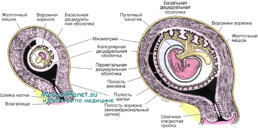
Its formation begins to occur after the fertilized egg is implanted into a specific wall of the uterus. Subsequently, a specific formation appears around it, which can be called a chorion. Later, its membranes begin to transform and are transformed into placental tissue.
Scientists have found that for the first time chorion appears in the body of a pregnant woman within 7-12 days after fertilization. Transformation into the placenta takes some time. On average, it is a few weeks. For the first time, placental tissue appears only at the beginning of the second trimester of pregnancy.
The name of the placenta acquired not by chance. This specific organ, which is formed only during pregnancy, has been known to doctors since antiquity. Agree that it is not difficult to notice it. During childbirth, after the birth of the baby, the placenta is born. This feature contributed to the fact that the placenta has long been called the afterbirth. It should be noted that this name has survived to the present.
In Latin, the term "placenta" is translated as "cake." This name almost completely characterizes the appearance of the placenta. It really looks like a cake. Often, doctors call the placenta also a “place for children”. Such a term is quite often used even in the medical literature.


Structure
Placenta pregnant has a heterogeneous structure. In fact, it is a unique body that must perform a huge variety of different functions. Any violations in the structure of the placenta can be very dangerous due to the development of pathologies. The presence of defects in the structure of the placental tissue causes a violation of the course of normal fetal development.
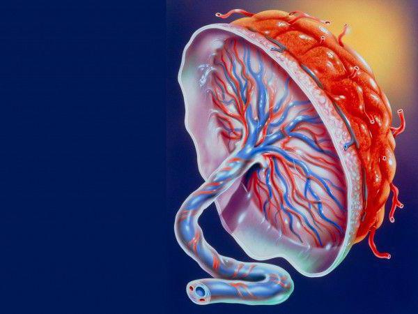
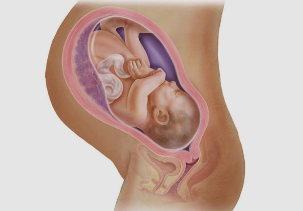
For reliable attachment to the walls of the uterus, the placenta has special projections, villi. Through them, and there is a reliable fixation of placental tissue to the wall of the uterus. This feature also determines the interaction between the small embryo, the placenta and the endometrium.
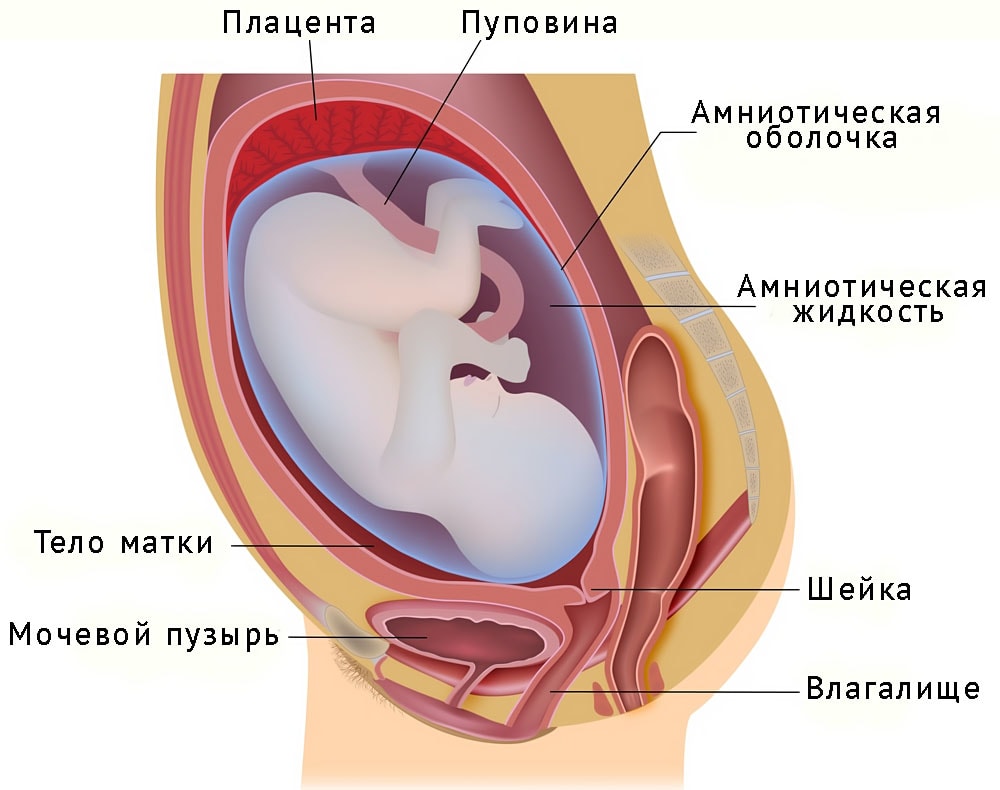
The umbilical cord is between the placenta and the fetus - it is a special organ that, in fact, connects the baby with his mother at the biological level. Such a unique relationship will persist until the very birth. Only after the birth of the baby, the umbilical cord is cut, which means the birth of a new person.

In the umbilical cord are important blood vessels - arteries and veins. Outside they are surrounded by a special substance - "Varton jelly." It has an interesting texture that resembles jelly.The main goal of this substance is reliable protection of umbilical cord blood vessels from exposure to various negative environmental factors.
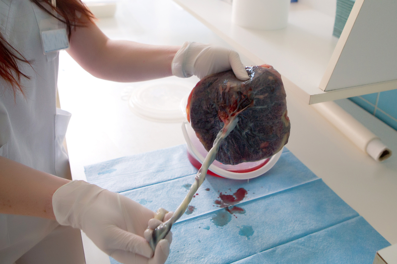
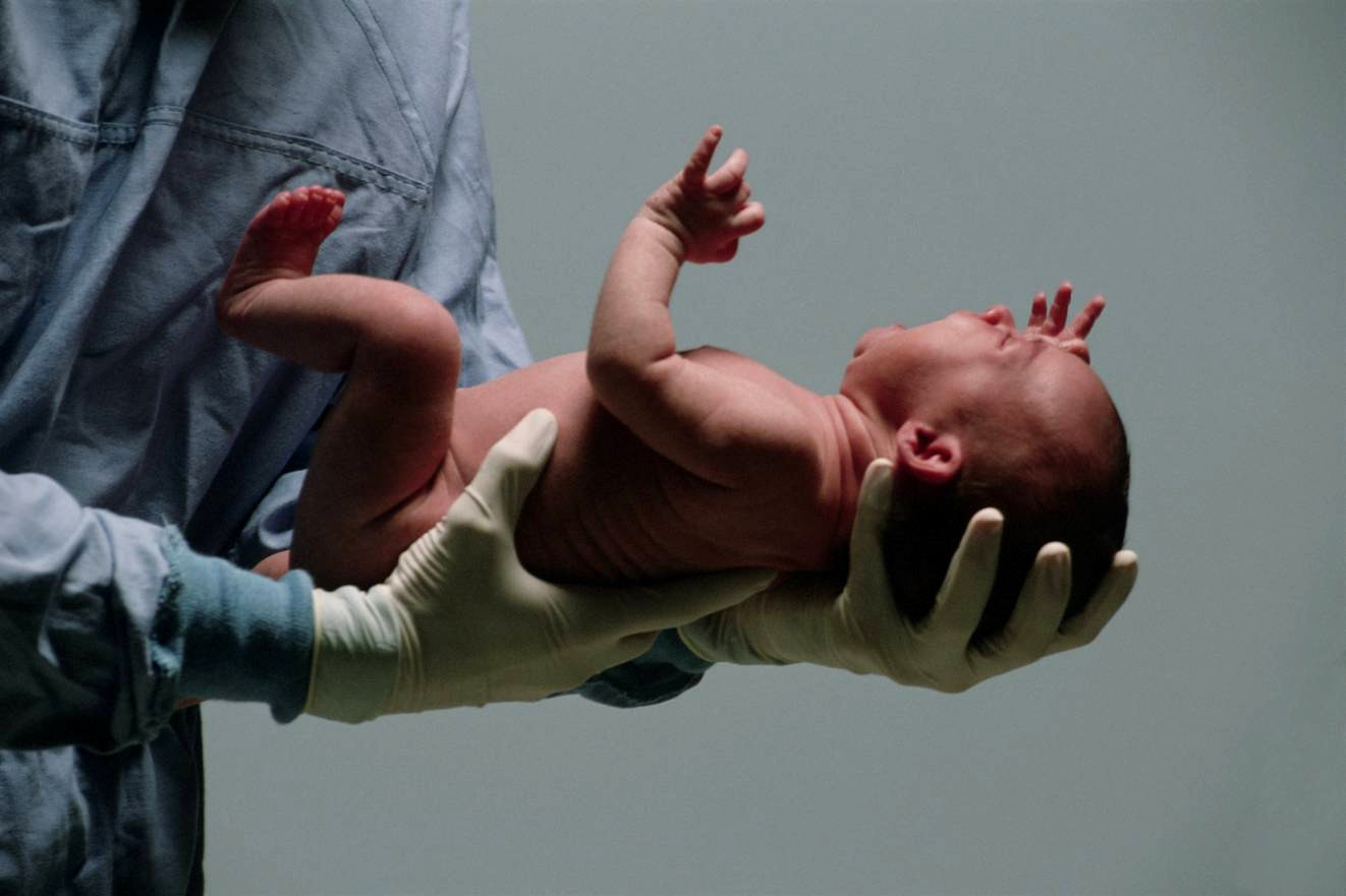
During the normal course of pregnancy, the placenta is retained in the female body throughout pregnancy. Her birth occurs after the birth of the baby. On average, the placenta is born 10-60 minutes after the baby is born. The difference of this time interval in different genera depends on many factors.
The entire placental tissue can be divided into 2 parts - maternal and fetal. The first one is attached directly to the uterine wall, and the second one - to the fetus. Each of the parts of the placenta has a number of unique anatomical features.
Mother part
This zone of the placenta is formed largely on the basis of the decidual membrane, and more precisely, its basal part. This feature determines the particular density and structure of the maternal part of the placenta. The surface of this area of placental tissue is rather rough.
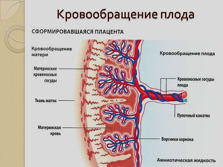
The presence of special partitions that are present in the placenta, provides separation of maternal and fetal blood flow. The placental barrier prevents the mother and fetus from mixing blood at this stage. A specific "exchange" begins to occur somewhat later. This is due to the active process of osmosis and diffusion.
Fruit part
This part of the placenta is covered with a special amniotic layer. Such a structure is necessary so that later in the uterus, a special aquatic environment will be formed in which the baby will “live” for several months of its intrauterine development.
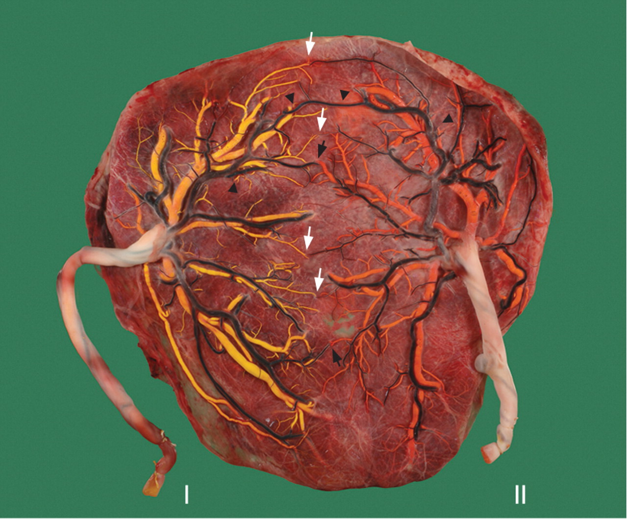
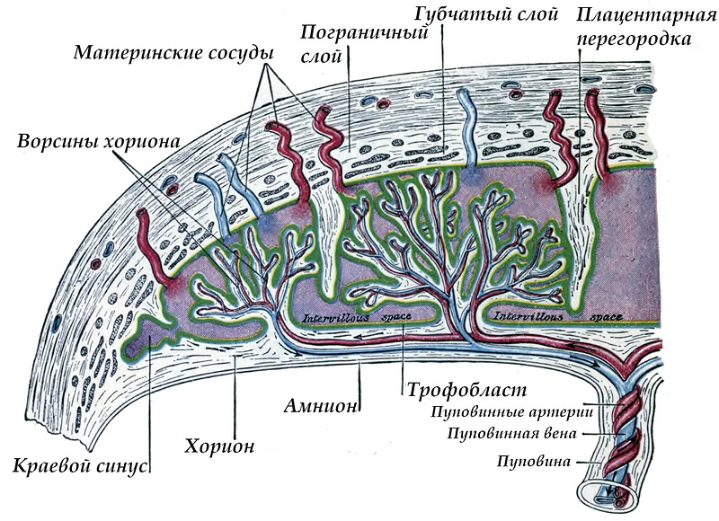
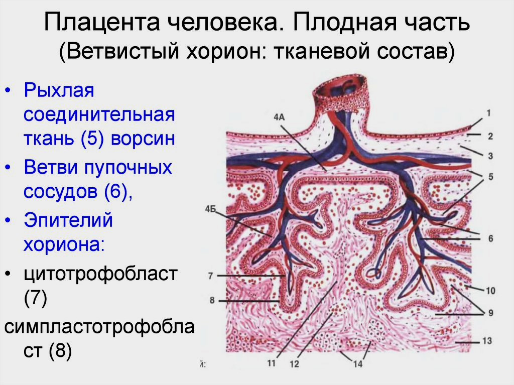
On the fetal side of the placenta there is a special chorionic formation, which ends in numerous villi. These villi are involved in the formation of an important element - intervillous space.
Some of the villi are called anchoring, as they are tightly fixed to the uterine wall, providing reliable fixation. The remaining outgrowths are directed into the intervillous space, which is filled with blood from the inside.
Decidual septa (septa) divide the surface of placental tissue into several separate parts — cotyledons. They can be called structural-anatomical units of the placenta.
The number of cotyledones changes as the placenta matures. When it finally matures, the total number of such structural-anatomical structures is several dozen.

Cotyledon
The main component of the placenta resembles in appearance the bowl. Each structural-anatomical unit of placental tissue has a large branch of the umbilical blood vessel, which branches into several small branches.
This structure provides a very important function of the placenta - the blood supply to the fetus with all the necessary substances for its growth and development. Abundant circulatory reticulum, which covers the cotyledon, provides blood flow in each individual area of placental tissue. This helps ensure uninterrupted blood supply not only to the placenta itself, but also to the body of an actively developing baby.
How is blood supply provided?
This question is very important, because without uninterrupted blood flow, the functioning of the placenta is impossible. The uterus, in which the baby develops, is nourished by the ovarian and uterine arteries. It is their doctors that are called spiral vessels. The branches of the ovarian and uterine arteries are in the intervillous space.
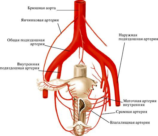
It is important to note that there is a pressure difference between the spiral vessels and the intervillous space. This feature is necessary for gas exchange and nutrient supply to occur. The difference in pressure contributes to the fact that blood from the arteries penetrates to the villi, washes them and then moves to the chorial plate.Then she gets into the maternal veins.
This feature of the blood flow provides a certain permeability of placental tissue. It is believed that the ability to penetrate various nutrients and oxygen gradually increases with each subsequent day of pregnancy. By 32-34 week, the permeability of the placenta is maximum. Then it starts to decrease gradually.
Weight
During pregnancy, the size of the placenta is almost always changing. Thus, by birth a healthy placenta weighs about 0.5-0.6 kg on average. Its diameter in most cases ranges from 16 to 20 cm.
The thickness of the afterbirth may be different. This largely depends on the individual characteristics, as well as on whether there are any pathologies of the formation of this organ. With each subsequent day of pregnancy, the thickness of the placenta increases.
Doctors believe that this increase ends only at 36-37 weeks of pregnancy. On average, after birth, the thickness of the normal placenta is approximately 2-4 cm.

Type of
Human placental tissue has a number of features that distinguish it from the placenta of other mammals. The human placenta is of the hemorrhoid type. This type of placental tissue is characterized by the possibility of the circulation of maternal blood around the villi in which the fetal capillaries are located.
This structure of the placenta has interested many scientists. Already at the beginning of the 20th century, Soviet scientists conducted a series of scientific studies and made interesting developments based on the properties of placental tissue. Thus, Professor V.P. Filatov developed special pharmaceutical preparations that contain in their chemical composition an extract or suspension of the placenta.
Currently, science has advanced greatly. Scientists have learned to work actively with the placenta. Stem cells, which have a number of important functions, are isolated from it. There are even cord blood banks where they are stored. The storage of stem cells requires certain conditions and responsible observance of a number of strict sanitary and hygienic rules.

For many years, scientists believed that the human hemochoric placenta is a sterile organ. However, numerous scientific studies have rejected this. Even in a healthy placenta, some microorganisms are found after childbirth, many of which live in the oral cavity of a pregnant woman.
How is it formed?
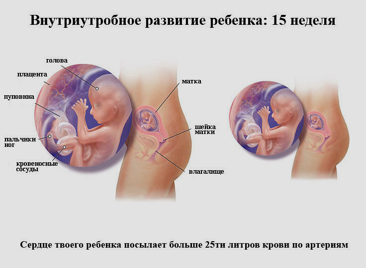
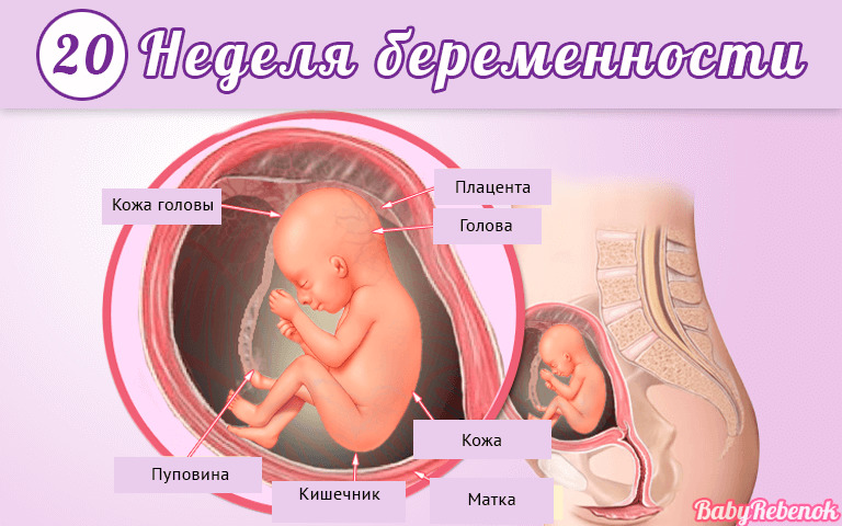
Placenta formation is a complex biological process. Scientists believe that the placenta is actively formed at 15-16 weeks of pregnancy. However, the term of the final development of the body may be different. So, only in the 20th week of pregnancy, the blood vessels start to function actively in the placental tissue.
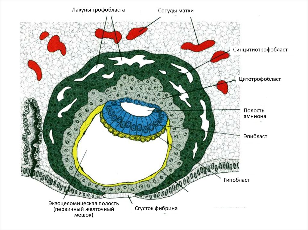
In most cases, the placenta is formed in the posterior wall of the uterus. Placental tissue is formed with the participation of a special embryonic formation - cytotrophoblast and the endometrium itself (the inner lining of the uterine wall).
The final histological structure of the placenta became known to doctors relatively recently - in the era of microscopic examination. In placental tissue, scientists distinguish several consecutive layers:
- Decidua - the first layer in the direction from the uterus to the embryo. In fact, it is a modified endometrium.
- Lanthans Layer (Rohr fibrinoid).
- Trophoblast. This layer covers the lacunae and grows into the walls of the spiral arteries, which prevents their active contractions.
- Numerous lacunaethat are filled with blood.
- Multicore simplastlining cytotrophoblast (syncytiotrophoblast).
- Cytotrophoblast layer. It is a layer of located cells that form syncytium and produce the formation of certain hormone-like substances.
- Stroma. It is a connective tissue in which the blood supply vessels pass.Also in this layer are very important cellular elements - Kashchenko-Gofbauer cells, which are macrophages and provide local immunity.
- Amnion. Participates in the subsequent formation of amniotic fluid. It is necessary for the formation of a special aquatic environment in which the prenatal development of the baby will occur.
A very important structural element of the placenta is its basal decidual membrane. It is a kind of barrier between the maternal and fetal part of the placenta. In the area of the basal decidual membrane there are numerous cavities within which maternal blood is present.
Functions
Placenta during pregnancy plays a very important role. The number of functions performed by this body is quite large. One of the most important of these is the protective or barrier function. The placenta is involved in the formation of the hemato-placental barrier. It is necessary in order to prenatal development of the fetus was not impaired.
The participation of the hemato-placental barrier involves the following anatomical units:
- endometrial cell layer (inner wall of the uterus);
- basement membrane;
- loose pericapillary connective tissue;
- trophoblast basement membrane;
- cellular layers of cytotrophoblast;
- syncytiotrophoblast.
Such a complex structure is necessary so that the hematoplacental barrier provides important functions of the placenta. Disturbance of the histological structure can be dangerous. In such a situation, the placental tissue simply cannot fully function.
Participation in gas exchange
Through the blood vessels, which are in large quantities in the placental tissue, the fetus receives oxygen, and also "gets rid" of carbon dioxide.
This happens through ordinary simple diffusion. At the same time, oxygen penetrates the body of the actively growing baby, and the exhausted carbon dioxide is released. This kind of "cellular respiration" occurs throughout the entire period of pregnancy. This unique mechanism develops due to the fact that the lungs of the fetus are formed rather late.
A baby in the womb does not breathe on its own. He will take his first breath only after birth. In order to compensate for this condition, such cellular gas exchange takes place.
Power supply
Despite the fact that a baby has a mouth formed as well as organs of the digestive system by a certain period of pregnancy, it cannot take food on its own. All the nutritional components that are necessary for the child's body for his birth, he gets through the blood vessels. Proteins, fats and carbohydrates enter the body of the baby through the arteries of his mother. In the same way, the baby receives water, vitamins and trace elements.
This feature of the fetal nutrition clearly explains why the diet of a pregnant woman is very important. For the full intrauterine development of the fetus, the expectant mother should carefully monitor what foods she consumes during the day.
It is very important that fresh fruits and vegetables are regularly present in the diet of a pregnant woman, as well as high-quality protein sources.

Isolation of unnecessary products exchange
The kidneys and the excretory system of the fetus begin to function quite late. While they are not well formed, the placenta comes to the rescue. Through placental tissue, the removal of unwanted metabolites worked out by the child’s body occurs. Thus, the body of the fetus "gets rid" of excess urea, creatinine and other substances. This process takes place through active and passive transport.
Hormone synthesis
Hormonal function of the placenta, perhaps, is one of the very important. During pregnancy, the placental tissue is even an organ of internal secretion, as it participates in the formation of biologically active substances.
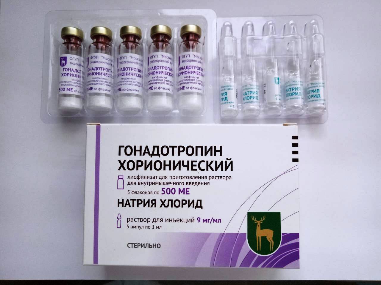
One of them is the most important hormone of pregnancy - chorionic gonadotropin. It is necessary for the normal course of pregnancy. This hormone ensures the proper functioning of the placenta, and also stimulates the formation of progesterone in the body of a pregnant woman. It is necessary during pregnancy in order to stimulate the growth of the endometrium and temporarily stop the maturation of new follicles in the ovaries.
Under the participation of the placenta, placental lactogen is also formed. This hormone is necessary in order to prepare the mammary glands for the upcoming changes - lactation. Under the influence of the placenta, the formation of another hormone necessary during pregnancy - prolactin. It is also necessary in order to prepare the mammary glands of the future mother for the upcoming lactation.
Scientists have identified that placental tissue can synthesize several other hormones - testosterone, relaxin, serotonin, and others. In addition to the active synthesis of hormones, placental tissue is involved in the formation of hormone-like substances, which are necessary for the normal course and development of pregnancy.
Fetal protection
This function of the placenta can be divided into several types. So, it can be mechanical and immune. Each of them is very important in the period of fetal development.
Mechanical protection of the fetus implies the protection of the child's body from any environmental influences. Placental tissue is a very delicate structure. It is located in close proximity to the fetus. With various injuries, the placenta "softens" the blow. This helps reduce the risk of damage to the fetus.


The immune protective function of the placenta is that the placenta is involved in providing the child's body with maternal antibodies. These special substances provide immunity of the fetus throughout its prenatal life in the womb.
Antibodies that enter the body of the baby from his mother through the blood, are immunoglobulins. Some of them calmly penetrate the placenta, getting into the children's body. Thus, the placenta helps to protect the baby from a number of bacterial and viral infections.
The entry of maternal antibodies also contributes to the prevention of the immunological conflict between the mother and the fetus. In this case, the maternal organism does not perceive the fetus as an alien genetic object. This feature helps to prevent rejection of the fetus from the uterus during the entire pregnancy.

It should be noted about the special role of syncytium - a special element of the placental tissue. It is involved in the absorption of a number of hazardous chemicals that can cross the placenta from the mother to the fetus. Thus, the placenta as it protects the baby’s body from the penetration into it of dangerous narcotic, toxic and other dangerous means.
It is important to remember that such selectivity of penetration can be individual. If the histological structure of the placenta is normal, then hazardous substances linger. If it is broken, then toxins and poisons can easily penetrate into the children's body, causing him irreparable harm. That is why doctors recommend expectant mothers during pregnancy to give up all bad habits.
Smoking and drinking alcohol, as well as drugs can cause the development of dangerous diseases in an actively developing fetus. It is much easier to prevent their development than to continue to try to cope with the pathologies that have arisen.
Maintaining a healthy lifestyle of the future mother is of great importance in the formation and normal functioning of the placenta.

Migration
The initial position of the placenta in the uterine cavity is a very important clinical indicator. Even during the pregnancy depends on how it will be located.
Usually, placental tissue is attached to the back or front wall of the uterus.Very rarely, it is attached only to one of the side walls. Bookmark placental tissue begins in the first trimester of pregnancy and is associated with the site of implantation of a fertilized egg.
Normally, a fertilized egg attaches to the bottom of the uterus. In this zone, there is a good blood flow, which is necessary for the full intrauterine development of the fetus throughout pregnancy. However, this situation does not always develop.
In obstetric practice, cases are recorded where the implantation of a fertilized egg occurs in the lower uterus. This is preceded by a huge number of various reasons. In this case, the fertilized egg can sink almost to the base of the internal uterine throat, where it attaches to the uterine wall.
The lower the implantation occurs, the lower the placenta is located. The growth of placental tissue in the area of the internal uterine throat doctors call previa. This dangerous pathology significantly worsens the course of pregnancy and may even cause the development of dangerous complications.

The initial location of the placental tissue may change. This most often occurs when the placenta is attached to the anterior wall of the uterus. The process of changing the initial localization of placental tissue is called migration. The displacement of the placenta in this case, as a rule, occurs from bottom to top. Thus, if the low position of the placental tissue was detected in the first half of pregnancy, then it may still change.
Usually, the migration of the placenta takes place rather slowly - within 6-10 weeks. It ends completely, as a rule, only by the middle of the third trimester of pregnancy.
The placenta, located on the back of the uterus, practically does not migrate. The probability of displacement of placental tissue in this position is extremely small. This is largely due to certain features of the structure of the uterus.
Norm
A healthy placenta is an important component of the normal course of pregnancy. The development of this unique organ of pregnancy occurs gradually. From the moment of formation in the female body to the birth, the placenta is almost constantly changing.
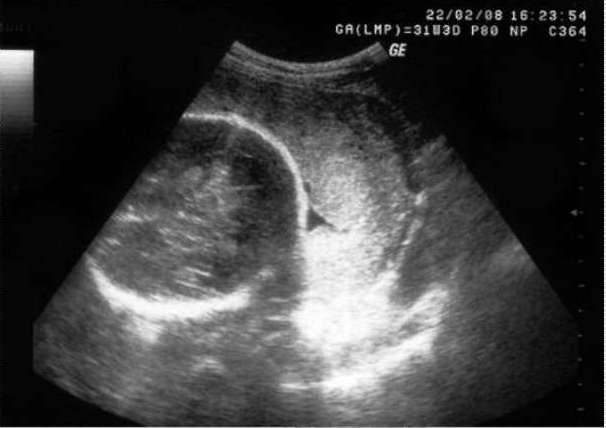
Doctors can evaluate the anatomical properties of the placenta, as well as identify various anomalies in its development, by performing ultrasound examinations. To do this, throughout the pregnancy, the expectant mother must undergo several ultrasounds.
With the help of modern devices, specialists can get a fairly clear visualization of placental tissue. During the ultrasound examination, the doctor may see the structure of the placenta, the presence of any diffuse changes in it, as well as emerging pathologies.

A very important clinical indicator, which must be determined by obstetrician-gynecologists during pregnancy, is the maturity of the placenta. At each stage of pregnancy, it varies. It's quite normal. At the same time, it is important to assess the compliance of placenta maturity with a specific period of pregnancy.
So, experts identify several options for maturity of placental tissue:
- Zero (0). Characterizes the normal structure of the placenta until approximately 30 weeks of gestation. The placenta of such maturity has a rather smooth and even surface.
- First (1). Characteristic of a healthy placenta in the period from 30 to 34 weeks of pregnancy. At maturity of the first degree, specific blotches appear on the placenta.
- The second (2). Formed normally after 34 weeks of pregnancy. Such placental tissue looks more prominent, specific striations appear on it, as well as small grooves.
- The third (3). It is the norm for a normal full-term pregnancy.The placenta, which has such a degree of maturity, has on its surface quite pronounced large waves that reach the basal layer. Also on the outer surface of the placental tissue appear merging with each other spots, having an irregular shape - salt deposits.
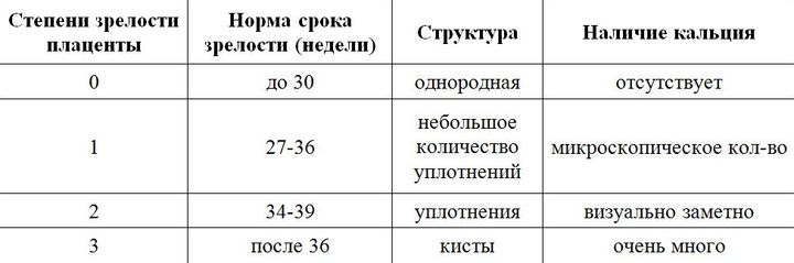
Determining the degree of maturity of the placenta allows doctors to orient in the term of the upcoming delivery. In some cases, the placental tissue matures too quickly. This leads to the development of a number of dangerous complications. In this case, the tactics of pregnancy must be revised by experts.
Pathologies
Unfortunately, abnormalities in the development and formation of the placenta occur in obstetric practice quite often. Such conditions significantly worsen the prognosis of pregnancy. Emerging defects in the structure of the placenta and contribute to the deterioration of blood flow, which is necessary for the full intrauterine development of the baby.
Currently, there are quite a lot of different pathologies of the placenta. One of the most dangerous of them is a strong increment of placental tissue to the uterine wall. It would seem that the stronger the placenta “grows” into the endometrium, the more secure the fixation should be, but in fact this is not quite so.

A strong increment of the placenta to the uterine wall is dangerous development problems with its separation during childbirth. In such a situation, the birth of a child, as a rule, proceeds normally, and the birth of an afterbirth is delayed. Such a clinical situation can be dangerous by the development of massive uterine bleeding.
Also, the long-term presence of the afterbirth in the uterus is a threat to the development of infection of the reproductive organs.
With a strong increment of placental tissue to the uterus wall, surgical gynecological intervention is required. In this situation, the doctors purposefully separate the placenta from the uterine walls.
Quite often scars form on the uterus. This happens usually in cases where various surgical operations were performed on it - cesarean section, excision of damaged tissues and others. Scarring of connective tissue leads to scarring.
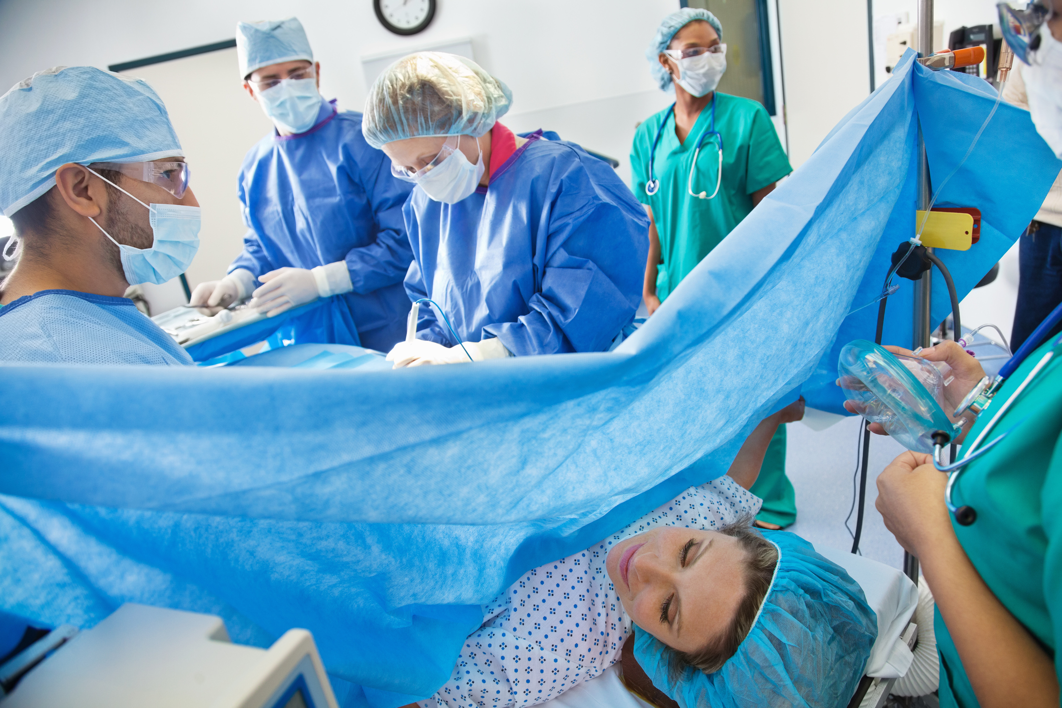
The growth of the placenta into the uterine scar is a rather dangerous pathology. In this case, dangerous complications may occur during natural childbirth. In order to avoid them, doctors often have to resort to performing surgical obstetric aid - cesarean section.
Strong descent of the placenta to the level of the internal uterine throat is dangerous development of its presentation. This pathology worsens the prognosis of pregnancy. With placenta previa, the risk of developing dangerous infectious diseases and premature birth is quite high. In order to preserve and prolong the pregnancy as much as possible, the expectant mother must strictly follow the recommendations made for her by the doctors.

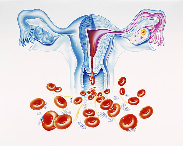
Placental abruption is another dangerous pathology that occurs in obstetric practice. It is characterized by the detachment of placental tissue due to certain reasons from the walls of the uterus. At the same time, as a rule, bleeding develops. If placental abruption occurs on a fairly large area, then this situation is extremely dangerous for the fetus. Massive detachment of placental tissue, accompanied by the occurrence of functional disorders in the children's body, can be an indication for an emergency cesarean section.
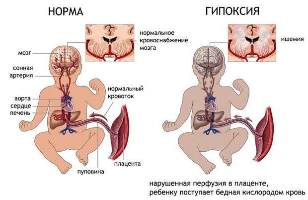
Another dangerous pathology is placental edema. The development of this condition can lead a variety of causes, including bacterial and viral infections. Prolonged edema of the placenta can lead to the development of placental insufficiency, fetal hypoxia, and also provoke premature labor. In identifying this pathology, doctors conduct a comprehensive treatment.
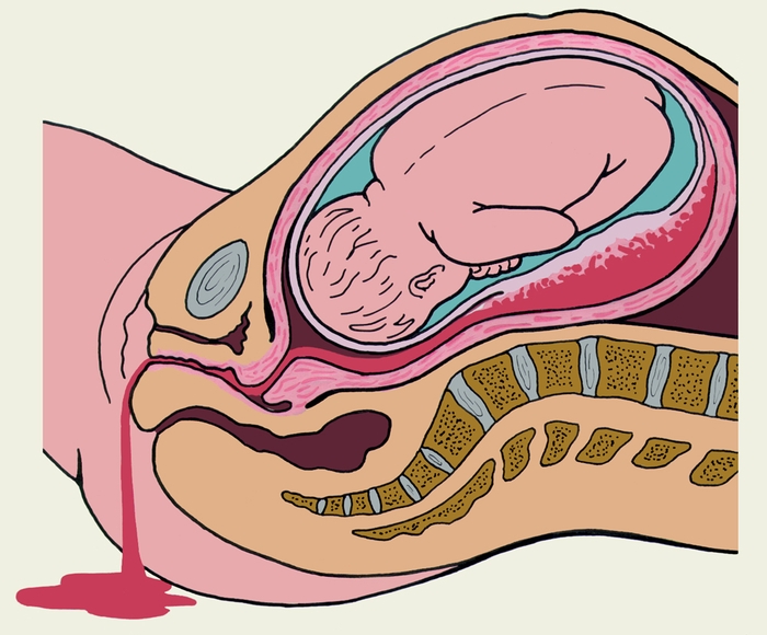

In the placenta is quite a lot of blood vessels. The placental tissue surrounding them is quite loose, tender.Strong mechanical effects can contribute to the fact that it appears small microdamages and even ruptures. As a rule, clinically such minor injuries do not manifest themselves for a long time.
If the breaks in the placental tissue are quite significant, it will contribute to the disruption of its functioning. In this case, the general condition of the fetus may be affected. Circulatory disorders can affect a baby’s heartbeat, as well as an increase in his blood oxygen deficiency.
Detection of defects and small hemorrhages in the placenta is possible only with the help of modern ultrasound examinations. Minor damage is usually determined retrospectively - after delivery during a visual inspection of the placenta.
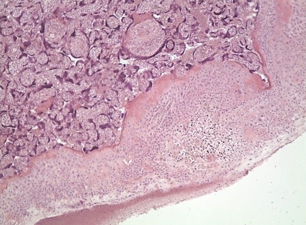
Structural changes can also be determined with the help of histological examination, which is performed after delivery. To conduct this survey, the afterbirth is sent to a special laboratory, where it is studied.

About what is the placenta, see the next video of Larisa Sviridova.


