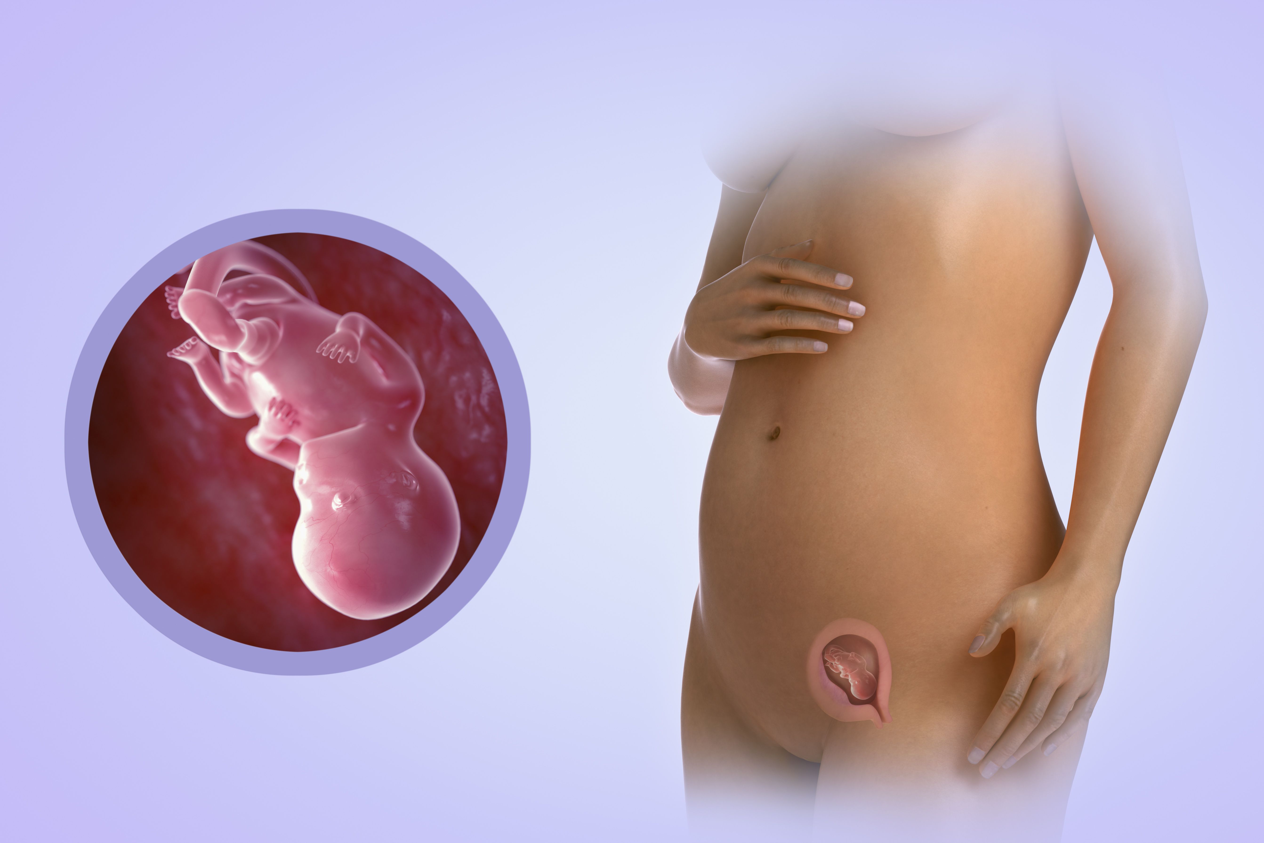
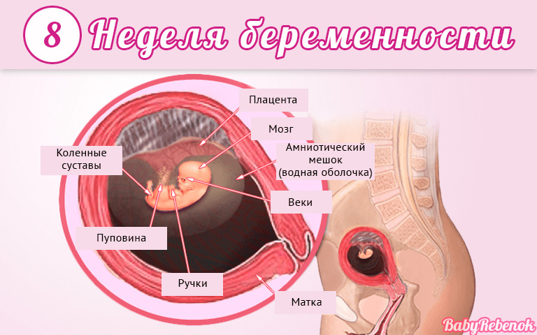
Fetal development in the 8th week of pregnancy

With each week of embryonic development, important biological changes occur in the baby’s body. The child is rapidly growing and developing.

How do doctors define term?
Doctors use in their daily practice a special system for calculating the age of the fetus. They measure in obstetric months and weeks. Thus, 8 obstetric weeks of pregnancy is equivalent to 6 weeks from the date of conception of the child.
Future moms more often use the calendar method. They calculate the age of the baby from the immediate date of conception.
To determine the obstetric period, doctors use the date of the first day of the last before the onset of pregnancy of the menstrual cycle. This method of calculation is more accurate and has already been used by doctors quite a lot. The entire period of childbearing in the obstetric method of counting is 280 days or forty weeks. It is best for future moms to use a system for calculating the child's age in common with doctors.

8 week of pregnancy is accompanied by the appearance of various sensations in a pregnant woman. She changes her mood and emotional background, there are nagging pains in her stomach. Many women start to get tired faster. Specific changes develop not only in my mother's body. In the first weeks of pregnancy, the child is actively developing the process of formation of internal organs.
Such a huge jump in intrauterine development is possible only during the first trimester of pregnancy. Several weeks have passed since the merger of the germ cells. During this time, a small organism has already formed from several cells.
What happens to the baby?
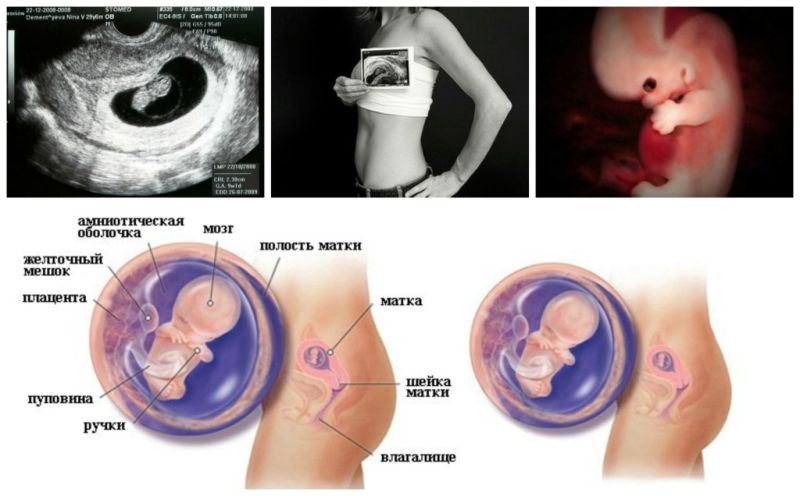
In this period of pregnancy, doctors call the child in her mother's abdomen an embryo. It is of such small size that it can only be determined by ultrasound examination. This period of intrauterine life is extremely important. At this time, the laying of all vital organs and systems is actively continuing. If it happens qualitatively, then the children's body will continue to function fully.
The size of the embryo at this stage is very small. It is 15 mm long. The weight of the child is very small: it is only 2 grams. In addition to the formation of the body, the internal organs begin to actively form in the embryo. Laying the kidneys, liver. A small embryo has already formed pancreas and stomach.
Interestingly, but even in this early period of pregnancy, the formation of gastric juice.
At this stage, the child has already formed a heart. Moreover, it is already beating. A small embryo weighing 2 grams has its own heartbeat. This is a true natural wonder.
The heart of the baby to this period of pregnancy has a four-chamber structure. Between the atria appears septum. Such a structure is normal for the entire human population.
The heartbeat of a child is an important clinical sign.It is determined during the ultrasound examination. To calculate it, the number of heartbeats per minute is determined. If the heart of the embryo beats too fast, in this case, the doctors set tachycardia. By reducing the number of contractions of the heart in a minute is determined bradycardia.
It is important that the heartbeat of the child remains within the normal range. This condition indicates that the cardiovascular system of the embryo functions well.
It is very important to regularly assess the baby's heartbeat over time. Significant deviations from the norm may indicate that various pathologies of the course of pregnancy are manifested in this way. This stage of embryonic development is also accompanied by the laying of the digestive and respiratory systems. Light will continue to be formed in the future. Their full functioning is possible only after the birth of the baby.
Transformed and bronchial tree. The bronchi forming it begin to branch out. In the future, their diameter and clearance will change.
Also, at 7-8 weeks of pregnancy, the child has a formation of the reproductive system. Sex organs are just beginning to form. But you can determine the sex of the fetus later. By this time of pregnancy the child has almost completely formed the nervous system. Also, by this period, the main anatomical elements of the brain were formed.
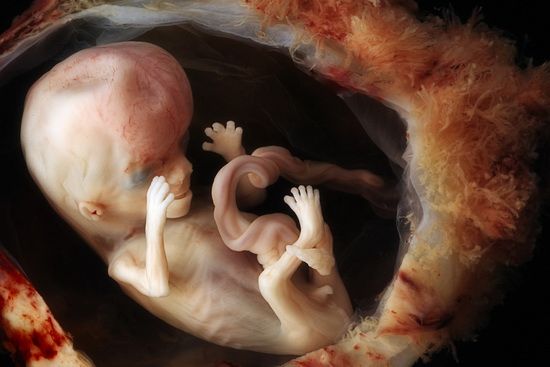
This stage of intrauterine development is accompanied by the active formation of the musculoskeletal system. The mobility of small children's joints is gradually increasing.
The child’s muscular apparatus is well developed, but this is difficult to notice due to the small size of the embryo. Not only the muscular apparatus of the body develops actively, but the smooth-muscular muscles of the internal organs are also formed.
The small embryo has a well developed chewing and facial muscles, muscles in the arms and legs, as well as the throats. The lower limbs at this period of intrauterine development are somewhat “lagging behind” the upper ones. The increase in dynamic functions leads to the fact that The baby’s nervous system continues to form and develop.. Gradually, the internal organs begin to respond to the effects of nerve impulses. This is necessary so that the digestive system continues to develop rapidly.
In the embryo, the diaphragm gradually begins to form. Glands of external secretion (sweat, salivary) continue their active development.
The first ancillary structures necessary for the development of the embryo begin to transform at this stage. The villi of the chorion gradually turn into the placenta. In the future, through the placenta, the baby will receive all the necessary nutrients for its intrauterine development. This stage is accompanied by the initial formation of the immune system. The child actively forms the thymus gland. In the future, this body of immunity will be filled with lymphocytic cells.
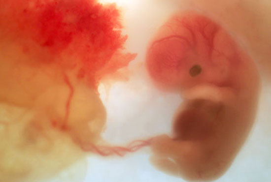
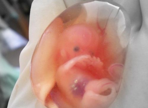
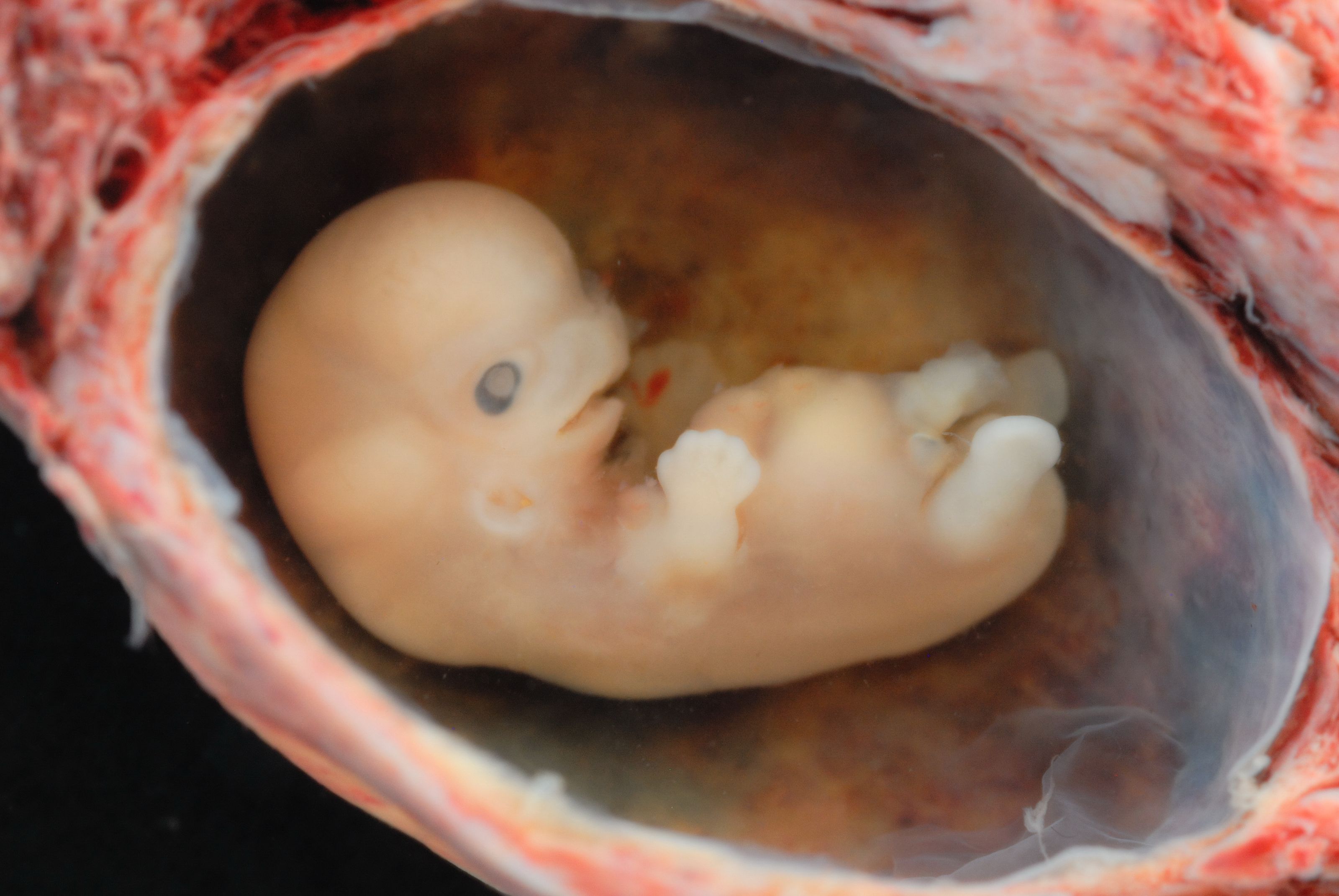
What does an embryo look like?
At such an early stage of pregnancy, a small embryo in its appearance already resembles a small person. His body has an elongated shape. Even the face of the fetus at this stage of its embryonic development is changing. Eyes become prominent. They look pretty big, because there are no eyelids. Also at this stage of gestation, the first beginnings of the retina and eye color pigments appear. Well visible eye sockets. The eyes are closer to the temporal zones of the children's head. In the future, the child’s face will be transformed, which will bring the eyes closer together.
The nose has a clearer contour, but still remains flat. Gradually begin to form the nerve endings of the olfactory analyzer. The nostrils of the baby are covered at this stage of its development with mucous plugs.In the oral cavity is an active formation of the gums. Even the beginnings of milk teeth begin to appear. Good contour of the upper lip.

In the language of the baby's taste buds are already formed. In the future, these nerve endings will change and improve, but the basic tab is already happening by this period.
Ears begin to form in a small embryo that is actively developing in the womb. The first rudiments of small toes appear on the arms and legs. At this stage, they are not yet merged with each other. After some time, they will separate. Active development of the nervous and musculoskeletal systems contributes to the fact that the joints become more mobile.
In some babies in this period of their development, during ultrasound, small movements in large joints can be seen. Usually this is manifested by small swinging movements with the hands.
In the future, the baby will develop and become more active.. With each week of pregnancy, the number of movements in a child increases. The child carries out small oscillatory movements in the mother's womb, but it is so small that the woman does not feel the movement activity of her baby during this period of pregnancy. By this period, the final formation of large blood vessels. They are necessary for the nutrition of the child during his intrauterine development.
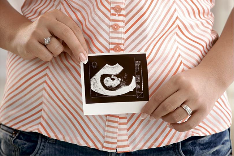
How to diagnose the development of the baby?
To determine the basic parameters of the fetus, doctors use ultrasound examination methods. They are assigned to all expectant mothers to confirm the fact of pregnancy, as well as the exclusion of ectopic form.
At this time, tubal pregnancy is a rather dangerous pathology. In this case, the development of the baby does not occur in the uterus, but in its appendages. In this case, the full development of the child is impossible. Tubal pregnancy always ends tragically. With the help of modern ultrasound, you can very accurately determine the location of the fetus. In multiple pregnancies, the position of each baby is assessed separately.
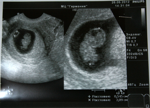
Despite the tiny size of the child, ultrasound doctors can determine the length of his body. So, The coccyx parietal size at this gestational age is approximately a couple of centimeters.
For the active and full development of the embryo necessarily need an aquatic environment. The diameter of the gestational egg, in which the amniotic fluid is located, at this stage is about 3 centimeters. When conducting ultrasound, the state of the reproductive organs of the mother is also evaluated. The structure of the uterus is evaluated, its tone is determined.
With the help of Doppler studies, specialists can determine the main indicators of blood flow in the main uterine blood vessels. Using this diagnostic method, one can assess how well the blood supply to the uterus is. Adequate blood flow is necessary so that the baby, who is in the womb, fully grew and developed.
To assess the growth of the fetus ultrasound examination is performed in the dynamics. In this way, the doctor can assess how well the baby is developing.


If a woman carries several children at the same time, then ultrasound may be required more. The multiplicity of their appointment is determined by an obstetrician-gynecologist.
Ultrasound on this gestational age can be done in different ways. The most commonly used is the transvaginal method. In this case, the study is carried out using a sensor that is inserted into the vagina. The transvaginal method of research at this stage of pregnancy allows to obtain more accurate and reliable results.. This technique has a number of contraindications. If they were identified, then transabdominal ultrasound is performed.
Many future mothers are worried about whether they will need any training before conducting a study. At this stage of pregnancy, as a rule, no special preparatory procedures are required. Often, future mothers begin to worry a lot before conducting a study. You can understand their excitement: they are very worried about the result and the presence of any pathologies in the child.

You should not worry: from the early weeks of pregnancy, the baby feels all the experiences of the mother. Calm and balanced mood during the diagnostic study is a must.
An ultrasound scan in the early stages of pregnancy allows for the timely detection of various pathologies in both the expectant mother and her baby. It is very important to carry out such an examination for women who have difficulty carrying babies and various gynecological diseases. If a woman has a burdened history of genetic and chromosomal diseases, then an ultrasound examination allows to detect their signs in the fetus in a timely manner.
About what happens to the pregnant woman and the fetus at the 8th week of pregnancy, see the next video.


