Ultrasound on the 8th week of pregnancy: fetal size and other features
8 week - an important period of pregnancy. There is an intensive development of internal organs, their tab has already been completed. The embryo is not at all like an embryo of an animal, every day acquiring new and new human features. That will show on this term ultrasound, we will tell in this article.

Purpose of the survey
Mandatory ultrasound at 8 obstetric week is not considered. Prior to the first scheduled screening first trimester, there are still about 3-4 weeks. However, even at this time the doctor may recommend a woman to undergo ultrasound diagnosis.
Many in this obstetric week for the first time turn to a consultation to get registered. Ultrasound may be necessary to clarify the pregnancy, if a woman, for example, does not know the exact period of conception or cannot remember the first day of her last menstruation.

There are other reasons why an expectant mother can be sent to the ultrasound diagnostic room at week 8:
- Confirm the fact of pregnancy, if on examination the obstetrician-gynecologist doubted this (the size of the uterus on palpation is too small).
- Make sure that the pregnancy is uterine, if earlier the woman had cases of ectopic pregnancy, miscarriages, missed pregnancies.
- Make sure that the pregnancy develops if the woman has spotting, pain.
- Clarify whether a woman has fibroids, cysts that could affect the delay in the absence of pregnancy.
- Find out the number of fetuses, if the woman already had multiple pregnancies, as well as in the case of high levels of hCG in the blood.
Survey method and preparation
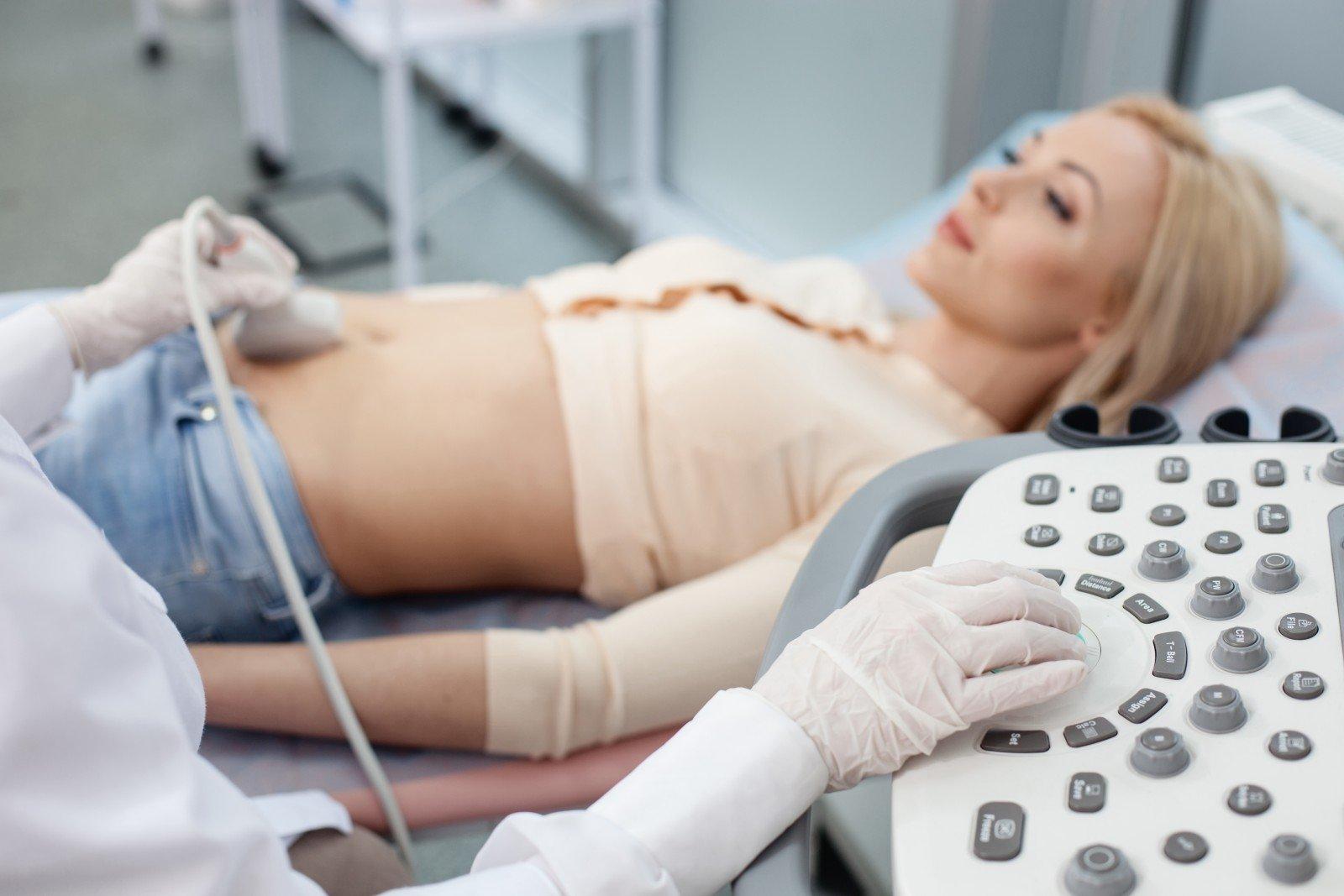
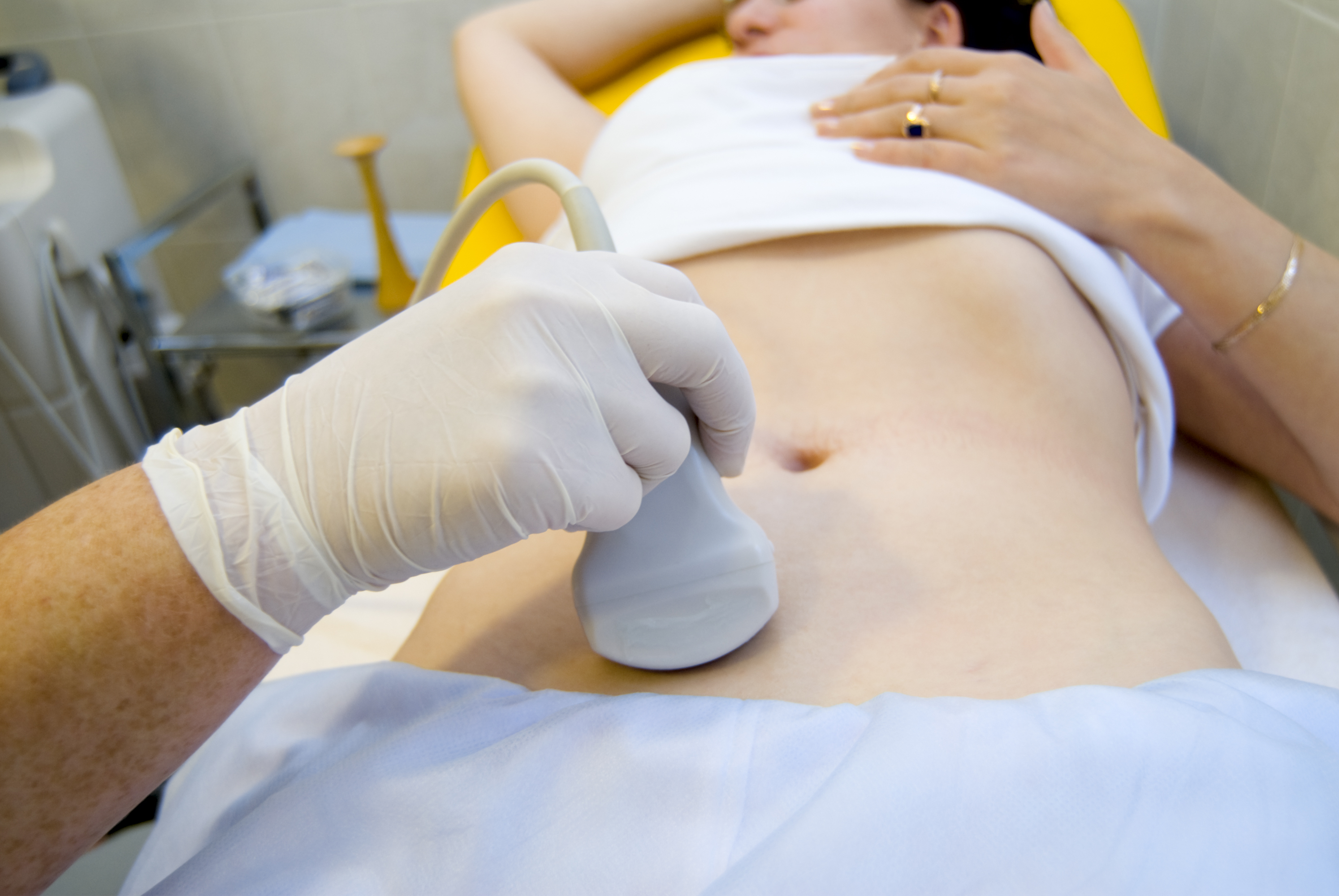
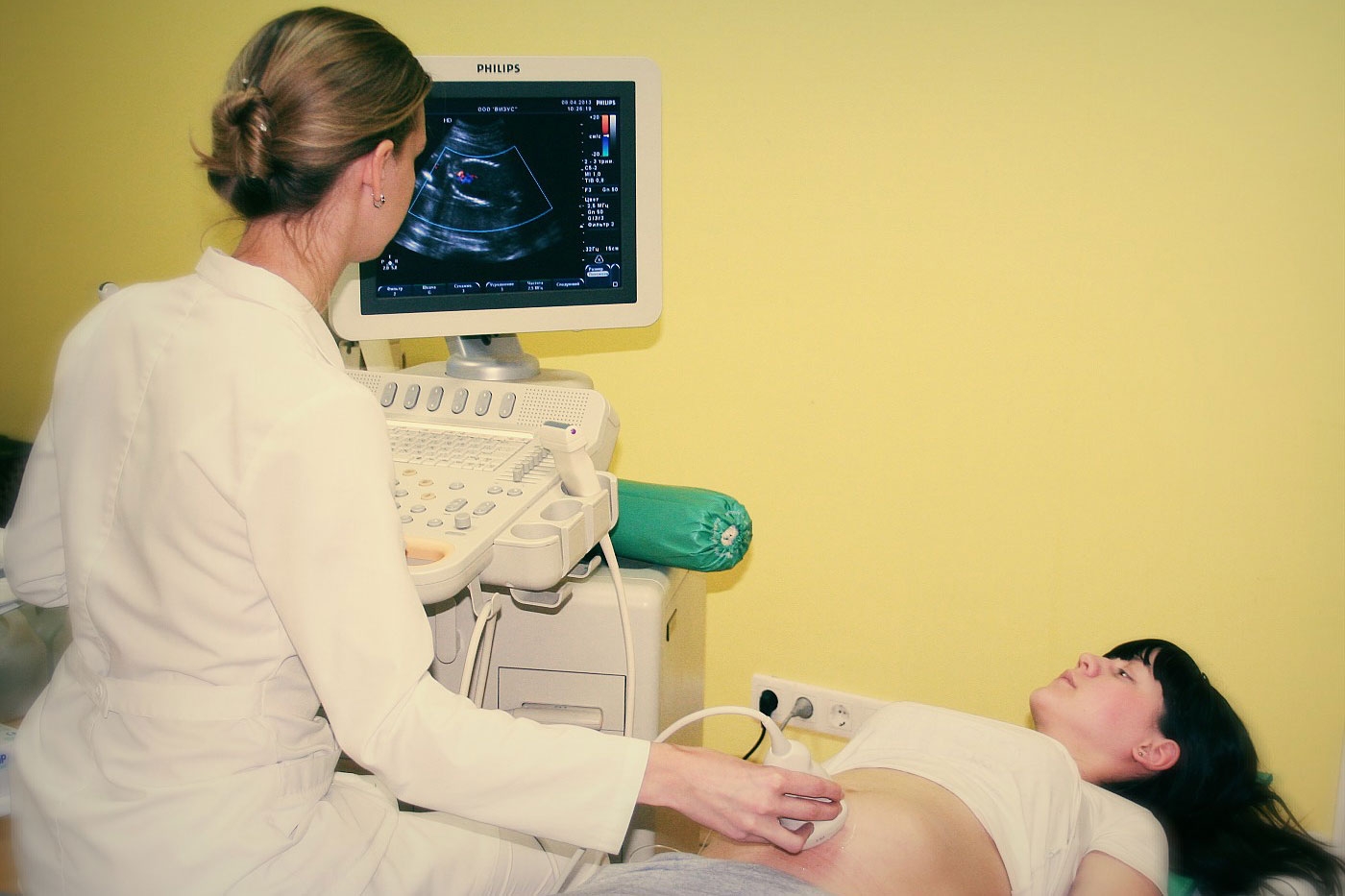
Ultrasound examination at such a short time (8 weeks by obstetric standards - this is only 6 weeks from the moment of conception) is carried out mainly by the transvaginal method. A woman is inserted into the vagina with a sensor in the condom. This method has a higher accuracy at an early date.than transabdominal ultrasound, in which the uterus and its contents are examined through the peritoneum.
If the ultrasound is planned, then it is desirable for the woman to prepare for the diagnosis.
A few hours before visiting the doctor should take "Espumizan"Or" Simethicone "to eliminate the accumulation of gas in the intestine, since swollen intestinal loops can squeeze the pelvic organs and distort the results of ultrasound.
Drinking plenty of water to fill the bladder is not required before undergoing transvaginal ultrasound. On the contrary, the doctor may ask to come with an empty bladder to get a clearer image.
The study of the pelvic organs with ultrasound does not cause unpleasant or painful sensations to the woman, does not harm the unborn baby. It lasts about 5-7 minutes.
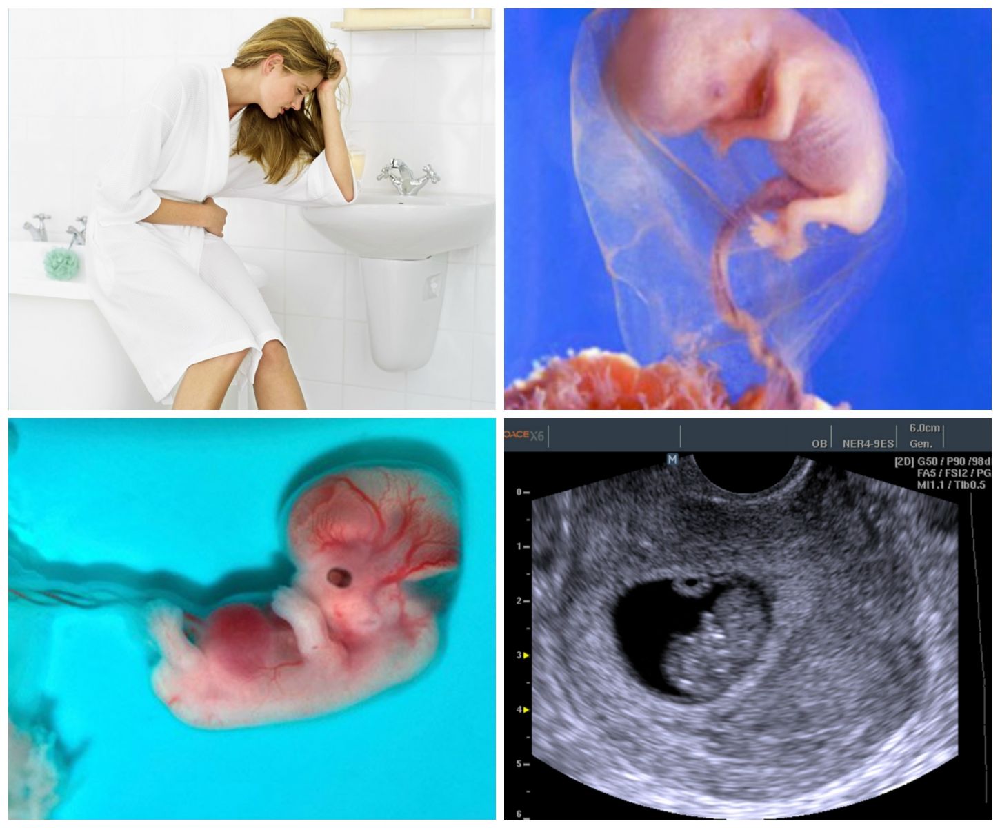
What will ultrasound show at week 8?
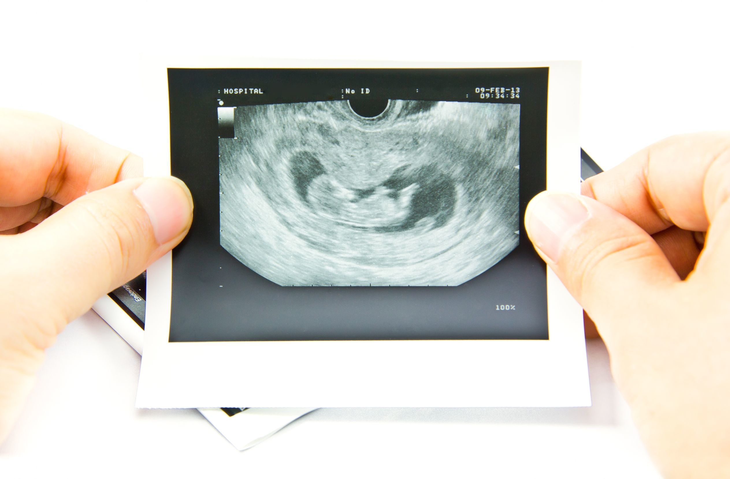
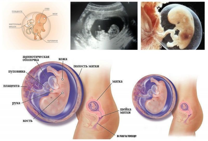
If a high-resolution device is installed in the ultrasound room, and the diagnostician will be so kind that it will show the future mother a monitor and explain what is happening there, then the woman will be able to see enough to understand that her little one has already passed a long evolutionary path. The ultrasound shows that the embryo has increased in size the head, it can be almost half of all sizes. The baby begins to form limbs, and the tail, which was before, no longer exists.
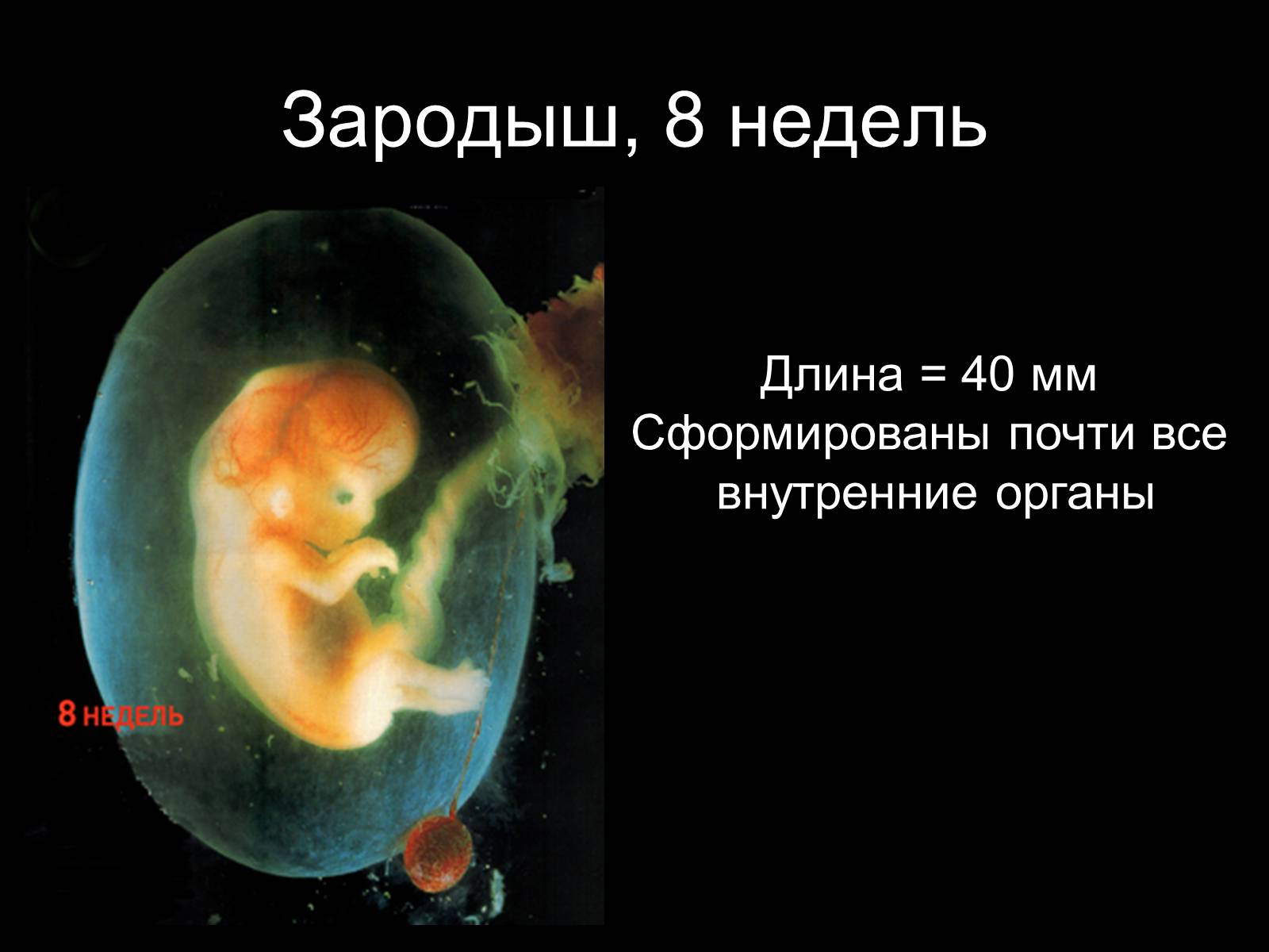
During this period, the formation of the eyes, eyelids, nose and lips, as well as the optic nerve begins, but all this cannot be seen on an ultrasound, the size of the unborn child is too tiny. But you can hear heartbeat crumbs and see the aortic valves contracting in his already completely four-chamber heart. An ultrasound doctor will be able to record and assess the baby's physical activity, which the woman herself cannot yet feel due to the small size of the embryo.

Week 8 completes the embryonic period of development, already in seven days, the baby will no longer be called an embryo; it will officially become a fetus. Also during this period, the testicles form in the baby’s abdomen, if it is a boy, or the ovaries, if it is a girl. However, it is not yet possible to see the floor on ultrasound, because the embryo does not yet have external sexual characteristics.

In addition, the doctor will assess the state of women's health, note whether there is a threat of miscarriage, interruption, separation of the ovum from the walls, whether the uterine walls are homogeneous in their structure, the state of the ovaries, tubes and neck of the genital organ.
According to the parameters of the ovum and the embryo, he will be able to adjust the exact duration of pregnancy.
Decoding results
From the doctor’s office of ultrasound diagnosis, the expectant mother will come out with an ultrasound examination protocol, which will indicate all the main parameters of her pregnancy at the current stage. Only specialists can figure out what abbreviations and numeric values mean. But after all pregnant women are very inquisitive, and they can not wait to learn about their baby as much as possible! Therefore, for informational purposes, we will describe how to decipher the ultrasound scan protocol, made on the eighth week.
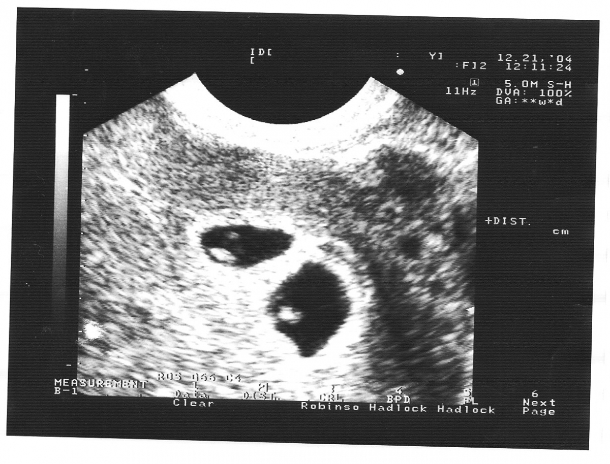
Fruit egg
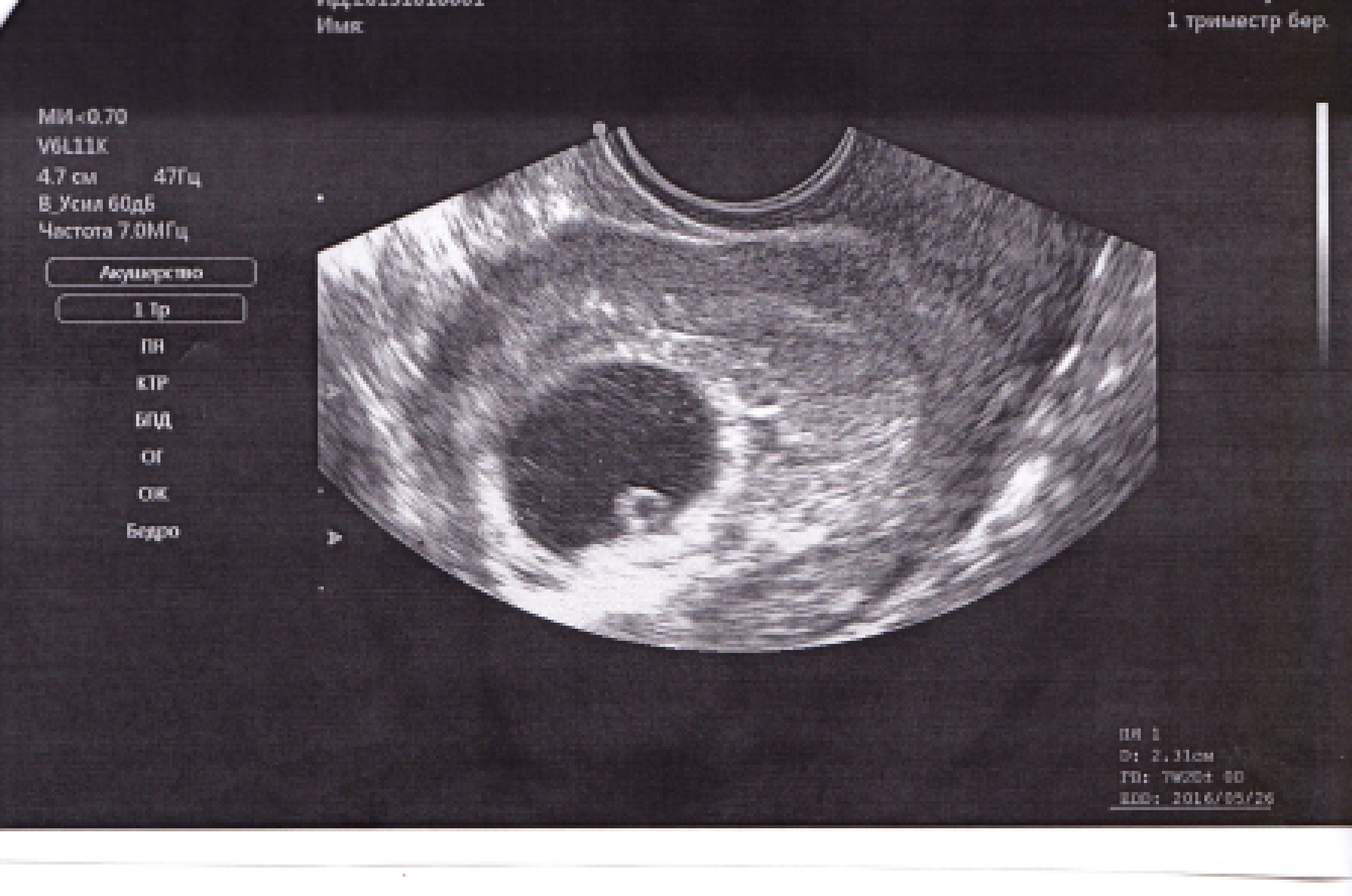
When there is no doubt that the pregnancy is present, the doctor writes that a fertilized egg is visualized in the uterine cavity. Careful examination of its shape and size. The main parameter is determined by the internal average diameter (this size in the conclusion is indicated as SVD).
A fetal egg is an indisputable confirmation of the fact of pregnancy, but it cannot be a parameter that determines the exact date, because the form of this formation is an individual feature. On average, the size of the ovum this week varies from 24 to 30 mm.
The average internal diameter of the ovum can be used to determine the duration of pregnancy, but this parameter is not essential for this purpose.
SVD in the 8th week of pregnancy:
The average diameter of the ovum inside, mm | Obstetric term |
19 (minimum value of 8) | 7 weeks + 0 days |
20 (minimum value of 9) | 7 weeks + 1 day |
21 (minimum value of 10) | 7 weeks + 2 days |
22 (minimum 11) | 7 weeks + 3 days |
23 (minimum 12) | 7 weeks + 4 days |
24 (minimum 13) | 7 weeks + 5 days |
25 (minimum 14) | 7 weeks + 6 days |
On the eighth week, the state of the yolk sac, a special organ, the “food storage” of the embryo, which exists only in the earliest terms and then disappears, is still assessed. Normally, the diameter of this bag at week 8 is 4.0-4.5 mm. Starting from week 10, it decreases and gradually disappears, transferring its functions to the formed placenta.
Embryo
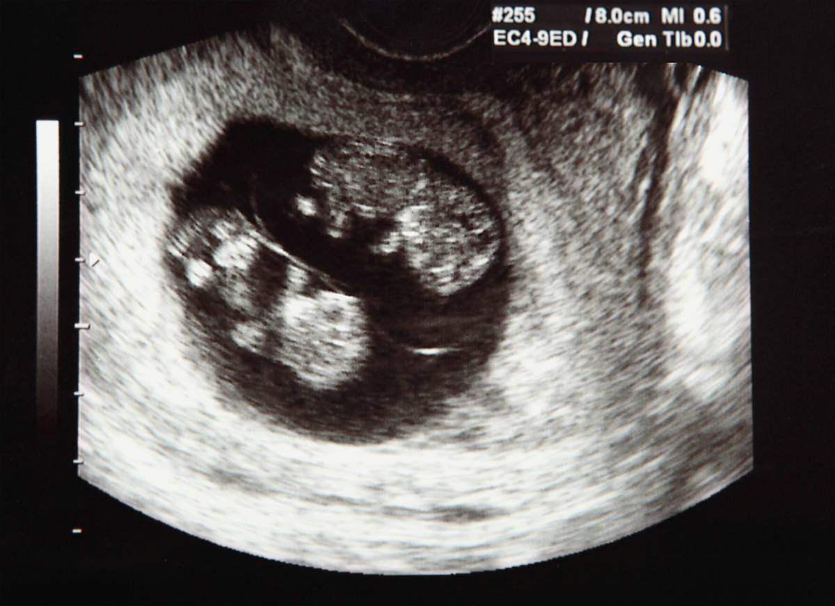
Normally, a doctor at week 8 determines a living embryo inside the ovum. The fact that the baby, whose size is only about 4-5 mm, is alive, can speak it heartbeat and the first movements. If the baby is showing signs of life, the doctor measures him from head to tailbone.And this is still the only “measure” that can be taken; other parts of the body and organs cannot be measured yet.
KTR
Much more about the term and development of the crumbs-embryo can tell this size. For the abbreviation KTR lies “Coccyx parietal size”, that is, the distance from the tailbone to the crown. If a snapshot is attached to the conclusion, then this segment in the first “photo” of the baby is indicated by the segment.

Table CTE at week 8:
Coccyx parietal size (mm) | Obstetric gestational age (week + days) |
10 | 7 + 0 |
10-10,5 | 7 +1 |
11,0 — 11,5 | 7 + 2 |
12,0 | 7 + 3 |
13,0 — 13,5 | 7 + 4 |
14,0 | 7 + 5 |
15,0 -15,5 | 7 + 6 |
16,0 | 8 weeks exactly |
Heart rate (HR)
To judge the state of the embryo allows the most "mysterious" value - heart rate. Many mothers guessing about the field of a child are trying to predict who lies behind the frequency indicated by the doctor - a boy or a girl. Experienced midwives do claim that boys 'hearts beat a bit slower than girls' hearts.
But this statement is relatively true only for long periods of pregnancy, when the midwife can hear by ear, not only the tempo, but also the heart tone of the baby.
In the early stages of the heart rate in boys and girls by gender does not differ. Normally, for the eighth week, the heart rate is 125 - 165 beats per minute.
Female reproductive organs
A close examination of the condition of the uterus, appendages, ovaries and cervical canal allows the doctor to draw conclusions about what predictions the pregnancy has and whether it is favorable. The size of the uterus, the thickness of its walls, the presence or absence of retrochorial hematoma (a sign of detachment of the ovum) are evaluated.
If everything is in order, then in the conclusion the dimensions of the uterine cavity are indicated, the lack of tone is emphasized, the cervix is described as completely closed, closed, the ovaries without features.
Ultrasound for multiple pregnancies
At week 8, the diagnostician, using a good scanner, perfectly sees the presence of two fetal eggs with two embryos or one egg with two embryos during pregnancy with twins. The above parameters are measured for each of the embryos.
In pregnancy, twins it is not always possible to immediately detect the second fertilized egg. Often it is not implanted in the uterus, but outside of it, therefore, when examining a woman with a probable multiple pregnancy, the doctor will study the tubes, ovaries and the space behind the uterus more closely.
Possible problems
During the ultrasound, the diagnostician will immediately see the presence of pathologies:
- Anembryony. This is a pathology in which there is a fertilized egg in the uterus, but there is no embryo in it. If at 8 weeks the doctor does not see the embryo, then he will prescribe a re-diagnosis after 10-14 days. If the second ultrasound does not detect the baby, the pregnancy will be recognized as not developing. A sign such as the absence of the yolk sac, if the size of the egg has already exceeded 13 mm, may also indicate the absence of an embryo. In the eighth week, the ovum already has a diameter of about 20 mm.
There is still time to wait for the appearance of the yolk sac. Its absence is recognized as a sure sign of anaembryony, if the diameter of the ovum has already exceeded 35-40 mm.
- Fading pregnancy. An existing embryo can stop developing for various reasons - both because of genetic mutations and because of an infectious disease that mother suffered at the very beginning of her pregnancy, due to the adverse effects on the maternal organism of radiation, alcohol, nicotine, narcotic substances . Such a conclusion can be made on the eighth week, if the heartbeat is not fixed, and the embryo itself does not make movements.
- The threat of termination of pregnancy. The thickening of the walls of the uterus suggests that the woman's reproductive organ is in an elevated tone. In this case, the doctor makes recommendations about the preservation of pregnancy at home or inpatient conditions. If a retrochorial hematoma is detected - a segment of the detachment of the ovum from the uterine wall, then the prognosis depends on the size of the hematoma.In the conclusion of the ultrasound, the doctor necessarily describes them and makes recommendations for treatment.

- Miscarriage. About miscarriage that has begun, a woman can guess the pain in the abdomen and bleeding. On an ultrasound at week 8, the doctor can confirm these concerns or disprove them. If the miscarriage has already begun, the equipment will record the development of uterus hypertonus, the fertilized egg will be deformed, as if flattened on top, uneven. Possible a large partial or complete detachment. If it came out spontaneously, then fragments of the fetal membranes may be found in the uterus, but this is not necessary.
- Lag in development. The strong lag of the embryo in development on the eighth week is fixed if its KTR and SVD are below the norm by 40-50%. If the heartbeat is fixed at the same time, then the pregnancy is taken under special control and given time to develop, and the dates are checked and corrected - it is possible that an error has occurred with their calculation.
A strong lag in size and the absence of signs of life are grounds for making a diagnosis of missed abortion.

Inspection accuracy
The accuracy of ultrasound as a diagnostic method is estimated at about 80-90%, but in the early stages of time accuracy is reduced to 70-75%. That is why when detecting problems in the eighth week, a second ultrasound scan is scheduled after one to two weeks to recheck the data.
The only exception is missed abortion and a diagnosed miscarriage, which carry big risk of mom's infection, the likelihood of sepsis. These problems require early surgical intervention.
In all other cases, no one rushes events. Time allows you to wait a bit and watch the crumbs in the dynamics. What can be seen at 7-8 weeks may disappear at 10-12, and such a development is not at all uncommon if a woman complies with all the recommendations of her doctor.


