Fetal development in the 19th week of pregnancy
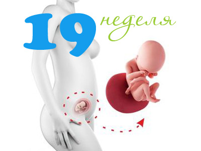
Each time period of pregnancy is a unique event, and the 19th week of pregnancy is no exception. At this stage of intrauterine life, quite important transformations take place in the baby’s body. Many of the internal organs have already been formed, while others are still continuing their active development.
Musculoskeletal system
Significant changes undergo bone structure. The small bones of the fruit become less fragile. Intensive development of the bones leads to the fact that the child continues to lengthen the arms and legs.
The muscles of the baby also undergo a number of changes. Improves their elasticity and ability to stretch. Such changes contribute to the fact that the fetus increases the number of possible movements committed by it. The motor activity of the baby is becoming more intense.
Toddlers can move, starting from the walls of the uterus, they are also able to turn their heads. The latter occurs due to an increase in physiological mobility in the cervical spine.
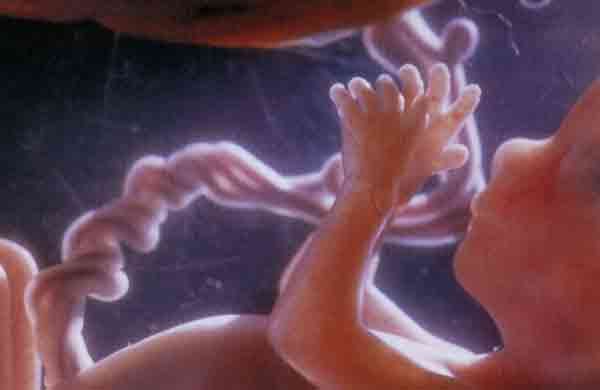
The child flexes and extends the handles, can touch his umbilical cord or face with his fingers. If twins develop in the uterus, then at this time they, as a rule, begin to actively “study” each other.
Heart and blood vessels
The circulatory system of the fetus at 18-19 weeks has a number of interesting features. So, a small heart of a child is able to pump almost 30 liters of blood. In its structure, it is quite reminiscent of an adult and consists of four cameras. Active contraction of the heart muscle contributes to the fact that the baby has a real heartbeat.
Doctors determine the frequency of the heartbeat, since this indicator is extremely important. During pregnancy, the baby’s heart is evaluated repeatedly. The obtained values of the heart rate (HR) of the fetus must be recorded in the medical records of the pregnant woman. This allows doctors to track the dynamics and draw conclusions about the well-being of the intrauterine development of the baby.
The heartbeat of the fetus should remain within the normal range. This indicates that the child’s heart is working normally, and his body does not experience hypoxia - oxygen starvation of tissues and internal organs.
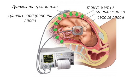
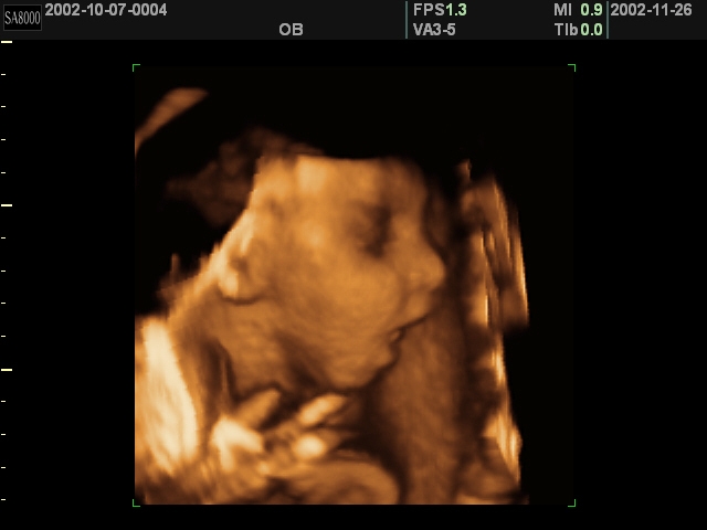
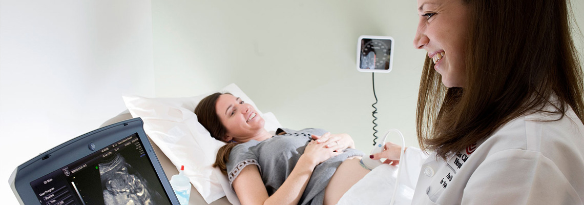
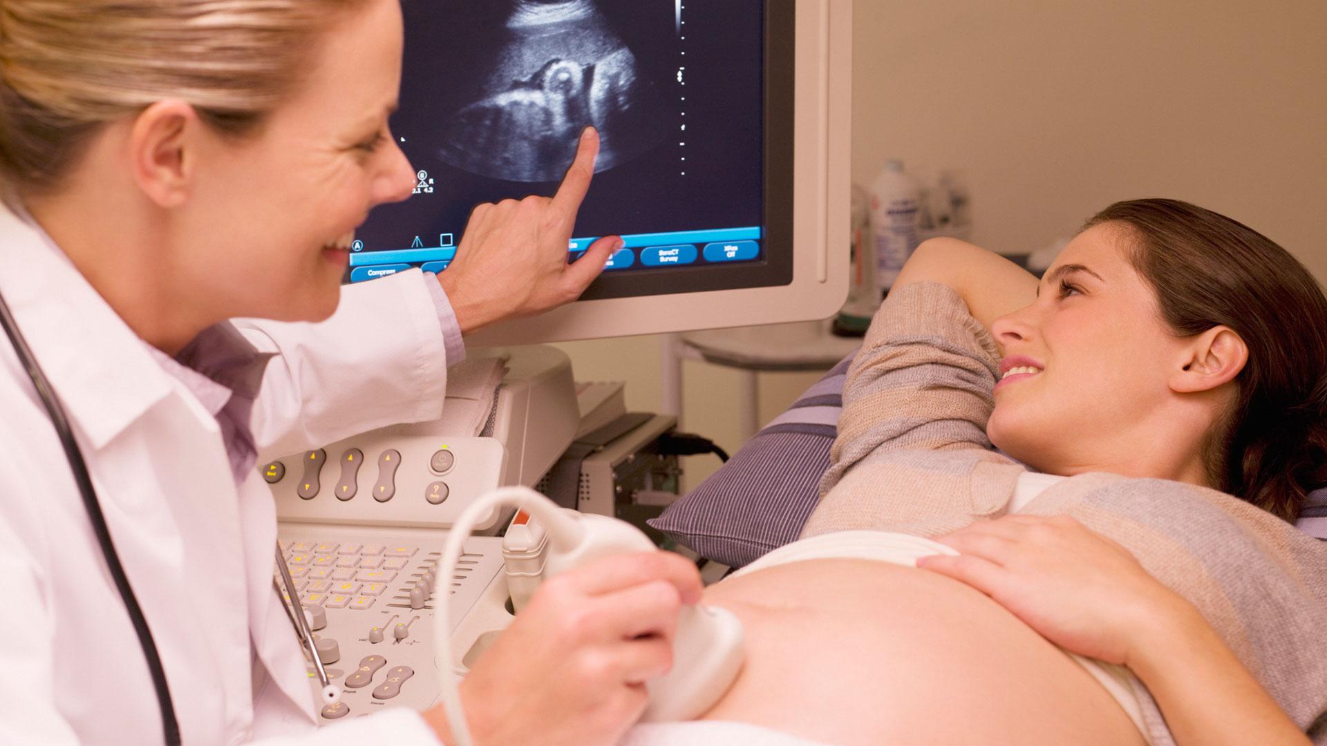
It is possible to determine the heart rate of the baby on this segment of its intrauterine life in different ways. Perhaps the most accurate is the calculation of the heart rate during ultrasound.
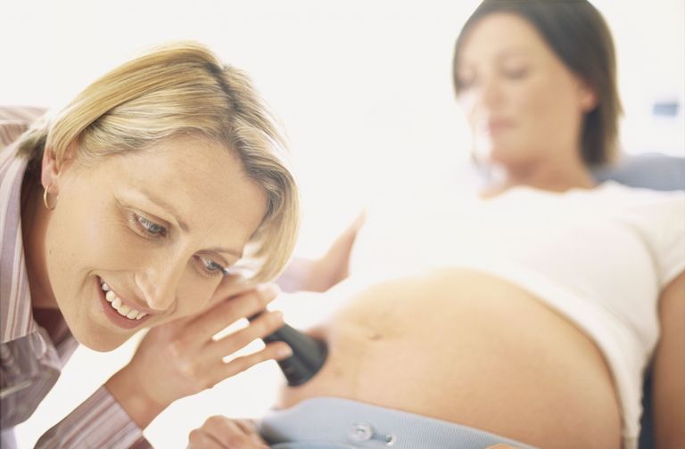
The obstetrician-gynecologist can also determine this clinical indicator, listening to how a small heart of the fetus works with the help of a special device - a stethoscope. The best point for listening to the heart rate is selected individually. It largely depends on how the fetus is located in the womb.
Often the heart rhythm is heard near the navel or slightly below. If a woman bears twins, then the heartbeat should be heard from each baby individually.Heart rhythm determination is an essential skill that every experienced obstetrician-gynecologist should possess.
The frequency of the baby’s heartbeat helps physicians to detect various intrauterine pathologies in the earliest stages of their formation in a timely manner. Each gestational period is characterized by its own fetal heart rate standards. For the 19th week of pregnancy, an indicator of 140-160 beats per minute is considered normal.
Active changes at this stage begin to occur in the blood vessels. Thus, their diameter gradually increases, and hence the lumen. The veins and arteries of the fetus are perfectly visible through its thin and delicate skin.
Kidneys and urinary organs
Interesting changes are beginning to occur in the urinary system. The kidneys of the baby are already formed, as well as the main urinary tract.
The child, being in the amniotic fluid, can swallow it, and later - and allocate. This feature is not only very important for the development of the entire urine excretion system, but also leads to regular renewal of the “aquatic” environment of the baby. Thus, the amniotic fluid may change several times during the day.

Nervous system
The number of special contacts between the brain cells in the crumbs begins to increase. This contributes to the fact that the baby appears first unconditioned reflexes, as well as significantly complicated behavior.
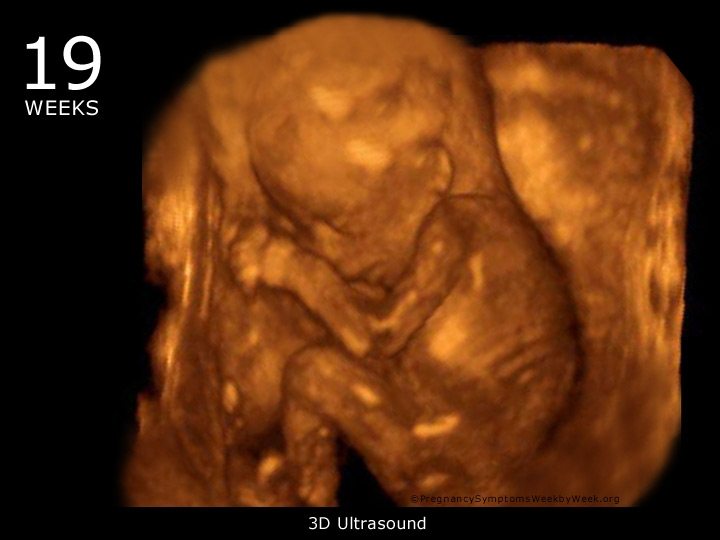
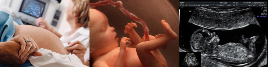
The first bright emotional manifestations, as a rule, are detected during the ultrasound examination. The kid can already smile at his mom and doctor, frown a little on his forehead or make a face. Such emotions cause future parents, as a rule, quite a lot of bright positive feelings. It is important to remember that their baby does not do it on purpose. Some kids turn away from the ultrasound sensor during the ultrasound, so it’s impossible to notice the emotional manifestations.
It also happens that during the ultrasound it seems as if the child is sleeping. At the same time he is in a calm state and does not make active movements.
At this stage of pregnancy, spinal cord development continues. This development contributes to the fact that the active movements of the baby are becoming more and more coordinated and streamlined every day.
First movements
The stormy activity of the baby in her mother's abdomen and its rather large size contribute to the fact that the expectant mother has the first sensations in her belly. This event for a woman is very touching and, as a rule, is remembered for the rest of her life.
It is important to note that Not all future mothers can feel their babies at this stage of pregnancy. It depends on many factors. Usually, rather active and mobile kids are pushed, calm children may not cause any concern to their mother.

Each pregnancy is a unique event. The absolute similarity of symptoms in this case never happens. For example, during the first pregnancy, the baby begins to push on the 18-19 week of pregnancy, and in the second - after 22. It is important to remember that this is a very individual feature that cannot be predicted in advance.
Body parameters
The size of the baby is a very important clinical sign, it must be guided by doctors who monitor the development of a specific pregnancy. Indicators of height and weight of the baby allow specialists to track the intensity of its intrauterine development in the womb.
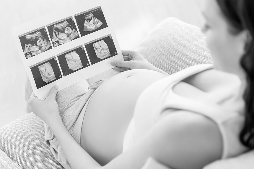
Each period of pregnancy is characterized by certain norms. These clinical parameters are usually determined during an ultrasound scan. The values obtained are reflected in the medical certificate of the specialist, which is issued to the hands of a pregnant woman after the completion of the test. The main parameters studied and their norms are reflected in the table below.
Clinical sign of the fetus | Normal values for the 19th week of pregnancy |
Growth | 21-25 cm |
Weight | 230-250 g |
It often happens that after an ultrasound scan, a pregnant woman goes into a panic or starts to worry that her baby weighs a little less than it should be by this date. Immediately it should be noted that this is not worth doing. The weight of the baby, like his height, is an individual parameter.. Not all children development occurs within the established age limits.
If the child’s weight or height is less than the values prescribed for a given period, the doctor will certainly pay attention to it. After this, fetal development requires more careful observation. As a rule, the weight of the child after a few weeks of pregnancy returns to normal.
Location
The position of the child in my mother's belly can be very different. Determine how the baby is located in the uterus, you can use ultrasound or clinical obstetric examination.
It is important to remember that the position of the fetus during this period of its intrauterine life is usually non-permanent.
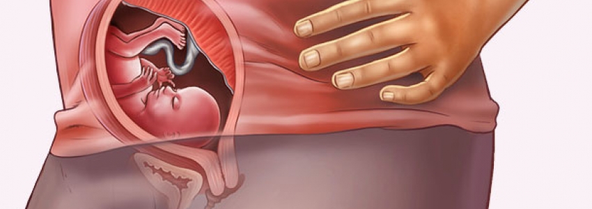
It often happens that an active and rather mobile fetus changes its position in the uterus. The child can be placed in different ways. Doctors consider one of the most functionally favorable locations of the fetus in the uterus to be a headache. In this case, the head of the baby is located in the direction of the inner throat of the uterus.
Head previa allows the fetus to properly move through the birth canal during labor. In this case, the head of the baby, which has the largest size for childbirth, moves first, which is more correct, according to experts. After the passage of the head through the birth canal, the rest of the child’s body is “born” much easier.
In addition to normal headache presentation, there are other, less favorable location of the fetus. So, the baby may have a transverse position in the uterus. In this case, his head is no longer located towards the internal fallopian pharynx. In this case, the independent birth of the baby without any consequences is hampered.
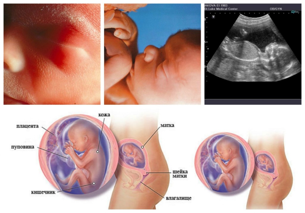
Baby look
The configuration of the baby’s body during this period of its intrauterine life varies somewhat. The body of the child becomes longer, the arms and legs are also pulled out. The proportions of the head vary somewhat. The kid already looks like a man, but only in miniature.
On the face of the baby's eyes are clearly different, they look quite large. Outside, the eyes cover the eyelids. The baby already has short cilia and even eyebrows.
Almost the entire body of the fetus covers the original lubricant. The secret of sweat and sebaceous glands is directly involved in its education. On the surface of the skin, it mixes with exfoliated skin cells, leading to the formation of a special lubricant. This lubricant is very important for the baby, as it is necessary to protect it from the effects of multiple negative factors and microbes.
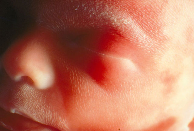

The child is already quite clearly visible contours of the nose and even the chin. Auricles are formed. The baby can already open and close the mouth. Also, the fetus has swallowing movements, due to which it swallows small portions of the amniotic fluid.
On small fingers, the child already has its own unique pattern - experts call this pattern fingerprints. On each handle and leg there are small nails.
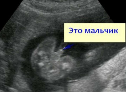
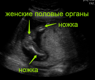
Can I find out the floor?
Determine the birth of a boy or a girl, at this stage of pregnancy is already possible. This is because the main external genital organs of the fetus by the 19th week of pregnancy have already formed.
Future parents are often interested in the question of whether errors are possible in determining the sex on this period. Errors and errors in establishing the floor are really still possible. - this largely depends on the experience and qualifications of the specialist who performs the ultrasound scan.
About what happens to the fetus and the future mother at the 19th week of pregnancy, see the following video.


