Screening ultrasound of the first trimester: terms and norms

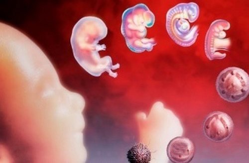
During the first trimester of fetal development many unique processes occur. During this period, the baby develops all the systems of internal organs. In order to timely diagnose various pathologies in such an important period, ultrasound screening is used.
What is it for?
The introduction of screening surveys in our country happened quite recently. This was facilitated by high maternal and infant mortality. In order to reduce these statistics, specialists from the Ministry of Health developed special recommendations. They provide for ultrasound several times during the entire period of pregnancy.
Every future mom will certainly be interested in the question of what screening is. In literal translation, this term means "screening". During screening, all pregnant women are detected. with the pathological course of pregnancy. Future moms, who have been identified any violations should be more closely monitored by doctors.
During this type of study, doctors define both the various developmental pathologies of a developing baby in the womb and the associated diseases of the internal genital organs of the mother. This is done on one study.
If at the time of the survey any deviations were identified, then the future mom is given recommendations. be sure to visit second and third screenings.
With the help of such a survey, it is also possible to determine the sex of the future baby - this can be done, as a rule, at 11-12 weeks of pregnancy. Experienced ultrasound specialists can see the sex of the child before, but often make mistakes.
Hurry up in order to find out who "lives" in the tummy, should not be.
The intrauterine development of a baby undergoes a series of successive stages. By the end of the first trimester, when the first formation of its genital organs occurs, and it is necessary to conduct a study to determine the sex.


The ultrasound carried out during this period of pregnancy also allows calculate the date of delivery more accurately. Usually this indicator is calculated in obstetric weeks. In this case, there is a definite difference between the gestational and obstetric terms. In order to eliminate confusion, experts recommend using the term "obstetric" gestational age.

The first trimester is not the best time to exclude all genetic and chromosomal diseases in the fetus. However, it is worth noting that during this period Some signs of these pathologies can be identified. For this, doctors and scientists have developed a whole range of different ultrasound criteria that are used to draw up a conclusion. They help doctors promptly suspect the presence of the first signs of genetic abnormalities in the baby.
Using screening ultrasound, it is also possible to detect certain impaired uteroplacental blood flow. This method will allow to determine the pathological contractions or other abnormalities of the blood vessels feeding the fetus. If necessary, an auxiliary method called dopplerography can be used. It allows you to define different pathologies of the blood flow more accurately. During the first ultrasound screening, specialists can also identify quite dangerous pathologies.
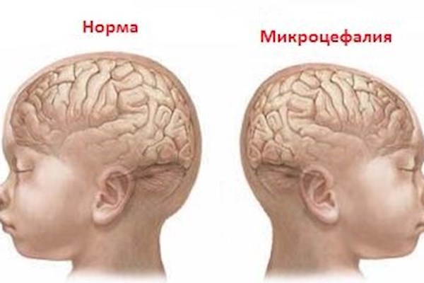
Rudimentary changes in the brain - an unfavorable clinical sign that requires timely diagnosis.
To identify emerging pathologies at this stage of pregnancy, sometimes an expert-level ultrasound is required.
A screening study is a necessary clinical test to identify dangerous conditions which can lead to the threat of miscarriage. Specialists with the help of special ultrasonic sensors look and identify various pathologies that contribute to the development of fetal hypoxia. This condition is quite clearly visible when using high-resolution devices.
Deadlines
Doctors recommend that screening be sure to all expectant moms, without exception. The second ultrasound screening study is also conducted, as a rule, absolutely to all expectant mothers. The third screening is usually prescribed for certain medical conditions in cases where it is actually required.

To obtain more reliable results, experts recommend ultrasound screening at 11-13 weeks of pregnancy. Usually this study is supplemented by a number of biochemical analyzes, which are given 2-3 days before the ultrasound.
Clinical situations are allowed where screening may be delayed for 1–2 weeks. However, the schedule offset for screening ultrasound should be agreed with the obstetrician-gynecologist.
Where can I do?
The obstetrician-gynecologist, who monitors the future mommy during the entire development of her pregnancy, gives directions for screening. This medical form indicates the personal data of the pregnant woman, the expected duration of the pregnancy, and also makes various notes about her state of health. In this direction also indicates the date and time of the ultrasound.

You can also study in a private clinic. For this, a referral from a doctor is usually not required. Future mom can sign up for such a study on their own.
Prices for screening ultrasound in private clinics vary. The average cost of such a study is 2000-4000 rubles. If the examination will be carried out on the equipment of expert class or a more qualified specialist, this diagnostic procedure will cost a little more. In large cities, the cost of the first screening ultrasound sometimes reaches 8,000 rubles or more.
Proper preparation
To obtain reliable and accurate results of the study, the future mom should be carefully prepared for screening. Usually, all recommendations are given to the woman at the next admission by her obstetrician-gynecologist.
Basic training in the early stages of all pregnant women, as a rule, is the same for all. However, there may be small differences. Usually they are associated with the filling of the bladder. Most women should not use much liquid directly before the diagnostic procedure. Especially if the examination will be conducted by the transvaginal method.
However, there are some exceptions. In these cases, doctors recommend a few hours before the ultrasound drink about 0.5 liters of fluid. The bladder filled with water pushes the uterus forward, making it more accessible for visualization. This special training is not required for all women. If she is still needed, the doctor will warn the future mom about it.
In the early stages of pregnancy, substantial preparation is not required. Usually, in order to obtain accurate research results, doctors recommend that future moms necessarily follow a special diet. In such nutrition all fat and fried foods are limited.

Dinner on the eve of the diagnostic procedure should be as easy as possible. Breakfast before the morning ultrasound should not be. You can drink only a little water.
For future mummies suffering from diabetes, especially with uncontrolled flow, such recommendations cannot be implemented. Skipping the next breakfast can lead to a critical decrease in blood sugar levels, which is highly undesirable for both mommy and her baby.

A few days before the screening test, pregnant women should exclude all fruits and vegetables from their menu. Also banned any varieties of cabbage, beans, kvass, and any carbonated beverages. All these products can lead to severe gas formation.
The intestines filled with gases will not allow the ultrasound specialist to conduct a full-fledged study. In this case, the phrase about ekonegativnosti will be present in the ultrasound report that the woman will receive after the examination.
Do not consume a lot of protein products for 2-3 days before the screening. Such food, especially in large quantities, also contributes to bloating. It is better that the food was as easy as possible, but high in calories and nutritious. For this perfect lean bird or white fish with cereal garnish.


The exclusion of physical activity is a very important stage of preparation for the procedure. Scientists have found that even the usual everyday activities that future moms do every day can lead to distorted results.
In order to minimize the risk of inaccurate research results, should limit the implementation of any physical activity. Expectant moms should replace them with fresh air walks at a moderate pace. They will benefit not only the female body, but also the baby, who is in her womb.

Any emotional stress also contributes to the violation of metabolic processes in the body of a pregnant woman. Experts have long revealed an interesting fact: pregnant women who are suspicious and worried about trifles during ultrasound the baby can turn away from the sensor or begins to move actively. In order to fully explore the fetus, doctors strongly recommend pregnant women not to be nervous and not to worry.
If the future mother continues to smoke while carrying the baby, then on the eve of the study, you should significantly limit the number of smoked cigarettes. Doctors have determined that nicotine and tar contained in tobacco cause a hypoxic state in the fetus. Also, these chemicals lead to impaired uteroplacental blood flow, which will be revealed on ultrasound.


Different methods
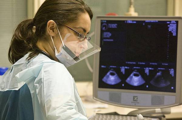
Screening tests are performed by ultrasound specialists. They have special training in this section of medicine. It is better that the first screening is performed by a qualified and experienced doctor who has sufficient experience in conducting such studies.
This will somewhat reduce the risk of possible errors and the conclusion of an incorrect conclusion after the survey. If, after an ultrasound examination, an obstetrician-gynecologist has any doubts about the unreliability of the results, then he can send the future mom to re-study already to another specialist.

There are certain situations when a screening study is not carried out in an ordinary antenatal clinic. Future moms who have severe comorbid diseases of the internal organs or pathologies of gestation can be referred to the perinatal center for examination. Specialists of this medical institution will be able to more accurately and accurately identify all the “hidden” pathologies or emerging anomalies of intrauterine development of babies.
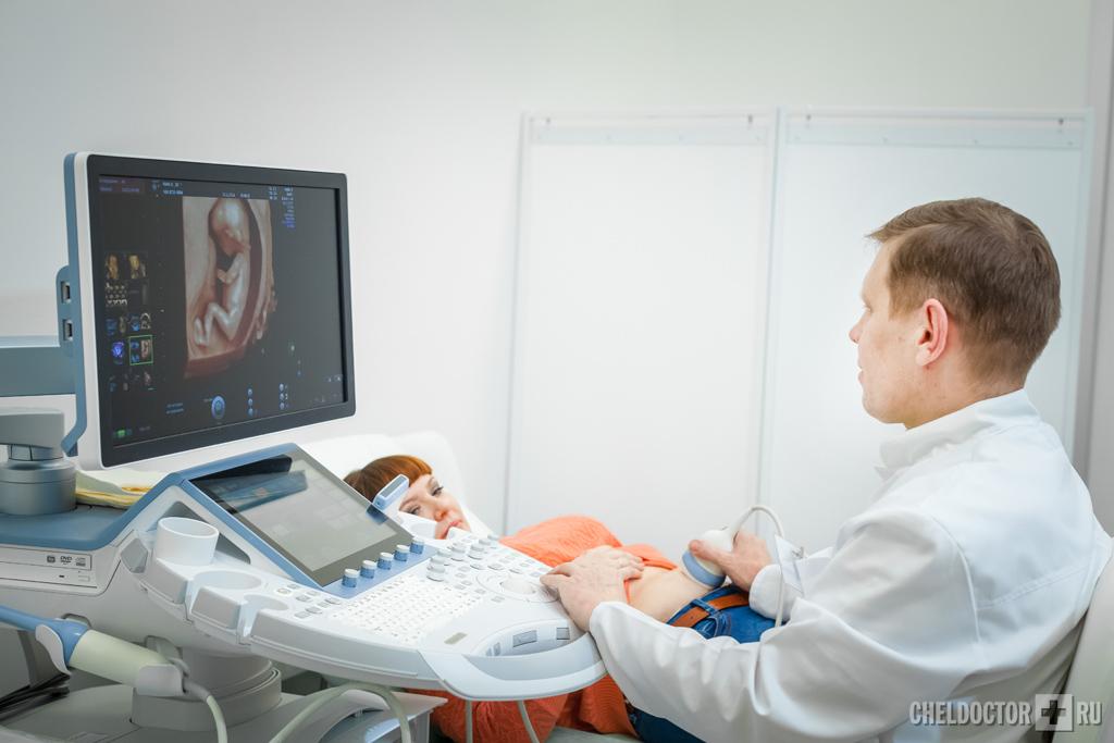
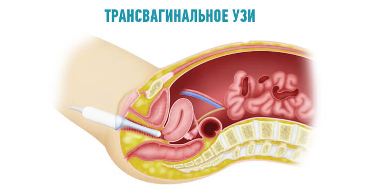
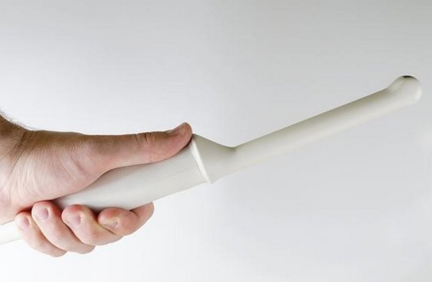
Currently, the study can be conducted by several methods.In the early stages in most cases used transvaginal. The technique of this procedure involves the use of a special ultrasound transducer inserted into the vagina. Afraid of this type of examination is not worth it. With the correct technique of execution, it will not give the woman any pain and discomfort.
The transvaginal method allows you to identify various pathologies of both the mother and the fetus quite effectively and accurately. For this diagnostic procedure, there are a number of contraindications, it is not suitable for women with exacerbation of colpitis or vaginitis. In these situations it is better to resort to an alternative method - transabdominal.
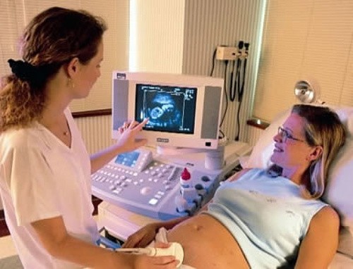
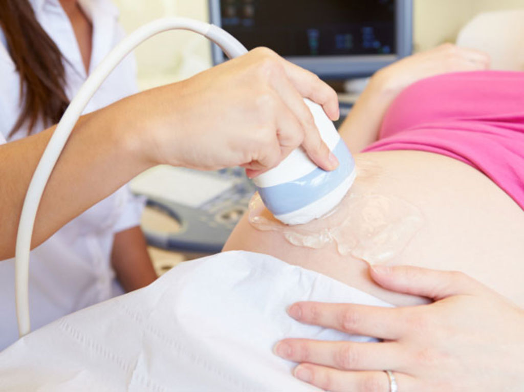
In this case, an ultrasonic sensor is used, with which the doctor drives a "pregnant" tummy. This method is also an excellent way to diagnose various intrauterine growth defects. Most expectant mothers prefer this method of ultrasound. As a rule, a screening study in this way is also carried out if during the diagnostic procedure the spouse of the patient is present.

For high-quality pictures, use a special diagnostic gel. It is transparent and slightly sticky to the touch. The gel is applied to the area of the future mom's tummy just before the procedure.
This substance helps ultrasound waves to better reflect and penetrate well into the internal environment of the body. The chemical composition of the gel is completely hypoallergenic and can not cause allergic manifestations.

What to take with you?
If the study will be carried out in the women's clinic, then from the future mother's house should grab a towel. It will be needed in order to lay it on a special couch on which the study will be conducted.
Usually, an ultrasound scan is performed on the back. Only with certain pathologies of the uterus in a woman, a specialist conducting research can ask her to turn on her left or right side. After the procedure, you may need paper napkins. They are needed to remove gel residue from the abdomen.

If the study will be conducted in a private clinic, you do not need to take anything with you. All necessary items are already included in the cost of the procedure and will be provided. A doctor performing an ultrasound scan can ask for medical records. For this, the future mom should not forget to take your card with you.
Evaluation of results
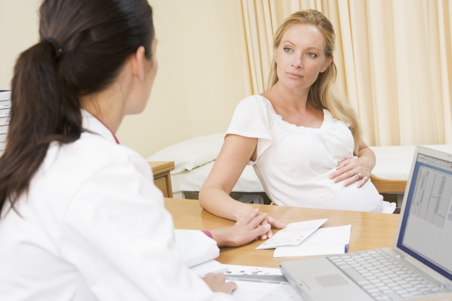
After the study, the doctor who conducted the screening examination will definitely give his opinion. It will only show the changes that the doctor saw during the study. Also in the conclusion should be indicated the size of all the parameters studied, which can be determined at this stage of pregnancy.
Expectant mothers should remember that such a conclusion is not considered a diagnosis.. It requires mandatory treatment of an obstetrician-gynecologist. Decoding can only be done by a doctor who is observing the future mommy and carrying out a clinical examination. It is he who determines the presence of pathologies and diagnoses.
In some cases, after the study, the doctor may refer the woman to an additional follow-up examination. Usually in this situation, some biochemical analyzes are performed. You may also need to consult a therapist or a "narrow" specialist.

Norms
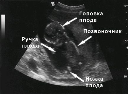
To assess the intrauterine development of the fetus at this time, doctors use special ultrasound indicators. They were developed by scientists taking into account the characteristics of the physiology and anatomy of the fetus. The use of these indicators helps professionals to timely suspect genetic and chromosomal pathologies, and also to identify developmental abnormalities in the baby in the early stages:
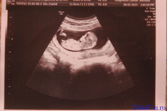
- Kopchik-parietal size (KTR) - This is a basic indicator that is used in prenatal research of the first trimester.Many experts prefer to use special tables in which normal values of this indicator are entered. With the growth of the baby, this parameter increases. This indicates a normal course of pregnancy and optimal intrauterine development of the fetus.
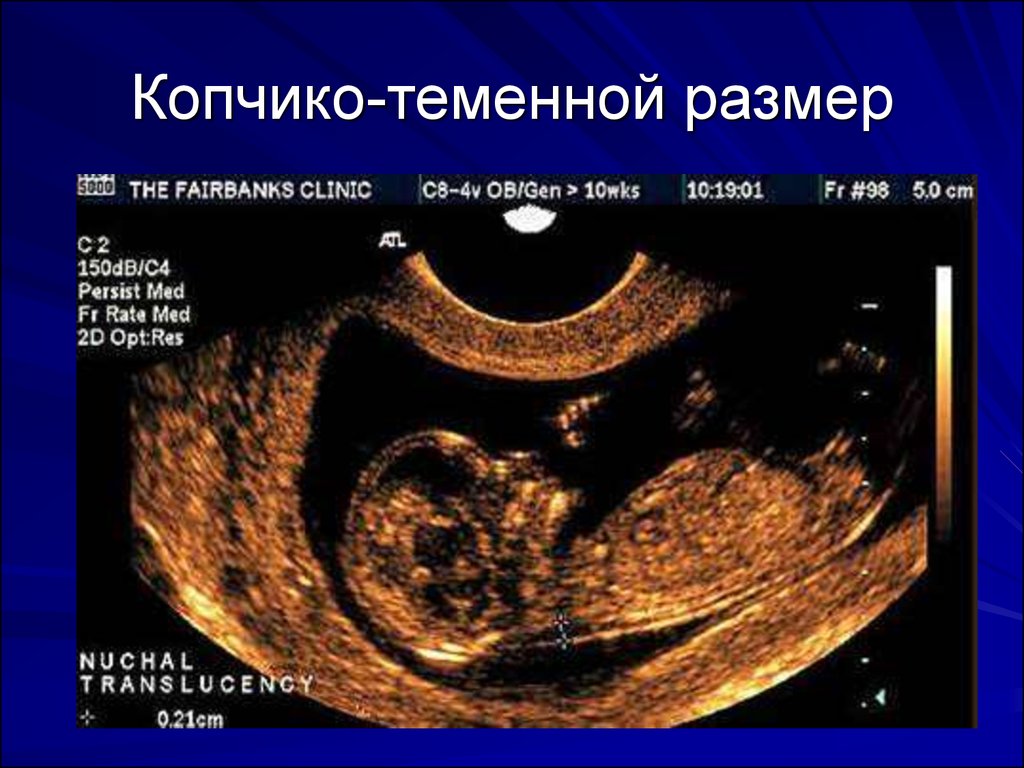
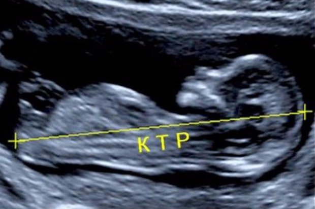
For a period of 11 weeks, this figure is 34-50 mm. It is important to note that these values are averaged. By week 12, the values of this indicator are already increasing to 42-59 mm. After 7 obstetric days KTR becomes 51-75 mm.
- Heartbeat - another estimated indicator. It shows how well the fetal heart muscle is contracting. Myocardium of the baby begins to work from the 6th week of its intrauterine development. This contributes to the fact that the doctor conducting the study can easily calculate the number of heartbeats per minute. Normally, this indicator by the 9th week of intrauterine development is already 130-140 per minute. Any deviations from normal values may indicate problems in the children's body. In this case, additional diagnostics may be required. Heartbeat is a very important indicator that must be evaluated.

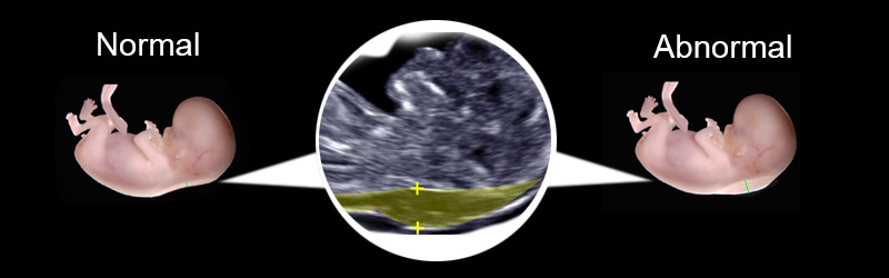
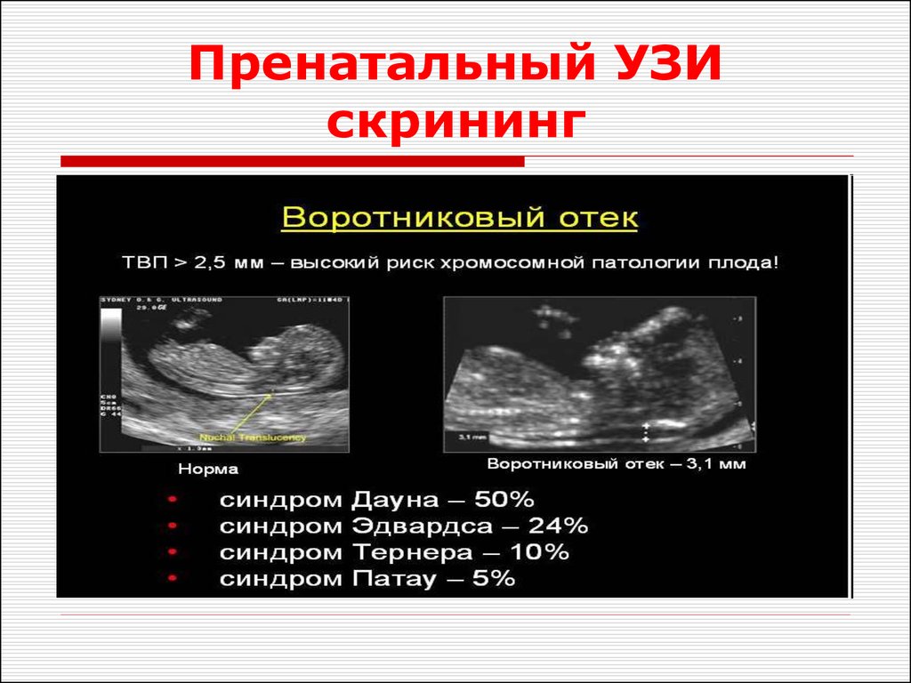
- Collar space thickness - Also very important ultrasound - a criterion for determining various gross defects of development. This indicator is used to predict the risk of various genetic indicators. At week 11 of fetal development, this ultrasound criterion becomes equal to 0.8-2.4. A week later, its values change to 0.7-2.7.
- At the time of the study, doctors can identify a rather dangerous condition called hypertonus. It can lead to spontaneous miscarriage or threatened miscarriage. In this situation, an obstetrician-gynecologist will send the future mother to hospitalization. To normalize the tone of the uterus requires complex therapy and the normalization of the day regimen.