Third trimester screening
The final stage of pregnancy is very important. At this time, it is necessary to identify pathology dangerous to the fetus. This article will help expectant mothers to understand why it is necessary to conduct this study.
What it is?
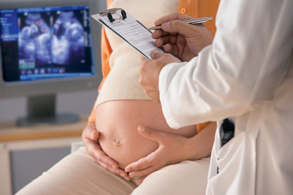
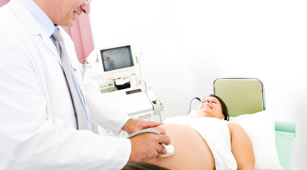
Screening of the 3rd trimester of pregnancy necessarily includes ultrasound, cardiotocography and Doppler. Only a comprehensive diagnosis helps doctors understand how pregnancy proceeds. Also, this screening is necessary to determine the tactics of childbirth.
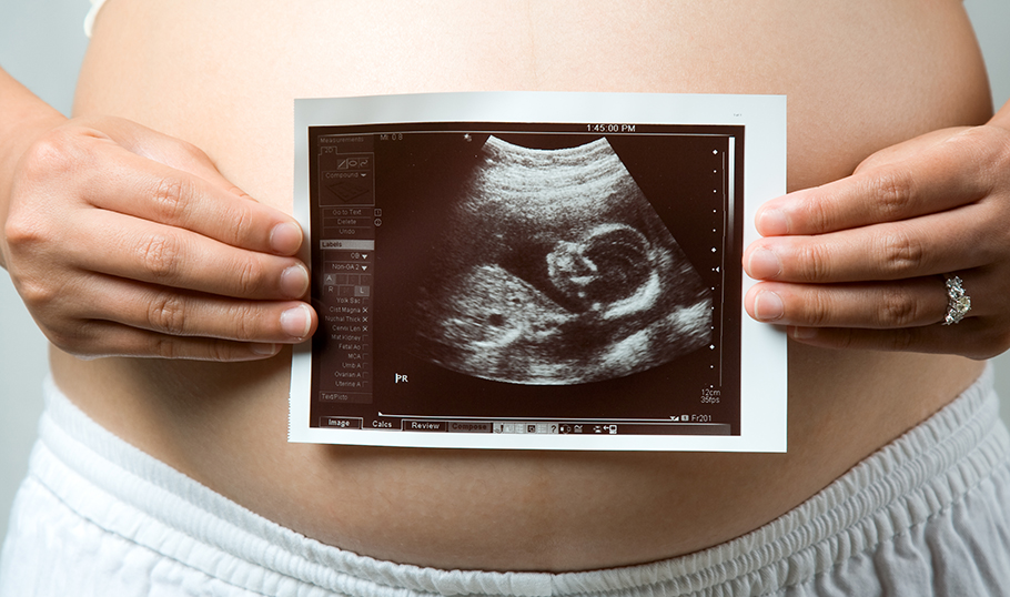
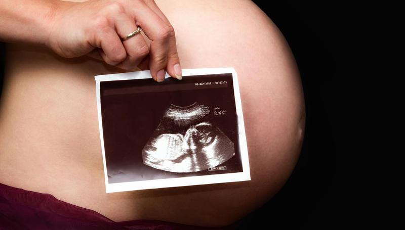
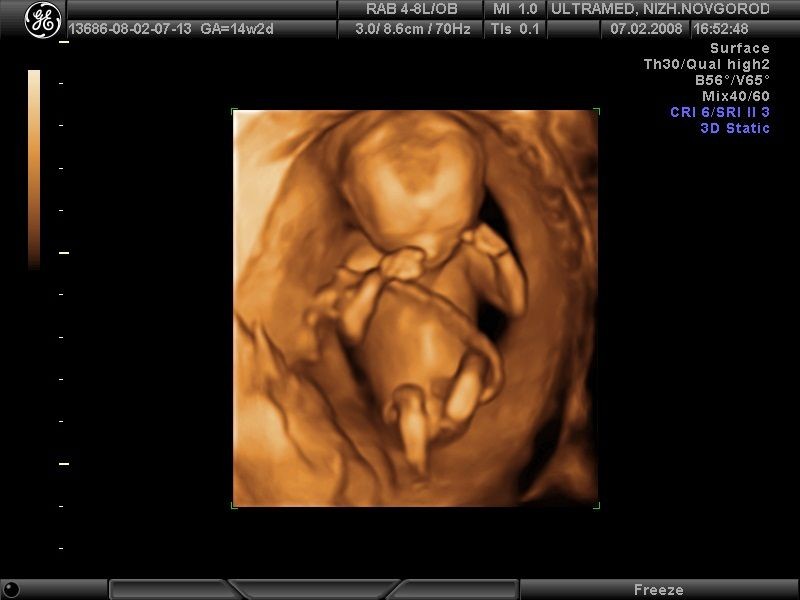
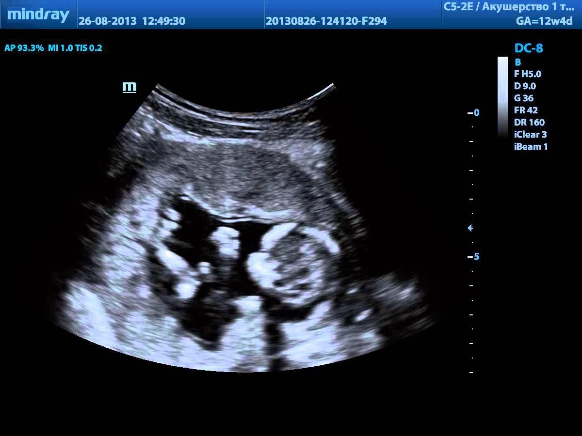
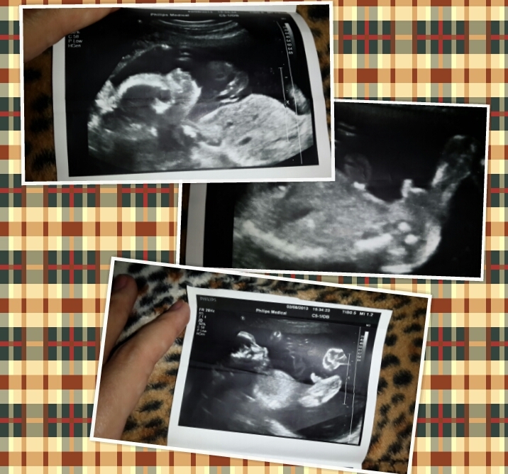
With the help of ultrasound, the doctor can evaluate the anatomy of the fetus and all fetal membranes. Also, this study determines the functioning and development of the placenta. With the help of ultrasound, you can establish the presentation of the fetus in the womb.
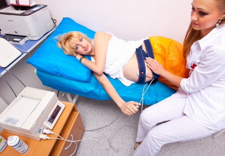
Heartbeat is checked using cardiotocography. This study also allows you to evaluate the work of the muscles of the heart. To conduct this study, several wires are attached to the future mummy's tummy. They can determine not only the number of baby’s heartbeats, but also establish the amplitude of contractile movements of the uterus.
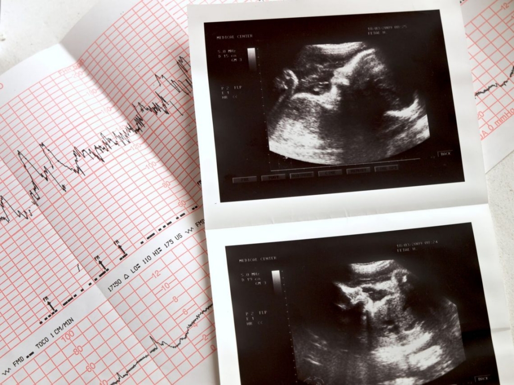
For the interpretation of the results obtained in the future it is very important to carry out the study correctly. For this, indicators that are determined both at rest and after activity are evaluated. Usually the duration of the procedure is 30-40 minutes.

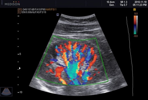
With the help of the Doppler test, it is possible to evaluate the functioning of the placenta. Also, this method perfectly identifies signs of placental insufficiency. Doppler in many ways resembles a conventional ultrasound. This test reveals any impairment of uteroplacental blood flow.
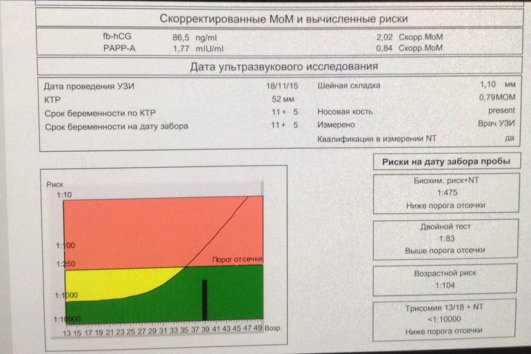
For the purpose of a comprehensive assessment of the final stage of pregnancy, additional biochemical studies may be required. These include hCG, alpha-fetoprotein, special markers for detecting Down syndrome, and placental lactogen.
Dates
Doctors recommend that such a complex diagnosis for pregnant women. at 30-34 week of pregnancy. In some cases, a slight offset of these terms is allowed. Usually such situations occur when the expectant mother has some serious diseases of internal organs or there are pronounced hormonal disorders.
Some mommies say that doctors sent them to the third screening a little earlier. Such a situation is possible if the observant pregnant obstetrician-gynecologist revealed any signs of deterioration of her state of health.
In this case, the list of analyzes may even be slightly extended.

Who should visit?
The third screening is not for all pregnant women. There are a number of medical indications that are required for the diagnosis complex:
- Doctors recommend that you be screened for 3 trimesters of pregnancy to all expectant moms who become pregnant after 35 years.
- Pregnant women who have a family history of chromosomal and genetic pathologies should also not miss the passage of this diagnostic complex.
- Also, the screening of the third trimester will be needed by expectant mothers who suffered from severe toxemia in the preceding weeks of pregnancy.
- If a woman who is carrying a baby has frequent miscarriages or difficulties in conceiving before the onset of this pregnancy, then diagnosis should not be missed. Quite often, various chromosomal or genetic pathologies result in spontaneous abortions, which manifest themselves in a similar way.
What shows?
In the third trimester of pregnancy can be identified already quite a lot of different indicators. With the help of ultrasound, you can determine not only the size of the head of the fetus, but also the main orthopedic disorders. Also with the help of this study, you can quite accurately establish its exact weight. Experienced ultrasound specialists can tell how much a baby weighs up to several tens of grams.
With the help of cardiotocography it is also possible to determine the filling of blood with oxygen. Decreased placental blood flow is a clear sign of such a violation. This method also reveals hidden signs of fetal hypoxia. In some cases, this symptom may be a manifestation of the entanglement of the umbilical cord of the neck of the child.

Doppler sonography is a method that gives doctors information about how the cardiovascular system in the fetus functions. Also, with the help of this study, doctors can identify various neurological disorders. This method can also diagnose a hypoxic state.
If the results are extremely unfavorable, then the doctor may suggest that the pregnant woman immediately go to the hospital. Such a necessary measure will help prevent irreversible adverse effects for both the mother and her future baby.
Biochemical screening significantly complements the findings of ultrasound. Increasing serum alpha-fetoprotein may be a likely sign of an established Down syndrome, Edwards syndrome, and some other genetic disorders. An increase in hCG in the blood can also be a very likely sign of genetic abnormalities.
The decrease in the concentration of gonadotropin in the blood is a physiological situation that develops at the very end of pregnancy. Nature invented it so that the process of intrauterine development was completed independently and as gently as possible. The increase in the last weeks of pregnancy of this indicator is a significant reason for early hospitalization in the hospital.

The detection of an excess amount of amniotic fluid is often due to the fact that the expectant mother suffers from diabetes. Also, the appearance of this clinical sign can be caused by various infectious diseases that a pregnant woman carries in the first two trimesters of her pregnancy.
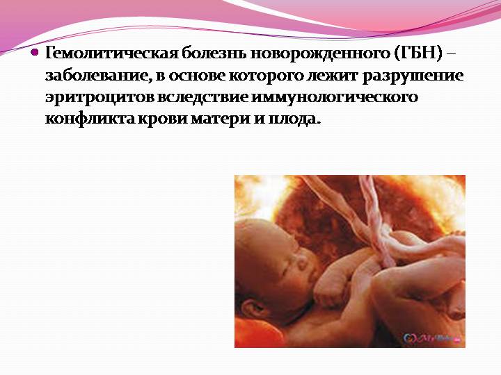
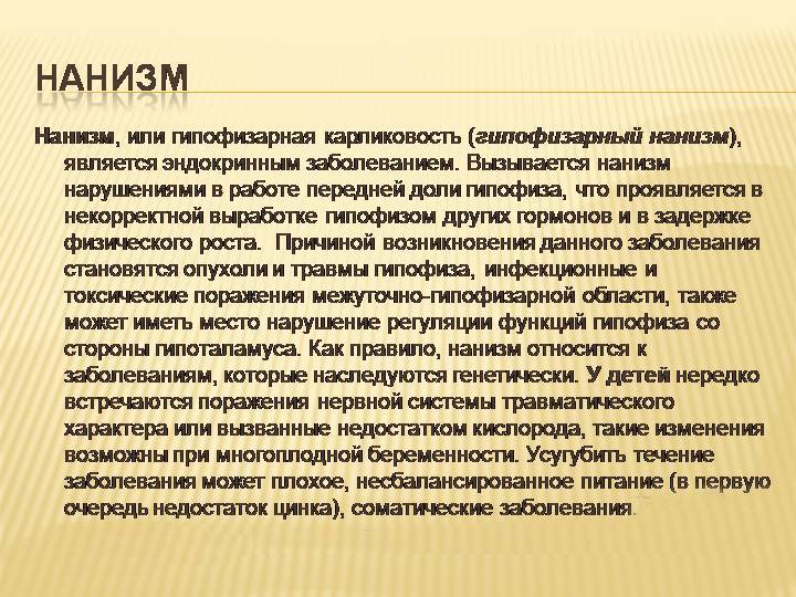
A significant increase in the abdomen of the fetus may be a manifestation of hemolytic disease or various liver pathologies. The appearance of this symptom is due to the accumulation of excess fluid in the abdominal cavity of the fetus. Reducing the size of tubular bones is often a sign of nanism. Also, this pathology is called a dwarf disease. This disease is quite difficult to treat.
What training is required?
Preparing for research is necessary. Proper preparation will help to get more reliable results:
- In order to eliminate inaccurate results, it is very important to follow a certain diet a week before cardiotocography and ultrasound. On the eve of the study should be eaten very easily.
- The use of fruits, vegetables and legumes for 2-3 days prior to the survey should be excluded. These products can lead to increased gas formation. Swollen bowel loops with gases quite often cause the appearance of echo negative. In this case, the interpretation of the results will not work.


- Also, 1-2 days before the study, pregnant women should exclude any physical activity.Nervous before the ultrasound also should not be. Future mom should keep calm and excellent mood. Walking in the fresh air on the eve of the study has a good therapeutic effect.
- Many doctors recommend an ultrasound scan on an empty stomach. However, this is not necessary. It is better to eat a little before this survey. You can also drink water. Bladder pre-filling may be required at later stages of pregnancy.

Norm indicators
To assess the final stage of pregnancy, a number of indicators are considered. To compile a comprehensive assessment, it is necessary to interpret several criteria simultaneously. Many of the estimated parameters are entered in special tables that contain their normal values.
Main criteria:
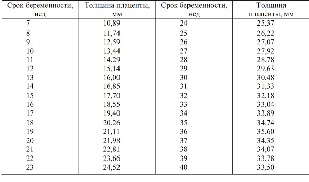
- Placenta Thickness - This is the main indicator that shows the development of this body. At the 32nd week of pregnancy, its values are 25.3-41.6 mm. The maturity of the placenta is also determined. Normal values of this indicator at this stage of pregnancy are defined as maturity of I or II degree.
- Amniotic Amount (amniotic) water in this period of childbearing a baby is 80-278 mm. The cervix at 32 weeks of pregnancy is still closed. Its size is 30 mm and more.
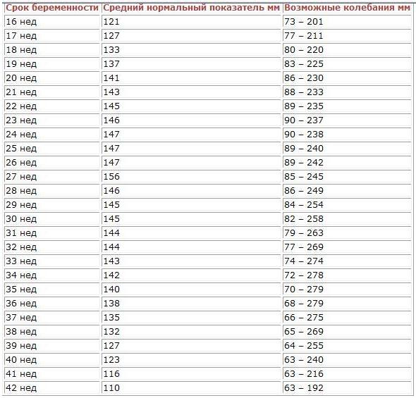
- Anatomical dimensions of the fetus are in this period very important. The size of the head of the fetus at 32 weeks of its intrauterine growth is 310-323 mm. The average weight of babies in this period of pregnancy is 1.8-2.4 kg. Values of abdominal circumference are 265-285 mm.
- Also to assess the development of the skeleton are considered and the size of some bones. The length of the humerus at week 32 of intrauterine development in a fetus is 55-60 mm. The femur is somewhat longer. Its values are 60-66 mm. The total length of the fetus is usually 43-48 cm.
- Cardiotocography indicators are scored. In the normal course of pregnancy, they are 8-12 points. Their assessment consists of a combination of some indicators. These include: heart rate and amplitude, basal rhythm and various deviations from it, as well as the speed of propagation of pulse waves.
- If the indicators of the future mother's cardiotocography fall into the range from 6 to 7 points, then in this case a mandatory additional consultation by an obstetrician-gynecologist. If this value falls below 5, hospitalization may be required.
- Doppler indices also very important. Normal blood flow in the uterine vessels is usually 0.34-0.59. This indicator is also called the resistance index. The blood flow in the blood vessels of the umbilical cord is 0.5-0.74.
- Together with instrumental diagnostic methods, doctors also evaluate biochemical screening indicators. Normal values of hCG at week 32 of intrauterine fetal development range from 2,750 to 78,000 mIU / ml. The norm of alpha-fetoprotein in this period of pregnancy is 100-150 units. The concentration of placental lactogen in the 32nd week of pregnancy is 3.2-10.1 mg / l.
These indicators are fairly average. In some cases, significant deviations are possible. The results are interpreted by an obstetrician-gynecologist.
In some cases, especially if there are doubts about the development of chromosomal or genetic pathologies, genetic counseling may be required.


When do false-positive results appear?
Incorrect diagnostics lead to the fact that the screening results can be considered unreliable. Quite often leads to incorrect interpretation of the obtained values multiple pregnancy. In this condition, the concentration of hCG significantly increases in the blood.
If screening is not carried out within the prescribed periods of pregnancy, the results also become unreliable. In vitro fertilization can also cause confusion with the establishment of diagnoses.This is largely due to a significant increase in gonadotropin or hCG in the blood.
In order for the doctors to consider the screening results to be correct, it is necessary to comply with all the regulated deadlines for carrying out such a study.
Delaying them can lead to overdiagnosis. In this case, mandatory monitoring of the state of the pregnancy course is required.
You will learn more about screening for the third trimester from the following video.


