Ultrasound in the 19th week of pregnancy: fetal size and other features

The 19th week of pregnancy is a calm and yet easy “equator” of gestation period. Before birth, it is still a long time, but the “interesting” position has already become noticeable to others. The stomach was rounded, and if the birth of twins is planned, then it cannot be hidden at all. This week a woman can wait for the second scheduled meeting with the baby on ultrasound. What the future mother can see, we will tell in this article.
Objectives of the survey
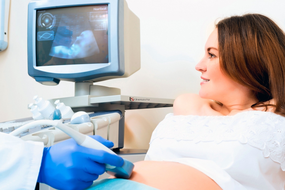
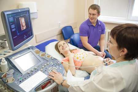
The second planned ultrasound scan, which is carried out on pregnant women who are registered at the clinic for women’s consultations, is usually appointed from 18 to 21 weeks. 19 week obstetric calculus - this is about 17 weeks of fetal life, exactly so much time has passed since the moment of conception.
Screening ultrasound scanning at this stage allows you to find out how a baby develops, establish whether he has developmental defects, and evaluate specific markers of possible pathologies.
The results of the ultrasound at this time complement the diagnostic laboratory picture, because somewhat earlier the expectant mother donated blood from a vein for biochemical analysis. Both results are cumulatively considered by a special computer program that calculates the risks of congenital anomalies.
In addition, an ultrasound scan at week 19 is prescribed to ensure the well-being of the child, if a woman has disturbing symptoms - nagging pains, atypical for mid-pregnancy discharge. Besides, An ultrasound scan may be recommended if the doctor has doubts about the correctness of the gestation period.

Method of conducting
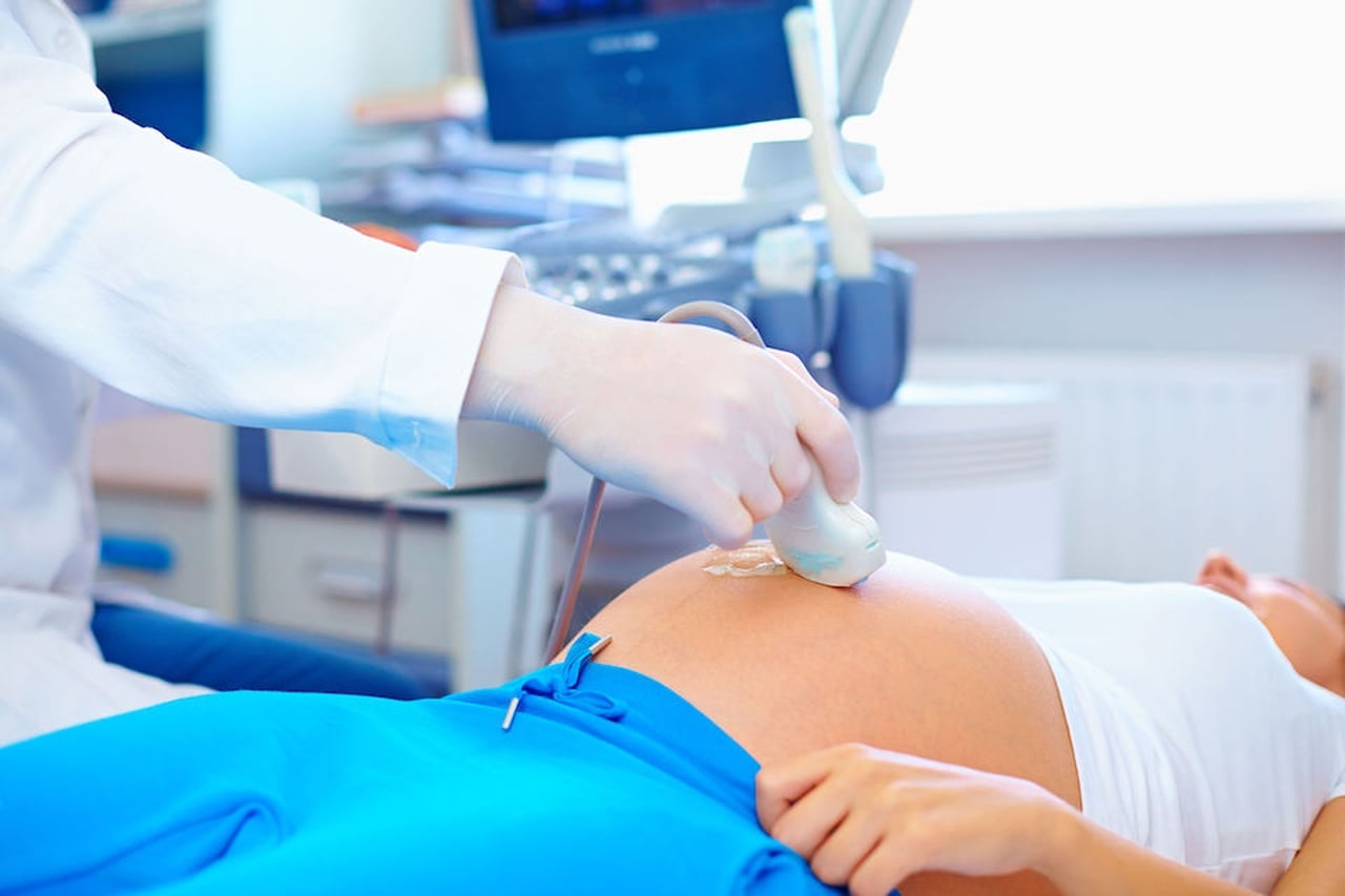
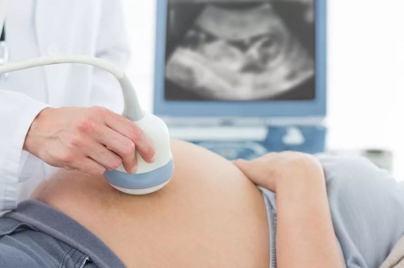
Ultrasound this week is performed abdominal - the scanner sensor is located on the anterior abdominal wall. No diet and reception of agents that reduce gas formation in the intestines are no longer required at this time, because the uterus is already quite large, and the amount of amniotic fluid allows ultrasonic waves to penetrate and reflect without problems.
To scan, a woman is placed on a couch in a supine position or on a side, if she bears twins, and the pressure on the inferior genital vein while lying on the back is already noticeably pronounced.
Vaginal ultrasound screening can also be applied., but only if the woman has a suspicion of threatened abortion. An internal examination with an intravaginal sensor allows you to accurately assess the condition of the cervix and cervical canal.
It is worth taking a clean diaper with you for examination to cover the couch, as well as paper napkins to remove excess gel from the abdomen.
What shows ultrasound
At week 19, my mother is waiting for a lot of interesting things, because her crumb has already grown noticeably. The size of the fruit at this time can be compared with a large potato. The growth of the baby is about 19-22 centimeters, and the weight is about 200 grams.
All organs and system of the baby are formed, now they are actively growing and improving. In the crumbs of the brain at week 19, special zones are activated, which are responsible for the five main senses - touch, smell, hearing, sight and taste.
The baby still looks thin, but from this week he begins to form subcutaneous fat, which will warm him immediately after birth until the thermoregulation processes are established. Thanks to him, the crumb will delight loved ones with puffy cheeks, dimples.
At week 19, the neck muscles become stronger, and the baby gains the opportunity to twist his head, “look around” on the sides. Small fingers on the arms and legs bend in all joints.
Secondary women already feel fetal movement, many women who have decided to become mothers for the first time are in a pleasant expectation of the first noticeable perturbations.
On the ultrasound monitor this week, the mother and the diagnostician will be able to see the baby who is actively moving, he still has enough space to even perform coups over his head and somersaults.
On the ultrasound you can listen and see how the crumbs of the heart beat hard and rhythmically. On this period, it is already possible to determine the sex of the baby with great accuracy.
An erroneous version of the diagnosis of sex during this period is unlikely, since the genitals are formed and clearly visible, and the baby, who is still not crowded, does not hide them, as happens in late terms in the third trimester.
In addition to the size of the fetus, the doctor will ask about the state of the placenta, which is already fully formed and performs the entire range of functions for nutrition and protection of the baby. With the help of ultrasonic waves its thickness will be measured, the location is estimated.
Decryption and rate
First of all, the diagnostician will tell you how many babies grow under the heart of a woman - one, two or three, and also assess the viability of each of them. Viability is determined by the presence of heartbeat and motor activity.
The doctor will also tell you in which position the child is in the head, pelvic or transverse position in relation to the exit from the uterus. To worry about the "wrong" location of the baby is not worth it. The child will change its position many times until the end of the gestation period.
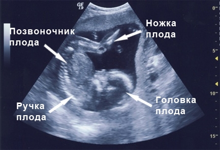
The main dimensions, which are calculated on ultrasound, are fetometric indicators. These include the size of the head - transverse, which is also called bipariential, and longitudinal - frontal-grinding. The doctor also measures the circumference of the head, tummy and the length of paired bones.
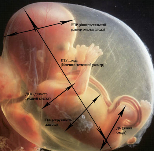
All these indicators are considered important signs of the normal development of the baby, as well as its compliance with gestational period.
Table norms fetometry at 18-19 week:
PDU, mm | LZR, mm | OG, mm | Coolant, mm | The length of the femur, mm | Shin bones, mm | Forearm bones, mm | Humerus, mm |
42-45 | 54-58 | 146-158 | 124-134 | 27-30 | 24-27 | 20-23 | 28-31 |
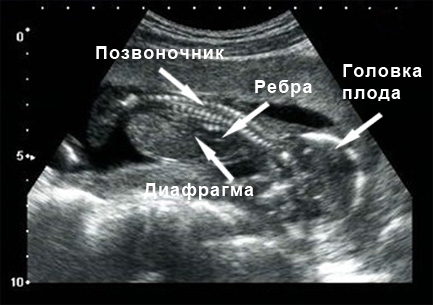
In the protocol ultrasound, the doctor describes the anatomical features of the child. At this time all internal organs are well visible, their presence and proportions.
The structure of the brain, the baby’s heart, stomach, kidneys, lungs, liver, gall bladder, intestines are subject to careful study. The term already allows you to see the malformations, if any. If there are no gross violations, the doctor writes that the internal organs are normal and there are no signs of developmental pathologies.
The thickness of the placenta this week is normally 20-21 mm. It should be zero degree of maturity with no signs of growth or thickening. Increased muscle tone of the uterine walls, as well as shortening of the cervix and the tight closure of the cervical canal may indicate threats of interruption.
Possible problems
The list of possible problems and issues that may arise in expectant mothers at week 19 is quite wide. We tried to answer the most common ones.
The parameters of the baby are behind the norm
It’s not worth worrying about and being nervous if the dimensions of the fetometry deviate by no more than two weeks. The reason may lie in late ovulation. Usually, in such a situation, a slight lag from the size of the average was noticeable even during the first ultrasound in the first trimester.
Sometimes the cause of deviations in size may be intrauterine growth retardation. A woman will definitely be shown to undergo additional diagnostics.
It is not necessary to consider each parameter separately, this is the key mistake of all future mothers.
If the ultrasound shows that there are deviations in only one parameter, this does not mean anything bad. It’s just that children grow up differently, “jumps”, and a slight difference can even out in a week or two.
Doctors look at fetal indicators of the fetus differently - for diagnosis, proportions rather than specific dimensions are of greater importance.
Why, after the ultrasound gave direction to genetics
Do not forget that the main purpose of this ultrasound is the screening of possible pathologies. If the diagnostician has some doubts about the outlines of the nasal bones, facial profile, pathologies of internal organs, he suggests that the child may have Down syndrome or have signs of achondroplasia, he will advise you to visit the genetics and do another ultrasound expert class.
A geneticist can offer invasive diagnosis to future mothers - cordocentesis or amniocentesis, in order to get results with an accuracy of up to 99%, which will allow to judge whether the baby has pathology or not.

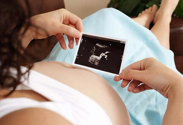
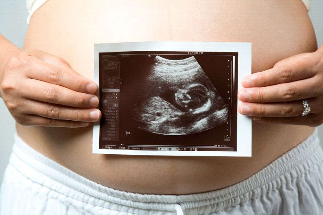
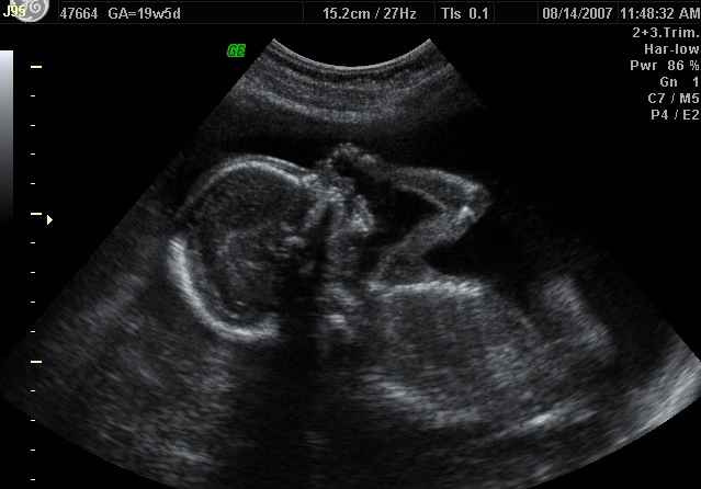
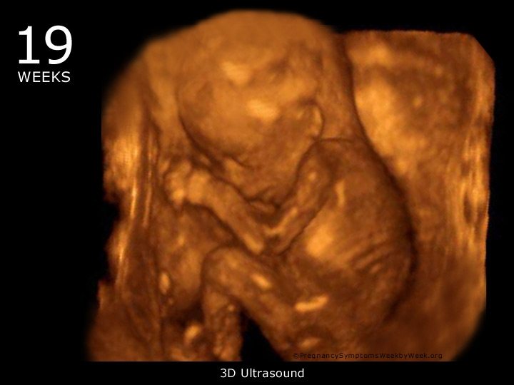
Snapshots
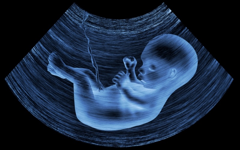
The usual two-dimensional ultrasound images this week give only a general idea of what the baby looks like. More details about the baby can be on the 3D-ultrasound.
Gender, defined and demonstrated to the future mom and dad during the passage of three-dimensional scanning, no doubt.
For information on how 3D and 4D ultrasound of the fetus takes place at 19-20 weeks of pregnancy, see the following video.


