The size of the liver by ultrasound in children
Modern methods of diagnosis of various liver diseases in children also include ultrasound. This examination has become routine and is used in a variety of clinical cases. Establishing the size of the liver is included in any clinical protocol for ultrasound.
Features of the structure and functioning of the liver
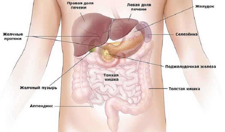
The liver is an organ that is responsible in the body for the performance of various functions. These include: the synthesis of certain hormones, detoxification of decay products and chemical toxins, participation in blood formation, the formation of bile, the maintenance of immunity, and many others. Determining the size of the liver is very important. Many pathological conditions, including very dangerous for the life and health of children, cause a significant increase in the liver - hepatomegaly.
For quite a long time, only the palpation method was used to determine the boundaries of this organ. It is carried out by doctors and to the present when conducting a clinical examination of the child. However, liver palpation and the definition of boundaries by this method is only indicative. The true size of the body can be determined only when using special instrumental types of surveys.

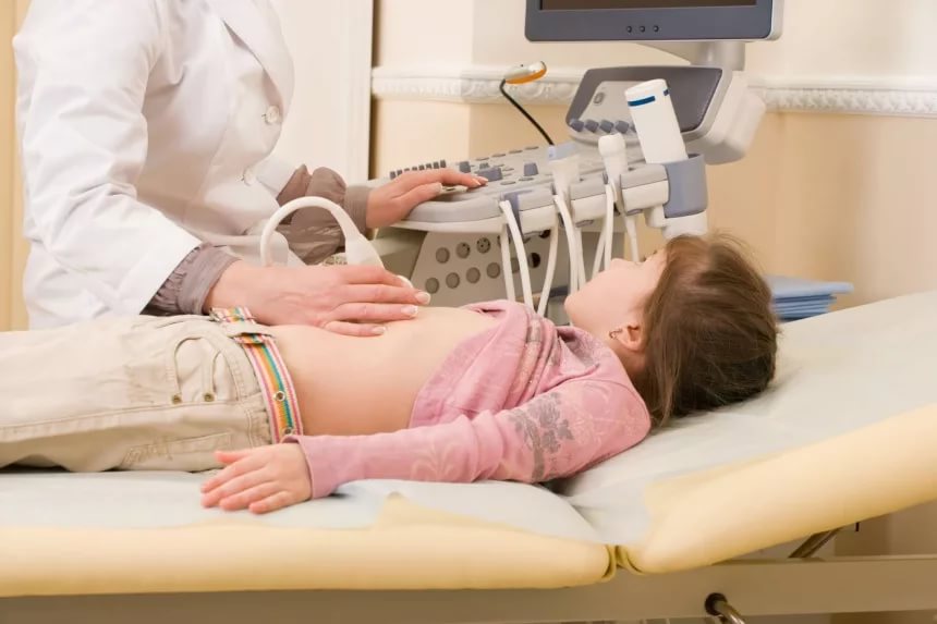
Currently, such instrumental tests include ultrasound. It is safe and does not cause any pain in the child during the procedure. Usually the duration of one study is 20-25 minutes. The time of the ultrasound procedure usually depends on the qualifications and experience of the doctor conducting the examination, as well as on the emotional state of the child. If the baby is nervous or starts screaming and crying, this can significantly complicate the conduct of the study.
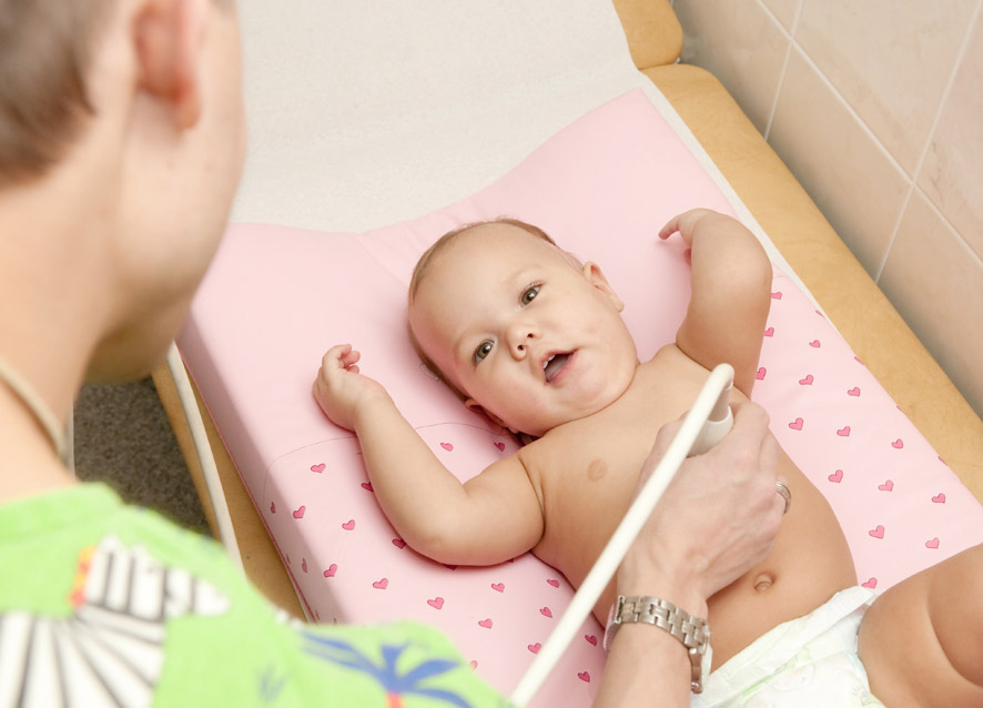
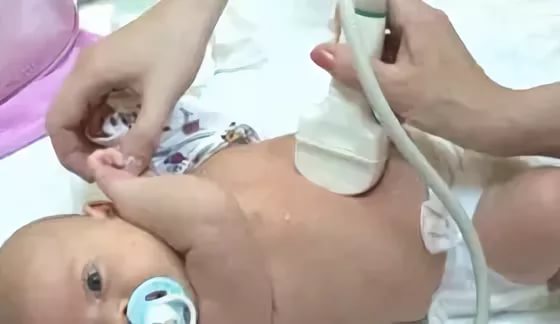
Ultrasound is usually performed in a special darkened room. The baby is lying on his back on the diaper-bed. The doctor lubricates the sensor with a special gel and begins to conduct research. During the examination, the doctor can see all the pathological changes in the liver tissue, as well as determine the size of the borders of the liver.
Ultrasound examination is actively carried out by children from 2 years. At an earlier age, there are certain clinical indications for ultrasound.

There are special tables that indicate the normal values of the size of a healthy liver. They are used by doctors of ultrasound diagnostics, working all over the world. Tables are made taking into account the age of the child. They allow doctors to evaluate the result and are also needed to establish the clinical signs of hepatomegaly.
The structure of the liver includes several anatomical structures - they are called hepatic lobules. Ultrasound allows you to set the parameters of the right, tail, left and square lobes of the liver. Also using this study, you can identify various pathological changes in all 8 segments. Ultrasound examination is also very effective for diagnosing the detection of various liver pathologies in newborns.
In addition to the structure of the liver tissue, the ultrasound doctor also conducts visual inspection of all adjacent anatomical organs with the liver. Experienced doctors can also evaluate the condition of the ligamentous system of the liver. Usually ligaments become visible when free fluid appears in the abdominal cavity.
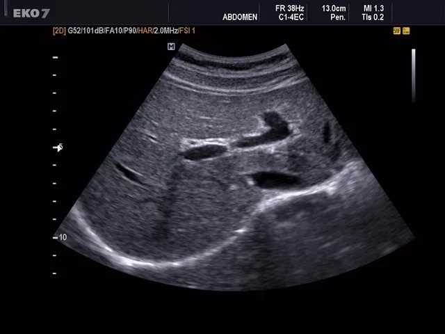
During the study, the doctor also conducts an examination of the blood vessels that feed the liver. For this, an additional doppler mode is used.

Currently, there are a huge variety of a variety of tables, which indicate the age parameters of the normal size of the liver. Below is one of them. To assess the condition of an organ, the dimensions of the right and left lobes of the liver are mainly used. The parameters depend on the age of the baby. It is important to note that these indicators have an approximate value and should be assessed comprehensively when setting a clinical diagnosis.
The size of the liver is normal (from top to bottom) is presented in the data in the following table:
Child's age | Right lobe, mm | Left share, mm |
1 year | 60 | 33 |
3 years | 72 | 37 |
4 years | 78 | 39 |
5 years | 84 | 41 |
7 years | 96 | 45 |
9 years | 100 | 47 |
11 years | 100 | 49 |
13 years old | 100 | 50 |
15 years | 100 | 50 |
18 years | 120 | 50 |
Training
To determine the exact boundaries of the liver and the true size of this body by ultrasound is quite simple. The high resolution of the instruments allows obtaining the most accurate and reliable imaging of the internal organs. Hundreds of thousands of ultrasound examinations are performed daily around the world.
To obtain an accurate result when reviewing the internal organs, it is very important to conduct preliminary training. Many parents often neglect this, which subsequently leads to the fact that the doctors of ultrasound diagnosis can not establish an accurate diagnosis. Preparation for an ultrasound is not difficult and it is easy enough to carry out independently at home.


The main condition for performing a qualitative study is the release of gas from the intestine. It significantly reduces obtaining a reliable result.
The intestinal loops swollen with gas do not allow the doctor to fully examine the liver and all its anatomical components. In order to reduce gas filling, a couple of days before the survey, all foods that increase gas formation are removed from the baby’s diet. These include: fruits and vegetables rich in coarse fiber, bread and muffins, sodas and kvass, milk, and a variety of pastries.
If your baby has chronic diseases of the digestive organs that cause pronounced gas formation, then you should consult with your doctor about which drugs should be used before the ultrasound procedure to reduce gas in the intestine. Usually, enzymes and enterosorbents are assigned to children. Before conducting an ultrasound examination, the baby should not be given gastro or fibrocolonoscopy.


Psychological attitude before the study is also very important, especially for the youngest patients. Younger children are better to turn the examination into a real exciting game. Older children and especially teenagers should discuss how the procedure will be performed. During the conversation, it should be noted that this study is absolutely safe and will not cause any pain in the child.
What is it for?
Ultrasound examination of the liver is carried out in a variety of pathological conditions. This method allows you to get fairly accurate and reliable results that help physicians to establish the correct diagnoses, and thus prescribe the optimal and effective treatment regimens.
The absence of radiation exposure allows the use of this study in both newborns and toddlers of older age.

Ultrasound of the liver is used to diagnose:
- Inflammatory diseases. As a result of pronounced inflammation changes the density and structure of the liver tissue. This leads to the fact that the liver grows in size. This symptom may be persistent or disappear after treatment.
- Neoplasms and malignant tumors. Today, it is the liver ultrasound that is used as a screening for these dangerous diseases. Using this method, it is possible to detect tumors in the liver at fairly early stages. Ultrasound also reveals the metastases of other neoplasms growing in other organs.
- Cystic formations. The presence of cavitary contents in the liver tissue is often a sign of a cyst. Such a pathological condition is often recorded in children during ultrasound. Detection of a cyst should be a pretext for a consultation with a doctor.
- Liver cirrhosis. This pathological condition practically does not occur in children's practice. Pathology is manifested by a marked increase in the liver. The prognosis of the disease is extremely unfavorable.
- Stagnant processesassociated with biliary excretion. Severe bile stasis can contribute to the development of chronic cholecystitis or hepatitis A. Stretching the bile duct indicates the presence of signs of accumulation of bile.
- Vascular pathologies. Liver hemangiomas are usually very well detected during ultrasound with an additional doppler mapping mode. The study allows to identify these dangerous pathologies at the earliest stages.
To learn how to prepare for a liver ultrasound, see the following video.


