4D ultrasound during pregnancy
Ultrasound diagnostic methods are constantly being improved. This is especially important when conducting ultrasound during pregnancy. This article will tell future mothers in more detail about the most modern method of ultrasound, called 4D.

What it is?
Doctors also call this type of examination “live” ultrasound. This newest method allows the specialists who conduct it to see not a static picture on the monitor of an ultrasound machine, but a full-fledged three-dimensional image.
This type of examination every day becomes more and more popular among future parents. Coming to a 4D ultrasound, moms and dads see their baby on the monitor, which usually moves or makes any movements with its arms or legs. Such research leads future parents. in real delight.
Immediately it should be noted that at this time their fetus does not feel such joy at all.

Initially, this research method was not designed as a fun or exciting procedure. Scientists have come up with it to identify difficult to diagnose defects intrauterine development. Also with the help of 4D studies, it is possible to identify various heart defects, as well as abnormalities in the cardiovascular system of the fetus.
During the examination, the doctor examines the uterus, with the fruit in it from various angles. The device processes the information received from the ultrasonic sensor and forms a three-dimensional image on the screen. Such an image is not static. Modern devices can simulate real movements that the fetus makes in the womb during the direction of the ultrasound sensor.
It should be noted that 4D ultrasounds can not be performed in all medical institutions. Ultrasound equipment used for such examinations is usually very expensive. Also to work on it requires some clinical experience and appropriate education.
It is quite difficult to enroll in experienced specialists who conduct such studies, as a rule, even in private medical centers.

When is appointed?
Future moms should remember that to conduct any ultrasound, including 4D, should be only for certain medical reasons. The normal course of pregnancy does not imply such a test.
Assigned to this study gynecologist. Usually perform 4D ultrasound is recommended for women with certain diseases of the internal genital organs or comorbidities. So, if the future mommy has cardiovascular diseases or there is a high risk of their development, then this research will be shown to her.

Doctors identify several most favorable periods of pregnancy, in which it is better to conduct this test. These include time intervals. from 20 to 24 and from 30 to 34 week intrauterine development of the future baby. Reviews of moms who have passed the research data on such dates also confirm this. They note that they have transferred this procedure quite well without any discomfort or adverse effects.
In some situations, research is required on expert-class devices.This is usually necessary if a pregnant woman has any complex pathologies that are almost impossible to detect with the help of conventional ultrasound diagnostics devices.
Also conducting research with the help of expert class equipment is required if the future mom bears several kids at once. Pregnancy after IVF may also be an indication for more accurate examination methods. Quite often, the use of expert ultrasound resorted to with surrogate motherhood.
How is it done?
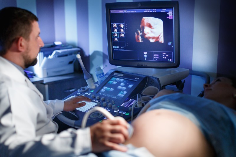
The method of conducting 4D research is somewhat different from the usual ultrasound test. The first significant difference is the time of the survey. A routine ultrasound scan usually takes from 20 to 30 minutes. For a 4D study, the doctor may spend about an hour.
As a rule, the patient being examined lies on a couch on the back. In the later stages of pregnancy, the doctor performing her research may ask her to roll over to the left side.
In this position, visualization of the uterus is significantly improved, as the pressure on the inferior vena cava decreases.
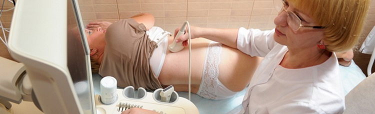
To obtain a high-quality image on the monitor, the patient's abdomen is smeared with a special gel. This transparent adhesive is necessary for better penetration and reflection of sound waves during the study. You should not be afraid of him. Its chemical composition absolutely harmless for both mom and her future baby. After carrying out this procedure, the remnants of the gel from the abdomen can be removed with a usual paper napkin or a handkerchief.
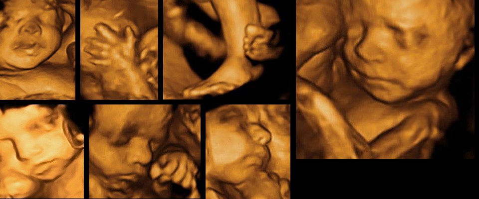
During the study, the doctor changes the position of the ultrasound sensor in different directions. This allows him to achieve on the monitor screen a fairly clear visualization of all parts of the spine and skull, as well as to examine the vital organs of the baby.
Also, with the help of 4D ultrasound, specialists can detect signs of various chromosomal and genetic diseases.

Investigation of the fetal blood flow is also an important issue that interests doctors watching the development of the future baby in the womb. With the help of carrying out volumetric ultrasound, it is possible to consider the blood vessels feeding the fetus. Also during the procedure, various pathologies present in the mother's body can also be identified.
Evaluate the work of the baby's heart is also possible with the help of this study. The uniqueness of this test is that you can inspect all the structures of the heart, its valves, as well as the coronary vessels. Careful consideration of such anatomical structures and the identification of pathologies helps to identify various heart defects in a timely manner. Usually in this case, repeated ultrasound is performed, which is performed in the first days after the baby was born.

Doctors say that with the help of 4D research, you can also identify various violations in the structure of the skeletonas well as emerging neurological pathologies. Such pathological conditions include hypoplasia, severe malformations of the fetal neural tube, as well as signs of skeletal underdevelopment.
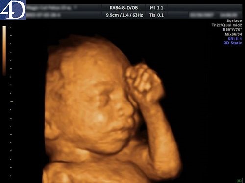
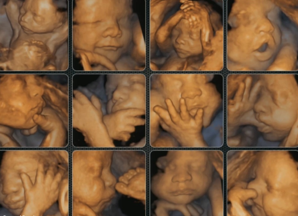
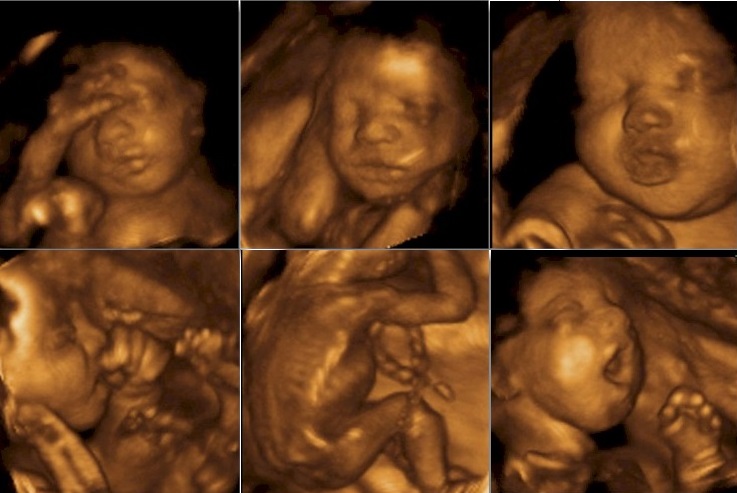
The image that is formed due to the reflection of sound waves of a certain frequency appears on a special monitor. During the study, he is seen by both the doctor and the expectant mother. AT third trimester of pregnancy visible not only active movements of the baby when he sucks his finger or twists his foot, but also his facial expressions.
While the ultrasound sensor is pointing at him, the child usually frowns forehead or smiles.
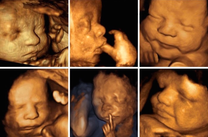
The appearance of a child of various emotions immediately amenable to interpretation. So, the future mommy believes that if the baby frowns, then he feels bad. It's not like that at all. The manifestation of emotions in the prenatal period of development - just a behavioral factor. A pregnant woman should not panic if she saw on the face of her unborn child some sort of grimace of displeasure or pain.This emotional background will be formed in the baby after his birth.
A significant advantage of this procedure is the ability to record images on various electronic media. Future parents can get a video or photo of their baby on a special disk. Also, a picture can be printed directly in the ultrasound room. Usually such a first photo of a baby causes tears of joy and genuine delight in future parents.

In some situations, unfortunately, get a high-quality photo or video does not work. This often occurs when a pregnant woman has signs of severe obesity. A large number of subcutaneous fat violates the conduct of ultrasonic waves, which can lead to a distortion of the result.
Previous operations on the uterus, which led to the appearance of scars on it, can also contribute to the fact that it will be impossible to obtain an image of the fetus.
High standing of the placenta is another clinical factor that will lead to the impossibility of recording the first intrauterine video of the baby.
If the pregnancy proceeds with shallow water, then in this case errors are also possible when trying to capture the image. In this situation, the picture becomes less clear, the outlines of the child become blurred outlines.
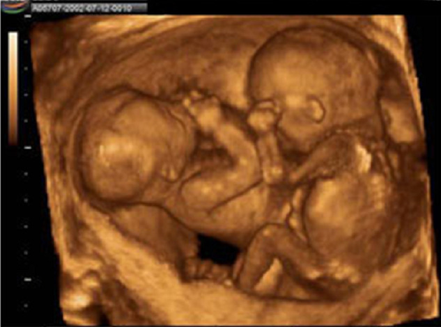
Using 4D research, you can also determine the sex of the child. The high resolution of expert-class devices allows you to get the most accurate result. Errors in this case almost never occur. In pregnancy, twins using this method, you can determine the sex of each baby.
Modern devices allow determine the intended weight and size of the fetus. Technical errors in such situations practically do not arise. Experienced ultrasound diagnostics specialists can determine the weight of the fetus with an accuracy of a couple of grams. This definition is very important, as it indicates how well the future baby develops in the womb.
Many moms do not know what kind of ultrasound method they choose. In this case, I would like to advise that such a decision should be made not independently, but together with your doctor. An experienced doctor will never order a study if it can do any harm to the unborn child.
Pregnant women should always remember that for screening various pathologies of pregnancy is enough to conduct and simple ultrasound. 3D or 4D studies are not significantly different. The difference in them is one - obtaining a spatial image.

Alleged harm
Obstetricians and gynecologists say that it is better not to do 4D ultrasound in the early stages of pregnancy. Research during this period of fetal development may be harmful. The initial weeks of embryo growth are accompanied by active organogenesis - the process of internal organ insertion.
The impact of ultrasound waves of a fairly high frequency and intensity can have an adverse effect on this process and lead to the development of undesirable consequences in the future. As a rule, these pathologies appear in a baby after birth.

In some cases, future mothers do such a study in the 12th week of pregnancy. As a rule, on their own. In this case, it should be remembered that if during the entire pregnancy no more than 2-3 ultrasounds are done, then it is not worth worrying about the risk of undesirable adverse effects. More frequent conduct of this study can lead to the fact that the child will manifest various deviations of the nervous and cardiovascular systems after birth.
There is no substantial need to perform 4D for a period of 12 weeks instead of the usual ultrasound. Even the usual research conducted in dual channel mode will show all the pathologies and abnormalities that are present in the baby.The normal course of pregnancy also does not require a “bulk” instrumental test.

The adverse effect on the fetus has a thermal effect. It arises, when the ultrasonic sensor touches the skin. The duration of the procedure also only contributes to a strong overheating of the tissues. Scientists have found that the frequent conduct of long-term ultrasound can lead to various neurological disorders in the baby after birth.
In modern devices, the thermal effect is practically minimized.
The developers of such equipment try to make this type of research as safe as possible for the body of a pregnant woman and for the fetus.

For more information about 4D ultrasound, you can find out further.


