What is hyperechoic focus in the left ventricle of the fetal heart and is it dangerous?

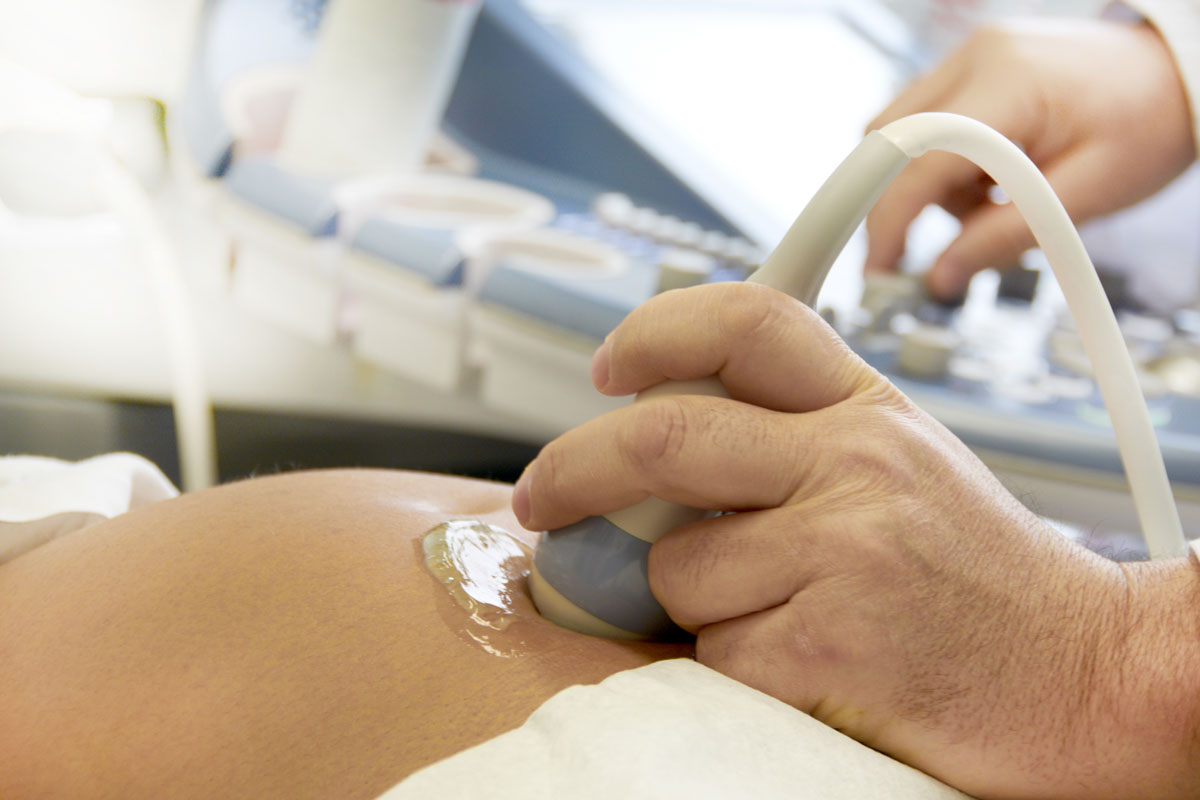
Ultrasound examination is one of the most informative and important during pregnancy. It allows not only to see the sex of the unborn child, but also makes it clear whether the development of the crumbs is normal, whether he has defects and diseases. But the terms used by doctors are not always understood by expectant mothers. A striking example of such terms is the detection of hyperfocus in the ventricle of the fetal heart.

What is meant?
Features of the structure of the baby's heart are interested in doctors on each ultrasound. There is nothing surprising in examining the structure of the ventricles, the atria. But the hyperechoic focus that interests us is always a random and unexpected finding. Especially no one looks out for her, because there is no disease or defect with such a name in medicine.
Hyperechoic is the area of the baby's heart, which appears on the monitor of an ultrasound scanner colored most brightly. This is a brighter spot of no more than 3 mm in size, which is noticeable in one of the little baby’s heart chambers.

The main thing that every woman who is preparing to become a mother needs to know is that the discovery of the GEF itself is not considered to be something scary and out of the ordinary. This is not a pathology, not an abnormal sign, not a defect, but only a small feature of the baby’s heart structure, which in no way can affect the functioning of a vital organ.
In the initial stages, hyperfocus is usually not detected, but often such a conclusion is carried out on ultrasound scanning after 17-18 weeks of gestation. Hyperfocus can disappear by itself, and already on the next ultrasound it will not be found, but it can be kept by the baby until the very birth, and at the same time the crumb will develop quite normally.
The brightly colored portion of the heart muscle may also be found in the right ventricle, but for most babies it is located in the left. 7 pregnant women for every hundred expectant mothers are faced with a similar phenomenon, and therefore it is impossible to call a rare hyperfocus precisely. Ultrasound machines of the highest quality often give the GEF as a hindrance, an error; if you re-examine it on a higher, expert-class device, the suspicions are not confirmed.

When is hyperfocus the norm?
The origin of the hyperechoic focus in the cardiac cavity of the fetus is still one of the most dubious and mysterious mysteries of science. It is most often associated with the fact that more calcium salts are deposited in a separate part of the heart muscle. It is believed that this occurs precisely when the baby starts intensive bone mineralization processes. This partly explains why the GEF is not found in early pregnancy, because mineralization is gaining momentum in the second trimester.
In this case, the hyperechoic region should not cause any anxiety and embarrassment - an amazing phenomenon, difficult to explain, but completely harmless.
There is another reason why the GEF site in the atrium can be detected - the presence of certain inclusions in the heart muscle structure itself. Often this is the way additional or false chords are seen on ultrasound. These areas of connective tissue are usually very thin, and if additional chords are found, they may well become noticeable. This happens with many people on the planet.By themselves, false, additional chords do not pose a danger to the life and health of the child, they do not violate the functions of the children's heart. It does not need treatment.
When is the GEF a danger signal?
Sometimes hyperechoic focus can be detected in babies with genetic pathologies. Previously, women were frightened of them, because he was considered one of the clearest signs of congenital gross abnormalities of the fetus, in particular, a sign of the presence of Down syndrome in the baby.
Today, GEF has a more loyal attitude, because an isolated “find” in a baby’s heart without other characteristic markers of trisomy in no way indicates that the baby has such pathologies, and also does not in any way increase the risk of chromosomal pathologies. therefore it is impossible to assume that the fetus has the same Down syndrome or another chromosomal abnormality only on the basis of a random finding in an ultrasound study of a hyperechoic focus.
A woman will be sent to a genetics consultation only when other signs characteristic of chromosomal abnormalities are detected - flattening of the nasal bones, thickening of the back of the cervical folds (collar space), deviation from the norms of quantitative indicators of certain hormones and proteins, which are determined in the blood of the future mother as part of prenatal screening.
You will also have to visit genetics if the GEF is detected in combination with other anomalies, for example, intestinal pathologies, anomalies of the structure of the extremities of the little ones, etc. If, in addition to hyperfocus, nothing is found, there should be no reason for excitement.

What to do when detected?
If an ultrasound scan in a woman reveals a hyperechoic focus in the child's left ventricle, the only thing that is needed at this stage is a thorough examination to see if the baby has other abnormalities or there is nothing besides the GEF.
To this may recommend the following.
- Expert-class ultrasound (there are such devices in every region in large maternity homes or in perinatal centers. 3D ultrasound can be done.
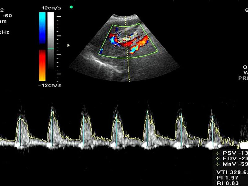
- Doppler ultrasound to assess the accuracy, speed and characteristics of blood flow in the heart and blood vessels of the baby.
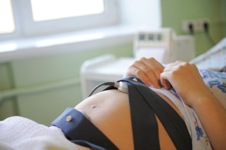
- Cardiotocography of the fetus, if the gestation period exceeds 29 weeks.

Even if the expectant mother has no reason to worry and the doctors will report an isolated GEF, such a mark will appear in her exchange card, but this should not be frightened again. This is reported, in particular, for a neonatologist at the maternity hospital, where a woman will give birth, but absolutely does not indicate pathology, abnormalities or illness. GEF does not need any therapy. At 33-34 weeks of gestation of the expectant mother with the previously established hyperechoic focus in the left ventricle of the fetus, a control ultrasound examination is performed that confirms or refutes its presence.
If this mysterious, brightly glowing spot on the ultrasound scanner screen remains on the heart muscle, it is assumed that these are unnecessary or false chords that do not in any way affect the baby’s well-being or health.
Just after the birth of the baby into the world, neonatologists in the children's ward of the maternity hospital will more carefully listen to the tones and noises in the baby's heart, if necessary, will make an ultrasound of the heart of the newborn.
There is no reason for the future mother with the GEF to worry and thus harm her child. - stress hormones, which she experiences while reducing the production of sex hormones, increases the likelihood of premature birth, the development of placental insufficiency. In addition, perinatal psychologists have long proved that the children in the womb perfectly feel the mood of the mother, and her excitement can harm the crumb much more, whereas the chords themselves or calcium deposits in the heart muscle do not cause any harm to the child.

For more information on hyperechoic focus in the heart of the fetus, see the following video.


