Ultrasound in the 9th week of pregnancy: fetal size and other features
At the 9th week of pregnancy, a woman usually registers with a antenatal clinic. This may require such a simple and informative procedure as ultrasound. What can tell ultrasound at this time, we will tell in this material.

Goals
The number of mandatory ultrasound procedures in 9 weeks is not included. Perinatal screening study is appointed only after a fortnight from 11 weeks. But the reasons for the examination at this time may be enough:
Unknown exact gestational age. Most often this happens in women with irregular menstrual cycles.
Pregnancy proceeds with complications. This may be bloody and blood-like discharge, the so-called "daub", pain, severe toxicosis.
- The mismatch of the size of the uterus obstetric period. The term physicians calculate from the first day of the last menstruation. Thus, 9 weeks is about 7 weeks from the moment of conception. The uterus is sufficiently enlarged so that an obstetrician-gynecologist can determine it during a manual examination.
If the reproductive organ is smaller, the woman will be sent for an ultrasound scan to exclude a non-developing pregnancy, tumors in the pelvis, which can "simulate" on the grounds of an "interesting position."
Pregnancy is twins or triplets, conception by IVF. The development of babies after in vitro fertilization requires enhanced control. If you have any questions the doctor carried out an ultrasound at any time.

Method and preparation
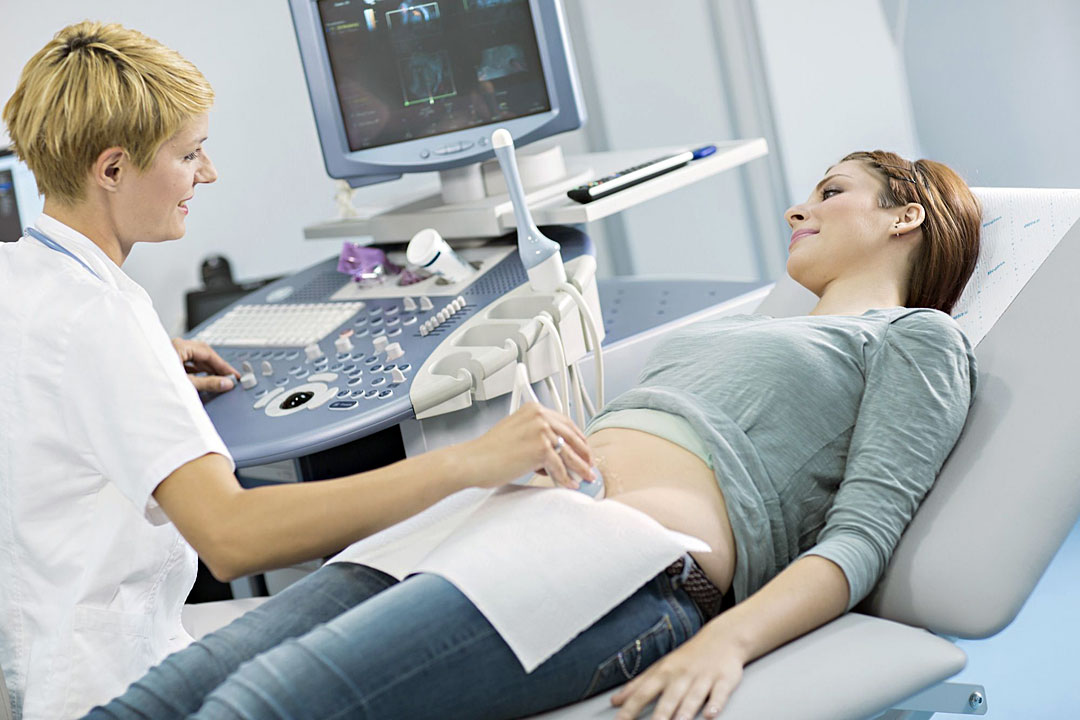
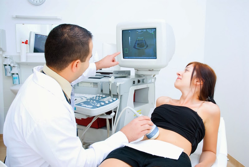
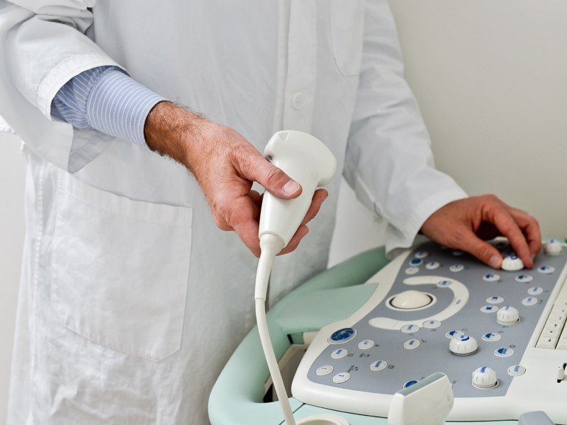
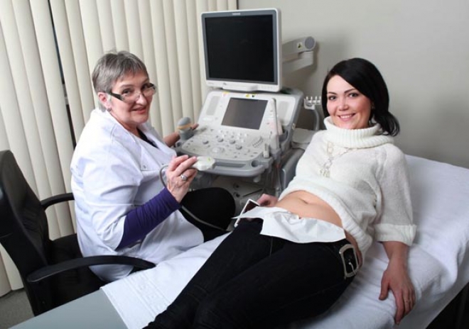
Ultrasonography at this time is carried out in a transvaginal way. Through the vaginal wall is easy enough to see all the necessary details. A vaginal sensor in a condom doctor examines on a couch where a pregnant woman lies, bending her knees, or on a viewing chair. The method in which the sensor is located on top of the abdomen, at this time can not be considered accurate and reliable, since the review of the uterus through the abdominal wall is still difficult.
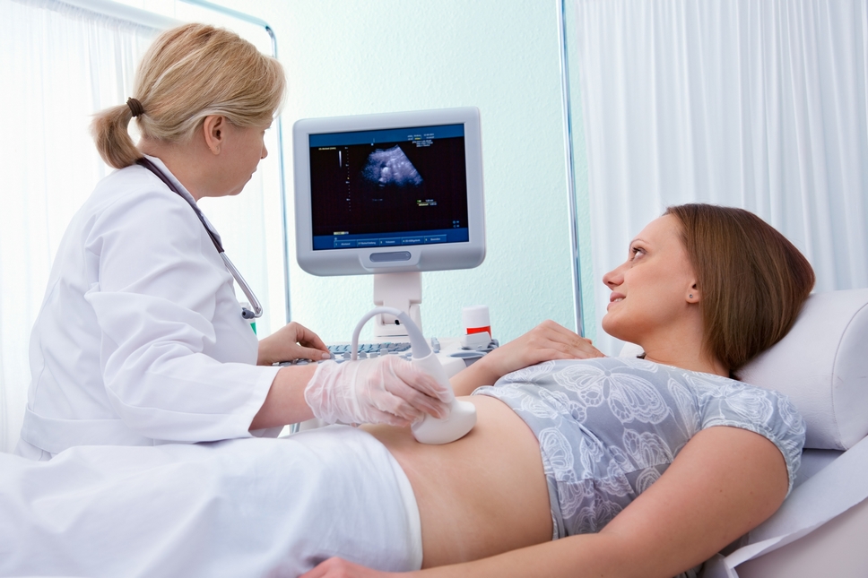
Preparation for ultrasound at 9 weeks implies that a woman needs to empty her bladder, and also to take drugs in advance that reduce the amount of intestinal gases, for example, «Espumizan"," Simethicone ". If the intestine is inflated, pressure on the pelvis will occur and the indications may be distorted. Ultrasound at 9 weeks lasts no more than 5-6 minutes, it does not cause discomfort to the woman, does not harm the child.
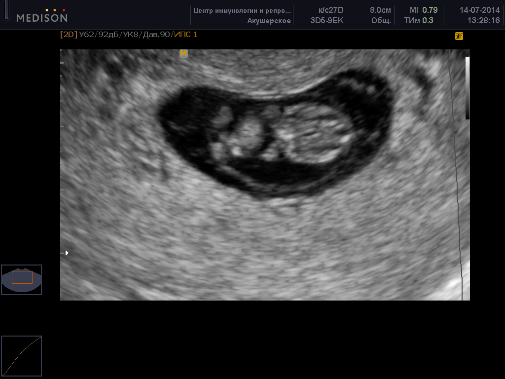
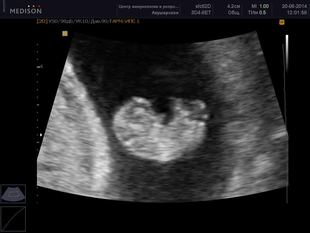
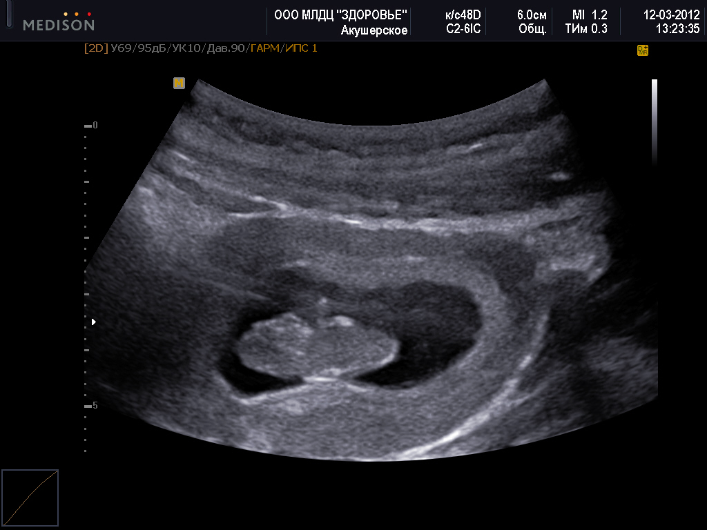
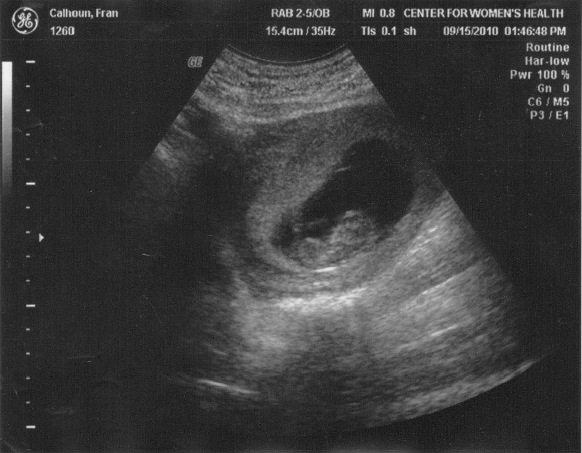
What can be seen on this date?
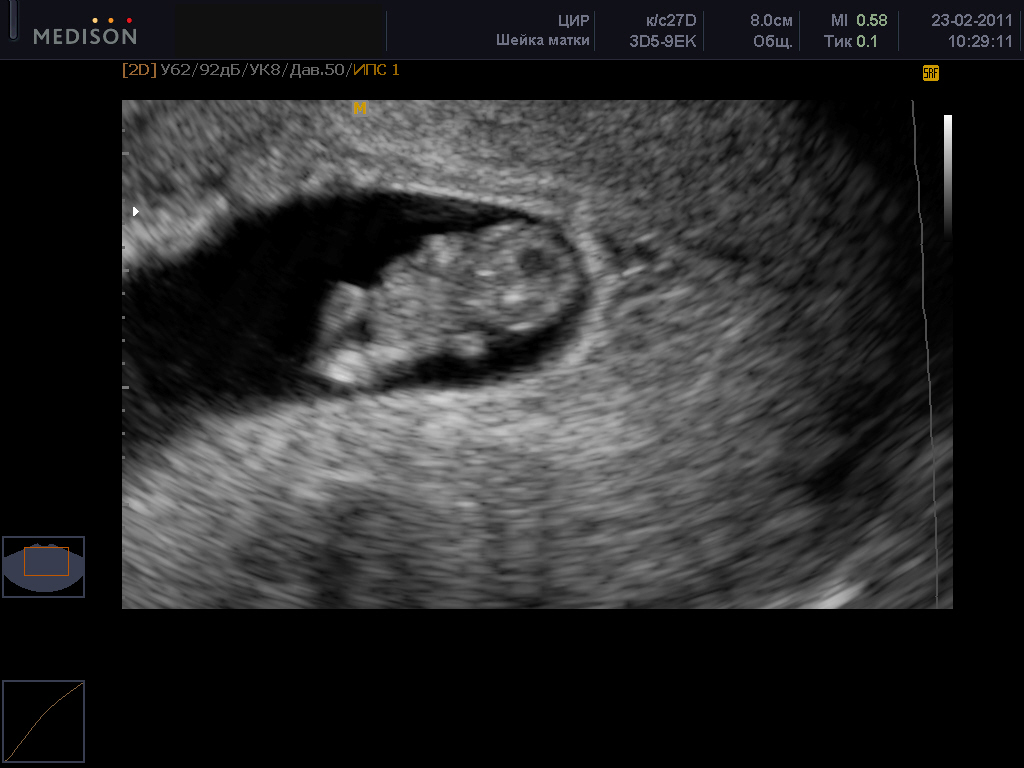
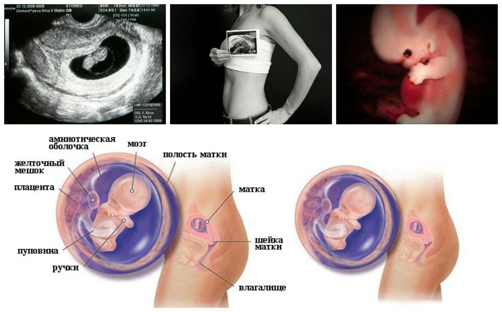
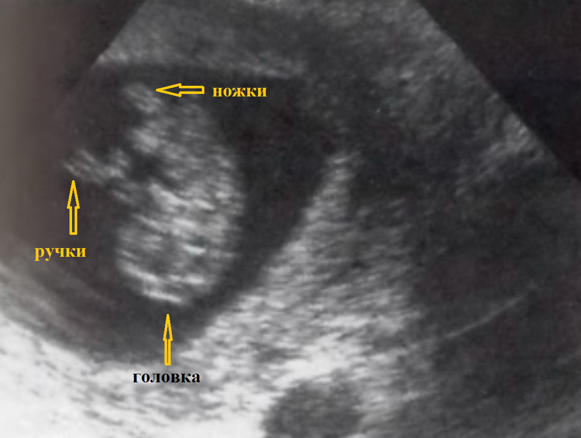
The baby at 9 weeks is very small, it weighs about 1.5-2 grams, and its average growth is about 29-30 mm. The baby is no longer an embryo, now all doctors respectfully call it a fetus, since the embryonic period of development is over. The toes on the hands and feet of the baby begin to form, the process of “shaping” the baby’s face begins - Formed facial bones on the head.
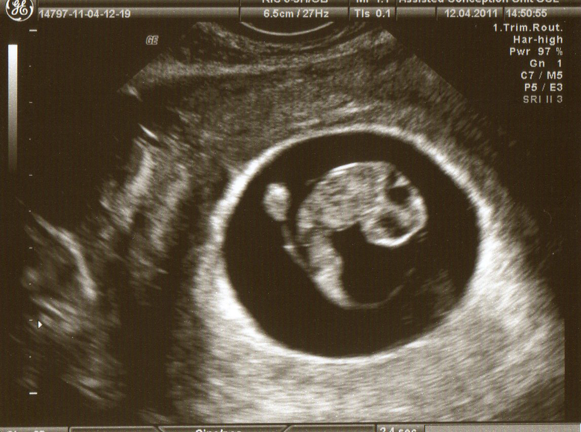
All this on ultrasound, of course, will not work. But it will be possible to listen to how his heart knocks, and also, if you're lucky, to see how the future son or daughter performs her first movements. In the womb it is still very spacious for him, and the expectant mother cannot sense his stirring.Starting this week, the fetus begins to respond to external stimuli, the formation of the ears, the reproductive glands, which are still in the abdomen and in boys and girls, continues.
A diagnostician in the ultrasound room will measure the growth of the baby, the size of the ovum in which it is located will assess the rhythm and frequency of contractions of the tiny heart. Special attention will be paid to the health status of a woman, because a lot depends on the work of her reproductive system. The received data will be entered in the form that the woman will give at the end of the inspection.
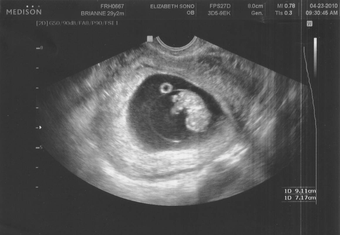
Decoding results
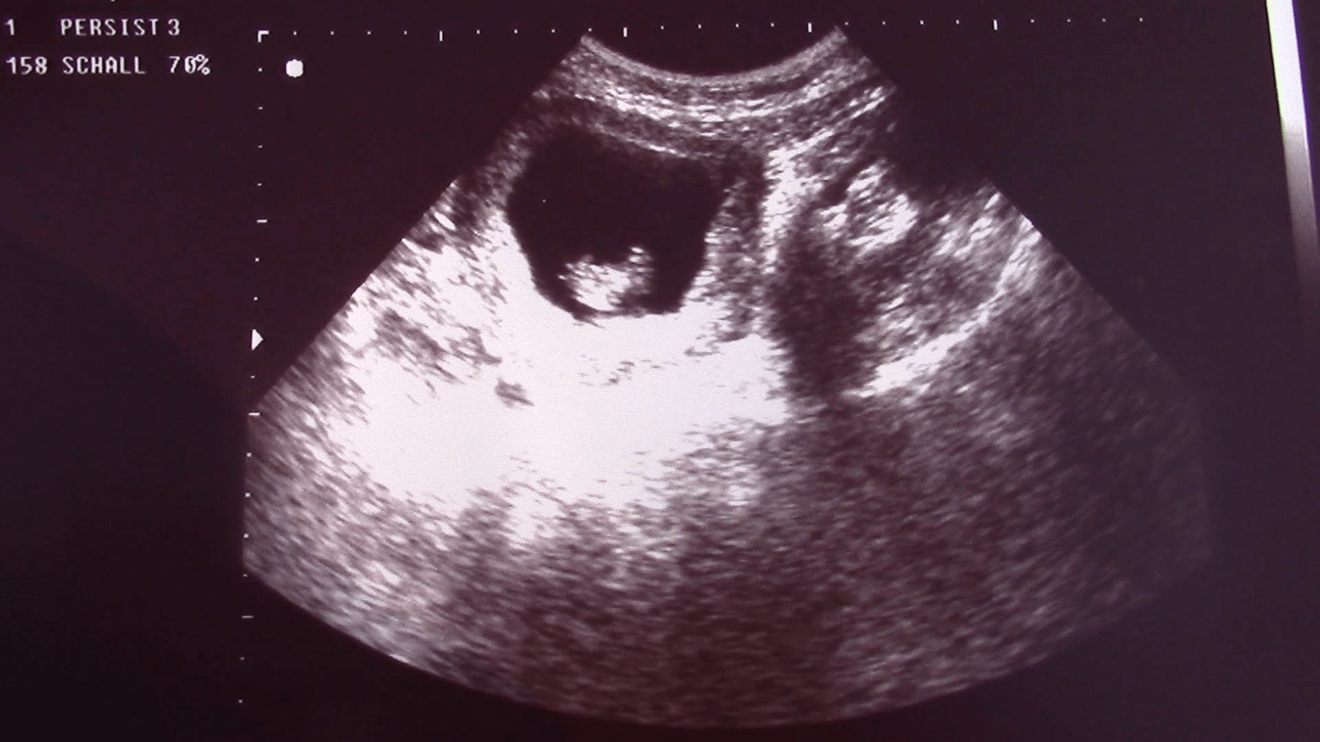
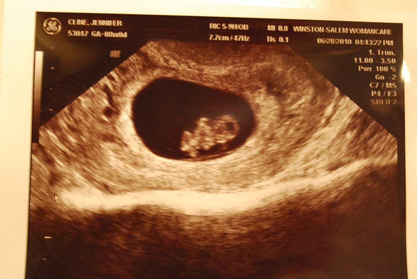
First, the doctor will describe the size of the ovum, evaluate its shape. During a normal pregnancy that proceeds without complications and threatened abortion, the ovum has even contours, it does not look deformed, crushed. Every week, the fertilized egg increases in size, however, it is measured only at the earliest dates.then the need for such a measurement is completely eliminated.
SVD - this is the name of the inner diameter of the ovum. At this time it may be different.
SVD at week 9
The results are compared with this table:
The size of the ovum by ultrasound, mm | Deadline (week + day) |
28 | 8 weeks + 1 day |
29 | 8 weeks + 2 days |
30 | 8 weeks + 3 days |
31 | 8 weeks + 4 days |
32 | 8 weeks + 5 days |
33 | 8 weeks + 6 days |
35 | 9 weeks exactly |

After measuring the ovum, the state of the yolk sac is noted. This is a temporary formation, which is a structure that provides crumbs of food, because there is no placenta yet. This bag is well defined at the earliest stages of pregnancy and gradually decreases in size until it ceases to exist altogether. At the 9th week of pregnancy, its average size is 5 millimeters.
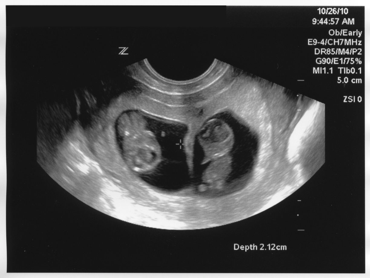
At this time, it is already possible to understand exactly how many embryos are in the fetal egg, as well as how many fetal eggs in the uterus. The seen doctor will describe in quantitative and qualitative terms - one or two fetuses, alive, with signs of physical activity.
KTR on week 9
Clarify the term, if it is not known for certain, and also understand how the baby develops will help KTR. The kopchiko-parietal size of the fetus is still its main indicator, the value of which is extremely great for diagnosis:
Coccyx parietal size (mm) | Obstetric term (week + day) |
17 | 8 +1 |
18 | 8+2 |
19 | 8+3 |
20 | 8+4 |
21 | 8+5 |
22 | 8+6 |
23 | 9 weeks exactly |
Heart rate (heart rate)
Normally, a fetal heart knocks at week 9 at a rate of 175 beats per minute. Allowable variations of this indicator from 155 to 195 beats per minute. Many mothers try to guess the gender of the future child precisely because of this indicator. see the genitals at this time is not possible.
There is an opinion that boys beat hearts a little slower than girls. From the point of view of medicine, there is no explanation for this, and the coincidences themselves, according to parents with experience, are 50x50. For a diagnostician, what’s more important is not “who is more like a heartbeat”, but whether it is rhythmic, whether there is a malfunction in the heart of the baby.

Additionally measured the thickness of the walls of the uterus. Its increase indicates the existing threat of miscarriage, the cervix is evaluated, the cervical canal is closed or slightly open, the endometrium is homogeneous, the size and position of the ovaries. Here it is not necessary to wait for complex and incomprehensible numbers, if there is a problem, then it is described in very concrete terms and explained by the pregnant woman herself.
Possible problems
The most likely problems that can be identified by ultrasound at 9 weeks:
Undeveloped pregnancy. At this time there are still risks that the baby will suddenly and for no apparent reason stop in its development. This can happen because of the illness that the woman suffered, taking medicines at the very beginning of pregnancy, genetic developmental abnormalities of the embryo, which turned out to be incompatible with the further life of the baby, as well as due to the poor state of the environment.
On the ultrasound monitor, the doctor will see a deformed fertilized egg, the fetus inside will not show signs of life, it will not have a heartbeat or physical activity.

Developmental delay. The doctor can make such a conclusion if the KTR of the fetus at the time of the examination is significantly different from the norm, but the fetus shows signs of life. Such a condition will require careful study of the yolk sac, as well as the fetus itself, developmental pathologies are not excluded. Mom will be prescribed supportive treatment with vitamins, vascular drugs, antispasmodics, magnesium and calcium drugs, and she will be advised to visit the ultrasound room in a couple of weeks to make control measurements of the fetus.


Lack of fetus. If the doctor finds a fertilized egg in a woman in the ninth week of pregnancy, but there is no fetus in it, the likelihood that something will change will tend to zero. The diagnosis of "anaembryonia" is made and a surgical cleaning of the uterine cavity from the structures of the ovum is prescribed.
Retrochorial hematoma. So called partial detachment of the ovum from the uterus. It is her most often found with bleeding in the ninth week. The forecasts are quite favorable. The future mother is prescribed bed rest, taking vitamins, antispasmodics, sometimes hemostatic agents. If the size of the hematoma is small, there is every chance of preserving the pregnancy.

Spontaneous abortion. In case of pronounced bleeding, blood clots, severe pain, ultrasound can be used to diagnose a miscarriage that has begun or occurred, as well as a miscarriage in the process. All these conditions are accompanied by a thickening of the walls of the uterus, hypertonia. Depending on the stage of the process, the ovum may still be in the uterus, go into the cervical canal or leave the uterus completely. This condition requires emergency medical care in a hospital.

Common Questions:
A list of them with likely answers is given below:
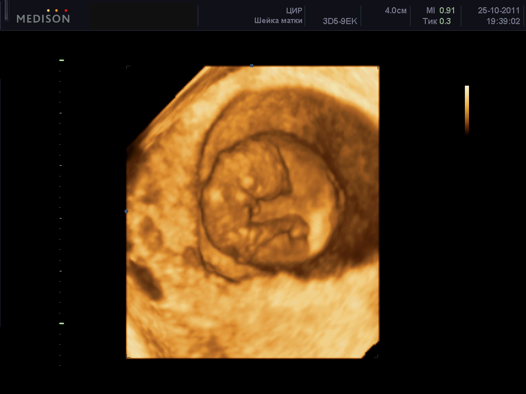
3D ultrasound at 9 weeks. This kind of ultrasound diagnosis at such an early gestational age is not performed.
Ultrasound with twins. The parameters of one fetus may differ from the norm, and the second - to be perfect normal. It is not accepted to consider this developmental delay if both fruits show all signs of life.
Ultrasound accuracy. On this period, the accuracy of ultrasound is about 85-90%.