Ultrasound in the 30th week of pregnancy: fetal size and other features
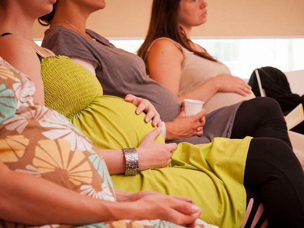
The third trimester of pregnancy has begun, and the woman is mentally preparing for the most important event in her life - the birth of a child. At week 30, she may be recommended ultrasound. Then we will talk about how the child looks now and what ultrasound indicators can tell about him.

Purpose of the survey
Ultrasound diagnostics does not belong to the category of routine examinations this week. Ultrasonography at 30 weeks gestation is prescribed out of plan. The first two screening studies are in the past, before the third at least a couple of weeks. Some of the future moms in this period are sent to the ultrasound diagnostics room of their own accord, to see if everything is fine with the child. Gynecologists even joke that The best sedative for a pregnant woman is a visit to the ultrasound doctor.
However, there may be medical indications for the examination. These include suspicions on the pathology of pregnancy. Weak and rare movements of the fetus, which are already considered daily at week 30, as well as too active movements, sudden jolts of the baby, can be a reason for the pregnant woman to be sent for an ultrasound scan unplanned.
The task of the examination in this case is to eliminate cord entanglement, hypoxia, and other unfavorable conditions that may pose a threat to the life and health of the child.
Ultrasound scanning during this period can be assigned to women who are carrying twins or twins, triplets, as well as women who have become pregnant through IVF. If the expectant mother has complaints of feeling unwell, pain, untypical discharge from the genitals for this trimester, she will also be recommended to undergo an ultrasound scan.
Ultrasounds are also subject to examination by pregnant women who have undergone the treatment prescribed by the doctor, in order to assess the condition of the baby and the effectiveness of the therapy. The lag in the growth rate of the height of the uterus' bottom, which is measured at each planned admission in the obstetrician-gynecologist's office, can also be the reason for issuing referrals for unscheduled ultrasound in order to eliminate the risk of intrauterine growth retardation and placental insufficiency.
Sometimes an ultrasound scan at this time is necessary to check the dates of pregnancy, to clarify the date of birth, because this week the paperwork for receiving maternity leave begins and the doctor must be sure that no errors occurred in the definition of dates.

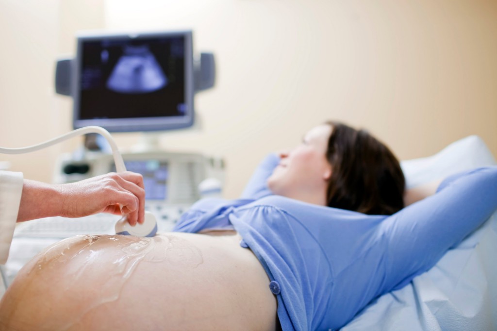
How is the research done?
At week 30, there is no need for intravaginal ultrasound diagnostics, in which the sensor of the device is inserted into the vagina. The review through the abdominal wall is not difficult even in obese women, so the study is carried out externally - transabdominal. No need to prepare for it. The woman had to drink water and fill the bladder only at the very beginning of pregnancy, now the fetal bladder is filled with a sufficient amount of amniotic fluid, which conducts ultrasound well, and the picture on the monitor is quite clear and understandable.
Filling the intestines, gases also can no longer affect the scan resultsbecause the large uterus pushed the intestinal loops into the background in the truest sense of the word.
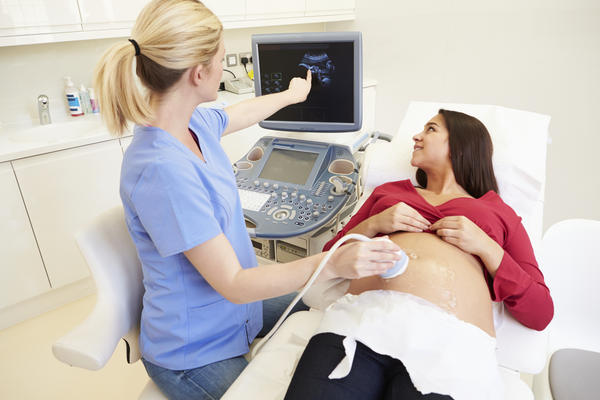
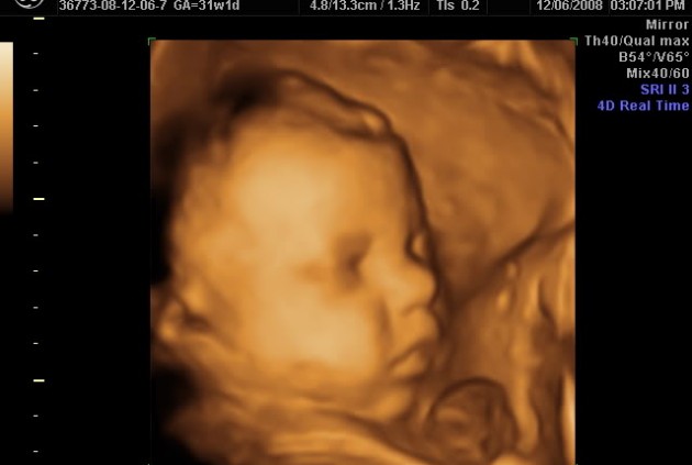
If a woman decides to visit at 30 weeks 3D or 4D ultrasoundShe is waiting for a very interesting sight. These procedures are carried out in the same way as the usual two-dimensional study; there are no specific features of these innovative diagnostic methods.
The only significant difference is the time required for the survey. Two-dimensional ultrasound will require no more than 10 minutes, while three-dimensional and four-dimensional studies require from 40 minutes to an hour. For any type of ultrasound this week is to bring along an exchange card, as well as a clean diaper and removable shoes.
What will the ultrasound show?
The future mom will be shown a pretty grown-up baby on the scanner's monitor. The size of the fetus this week is impressive - the growth of the crumbs approached 40 cm, and the weight is in the range of 1.5 kg. From the beginning of the third trimester, the baby is actively gaining subcutaneous fat, the folds and wrinkles on his skin gradually smooth out, puffy handles, cheeks and dimples appear so cute for the mother's heart.
The small lungs from this week begin to produce a surfactant - a substance necessary for normal functioning of the respiratory system. It is the surfactant that will not allow the lungs to stick together during the expiration, and its concentration is very important so that the child, if he is born premature, can breathe.
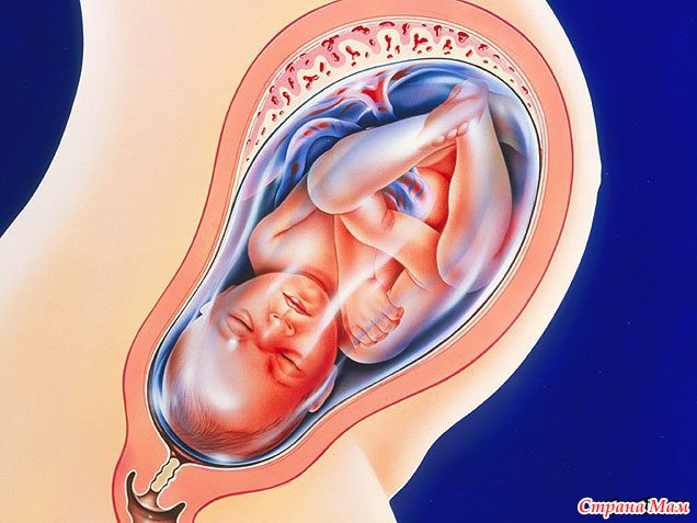
The movements of the child are no longer as active as in the second trimester, when the mother could observe them during the passage of the second screening ultrasound examination. The reason is cramped. A child who has noticeably grown becomes uncomfortable and cramped in the womb, he already seeks to adopt a fixed compact and most convenient position for himself.
Tumbling and swimming in the amniotic fluid of the baby can no longer.
The heart of the baby in the 30th week knocks very clearly and rhythmically. Hearing his mother will be able to, doctors provide pregnant women with this opportunity. Frequency heartbeat OK at this time - from 130 to 170 beats per minuteand, according to experienced obstetricians, in boys, the heart beats somewhat less frequently than in girls, the difference is approximately 10 beats per minute.
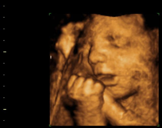
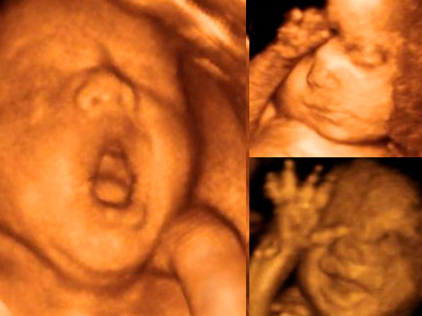
The child has very well developed mimic muscles, so the baby on the ultrasound can grimace, yawn, squint, frown and smile. If you're lucky, mom will be able to get a picture with such a funny attractive face. The vision continues to develop, the baby perfectly distinguishes light and darkness, his hearing allows him to distinguish the voice of the mother from the voices of other people. The baby continues the formation of the nervous system, in the brain every day there are all new grooves and gyrus - the baby is getting wiser by leaps and bounds.
In most boys, the testicles descend from the peritoneum into the scrotum, and if this has not yet happened, then there is still time. The child has all the organs and systems formed, they work. The stomach digests the amniotic fluid that the baby swallows, the intestine accumulates the original feces meconium, the liver at this time begins to accumulate iron, so that the child does not have anemia after the umbilical cord with the mother stops and an independent existence begins.
The child has small nails on the fingers and toes, the skin ceases to be saturated red and begins to take on a more familiar human tone.
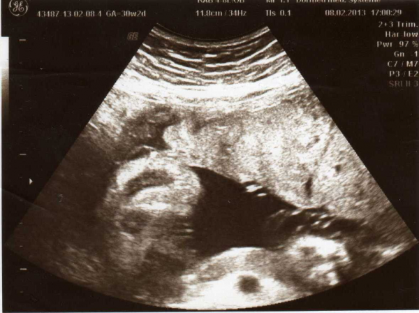
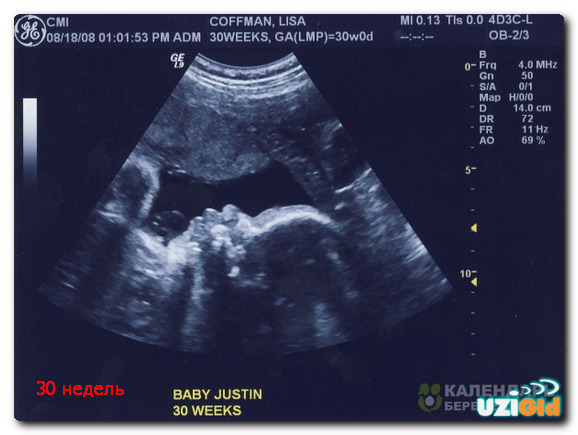
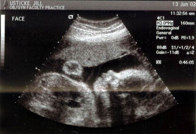
On ultrasound at this time, the mother will not be able to see her unborn child fully in full growth, since the sensor review does not allow for such a large picture. But the doctor will be able to show separate parts of the baby’s body with sufficiently high definition.
Decryption and norms
The norms included in the tables, with which diagnosticians and future mothers are checked, may differ from one child to another during this period.The fact is that on such a long time the crumb is already similar to its parents, it inherits the growth and susceptibility to a certain type of body build, and therefore one child may have longer legs, and the other will have an anticipated weight ahead of the average values.
Fetometric measurements at this time give only an approximate picture of the compliance with the terms of gestation, to a greater extent doctors on fetometry try to imagine proportional to the addition of the baby.
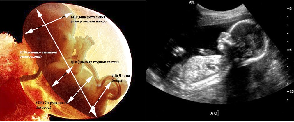
As in previous weeks, the frontal-occipital and bipariate head size is measured, the length of paired bones - femur and humeral, lower leg and forearm. In numerical terms, the diameter of the chest and the circumference of the head and abdomen are determined. The table of fetometric indicators at the 29-30 week of pregnancy is as follows:
BPR, mm | LZR, mm | The length of the femur, mm | The length of the bones of the tibia, mm | Forearm length, mm | The length of the humerus, mm | The diameter of the chest, mm | Head circumference, mm | Abdominal circumference, mm |
76-78 | 94-97 | 53-56 | 76-79 | 44-46 | 51-53 | 76-79 | 275-285 | 253-264 |
The position of the fetus in the uterus, which is found on this ultrasound, can persist until the birth, because the grown child is now quite difficult to roll over, but it is still possible, so worry about pelvic or transverse presentation is not worth it. The child has about 10 weeks to take the head position, which is considered optimal for the generic process.
If this does not happen, doctors a couple of weeks before the birth, they will decide on the delivery tactics. The amount of amniotic fluid at this time is 64-90 mm, there is a tendency to reduce their number, because a place in the uterus is required for a growing child. The umbilical cord with its normal development up to 30 weeks has a zero degree of maturity, and after this period becomes the first. The thickness of the “children's place” in this period is normal - 29.6-30.4 mm.
An ultrasound scan allows you to see the cord entanglement, if any. The umbilical cord itself in a normal pregnancy has three vessels. The blood flow in them at week 30 can already be measured during the passage of an ultrasound with a Doppler, vessels with different filling and intensity of blood flow on the monitor will be painted in a different color.
The doctor, with the help of the USDG, will be able to understand whether the child is getting enough food, if there is enough oxygen, or if he has signs of hypoxia. Determining gender from week 30 becomes difficult. The big baby is “going” into a ball, it presses the legs, therefore it is impossible to look at intimate places.
But if the crumb shows off its external genital organs, there will be no doubt, they are already quite well developed, and the mother will be able to consider herself who should be born - a boy or a girl.

To notice any warning signs of possible preterm birth, the doctor may examine the uterus of a pregnant woman with a vaginal probe to assess its size, the degree of closure of the cervical canal. At this time it should not be ajar.
If the walls of the uterus are in good shape, and the state of the cervix does not inspire confidence in the doctor, he can hospitalize the expectant mother to the department of pathology of pregnant women of her maternity home so that in the hospital try to prolong the pregnancy. The baby can already survive if premature birth occurs, but it is better if he waits at least another 7 weeks.

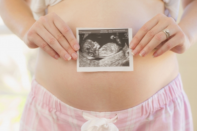
Snapshots
Two-dimensional ultrasound images allow you to see the profile of the baby, his arms, legs, but the traditional ultrasound, alas, does not allow even approximately imagine how the baby will look after birth. On 3D ultrasound, you can get more interesting pictures, looking at that and the future mother, and the future father will be able to easily understand who the child looks like.
At 30 weeks, the pictures are quite clear., a little later it will be quite difficult to get a clear image on the ultrasound, as the amount of amniotic fluid is gradually decreasing, respectively, the visualization will deteriorate.
Pictures should be asked for in electronic form on any information carrier, since paper “photos” that crawl out of the ultrasound machine are fragile.
You can learn about what happens with mother and baby at 30 weeks of pregnancy, from the following video.


