Do ultrasound in 2-3 weeks of pregnancy?
The early gestation period is quite an exciting time. It is at this time that the future baby starts laying the vital organs. This article will help expectant mothers to understand whether it is possible to carry out ultrasound in the initial weeks of pregnancy.


When is it held?
In most cases, the first ultrasound carried out during pregnancy is performed only at 10-12 weeks. Doctors did not establish this period by chance. It is believed that the effects of ultrasound at an early stage of embryo development can lead to various adverse effects. The most dangerous of them are the formation of various disorders of intrauterine development.
To assess the duration of pregnancy, doctors use a special term - “obstetric term”.
It is counted from the first day of the last menstrual cycle. Usually this period differs significantly from gestational, which is used by doctors of ultrasound diagnostics. This term, for obvious reasons, will be less obstetric.

It should be noted that at present the situation with the establishment of the duration of pregnancy has improved significantly. Modern ultrasonic devices used in the work, immediately set obstetric gestational age. It is determined by the introduction of the initial parameters, which the doctor indicates before conducting the study. In this case, in the conclusion after the test, the doctor indicates the already obstetric period of pregnancy. This is much more convenient, especially for obstetrician-gynecologists in their work.
In order for future moms not to get confused with the use of these concepts, they should use obstetric gestational age. This will allow pregnant women to speak the same language with their obstetrician-gynecologists.

Carrying out ultrasound in 2-3 weeks is carried out in most cases to exclude or confirm the fact of pregnancy. However, this type of survey is not a “gold” standard for this. Confirms the presence of pregnancy in the female body at this stage, a clinical examination, which holds a gynecologist, as well as a special biochemical marker - hCG. The increase in the blood of this hormone in most cases indicates the possible emergence of a new life in the female body.
Ultrasound examination conducted by a woman who has a delay of the next menstruation, quite often reveals not pregnancy, but various pathologies. As a rule, the polycystic or ovarian cystic masses existing in the patient lead to the development of this situation. Hormonally active neoplasms in this case lead to an imbalance of female sex hormones, which ultimately contributes to the delay of menstruation.

How is it done?
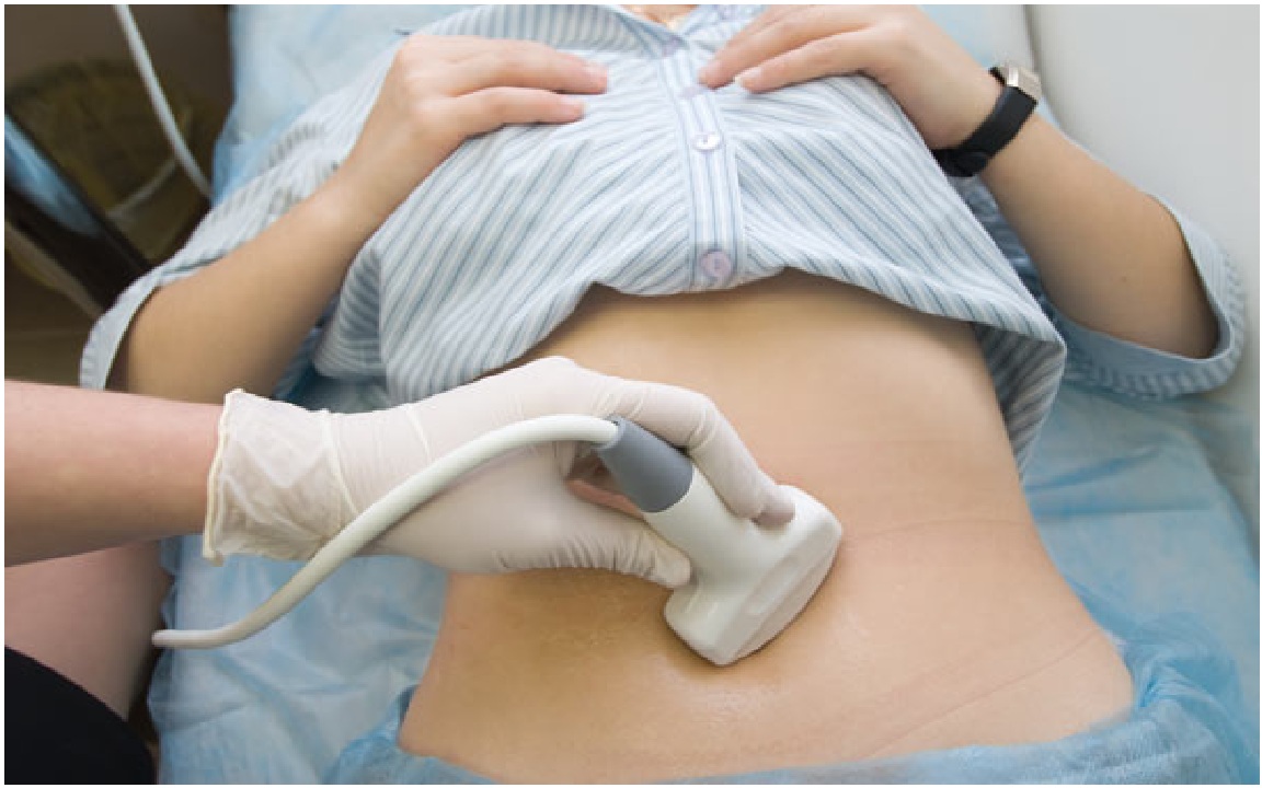
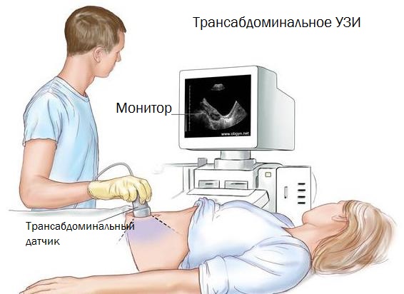
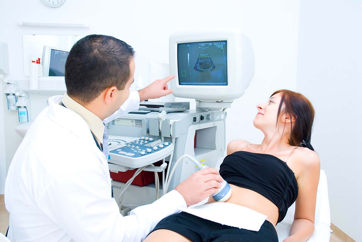
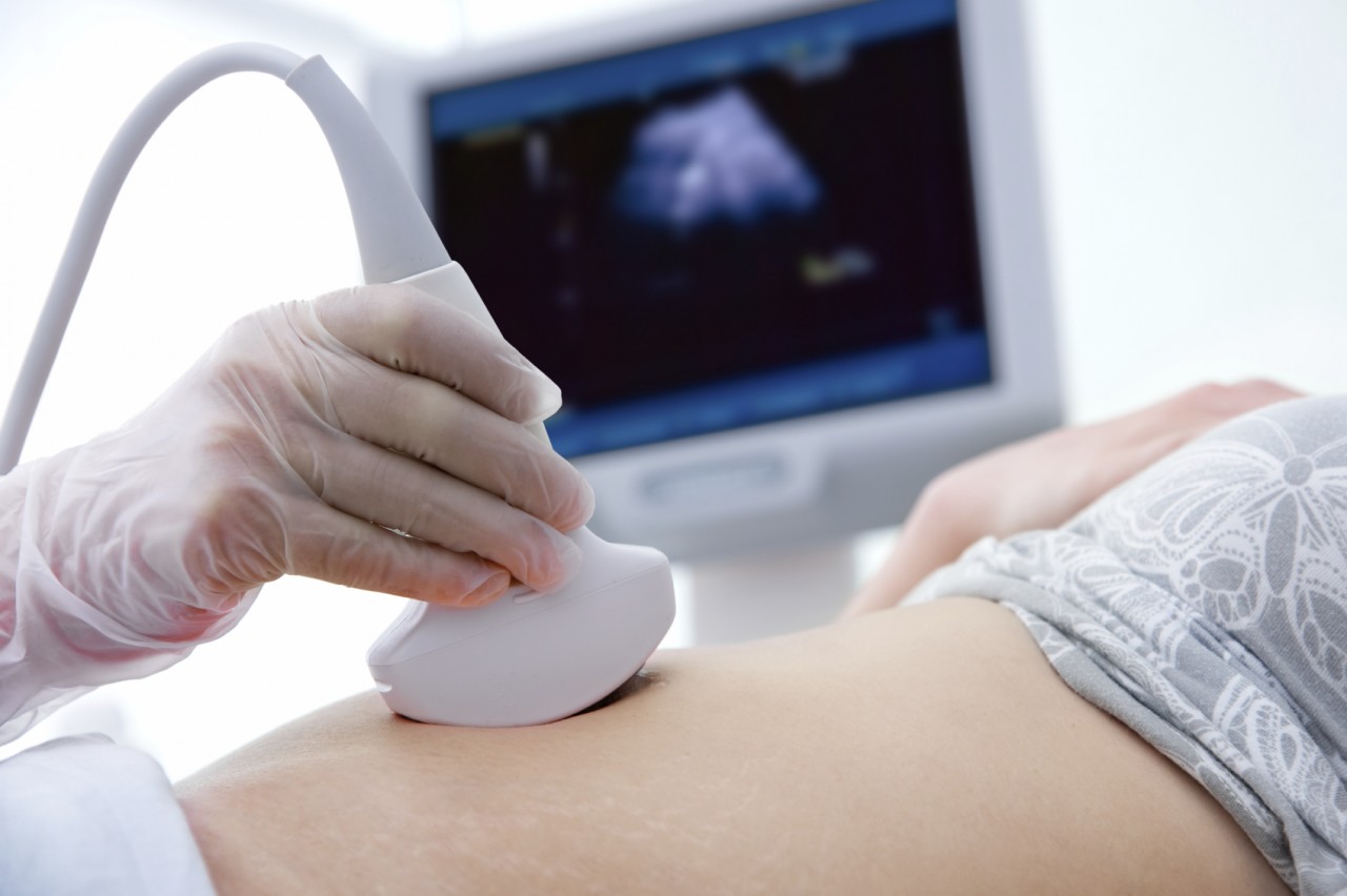
To conduct the study, doctors use special sensors. Ultrasound can be performed in two ways - transabominal and transvaginal. Each of them has its own indications and contraindications. These methods allow us to identify what the uterus and other reproductive organs look like in the future mother.
Chooses which method is better to use in each specific situation, gynecologist.
There are cases when the date of the alleged conception is set incorrectly.In this situation, during the ultrasound examinations of the doctor, it is determined that the child is more than the expected period of 2 weeks. Experts can also establish that the fetus is 2 weeks behind in development. In such a situation, doctors prescribe a test study, which is carried out in a few days.

To conduct the study, doctors use special sensors. With the transabdominal method, the examination is carried out through the skin of the abdomen. A transvaginal probe is inserted into the vagina. Many experts believe that the study of this method in the early period of pregnancy is more informative.
During the ultrasound, the doctor can take pictures. They are supported by a medical card. Take such pictures in cases where a woman has any abnormalities or pathology. With repeated research specialist ultrasound can repeat this picture. This is necessary in order to track the dynamics of the development of this pathological condition.

What can be determined at this time?
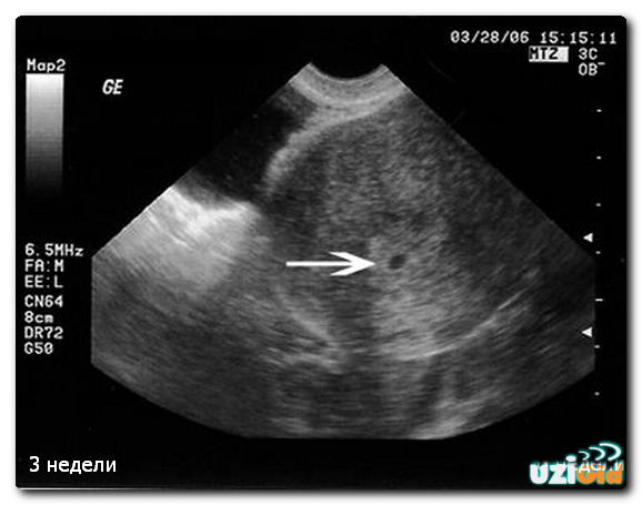
Ultrasound, carried out in the early period after conception, can only show the presence of the egg, which remained in the uterus. This is the time when a small fertilized egg can leave the reproductive organ along with blood flow. In this case, the woman may not even understand that she was “pregnant” for a couple of weeks. The delay of menstruation after unprotected intercourse is perceived by her, as a rule, as a confused hormonal background.
Obstetricians say that pregnancy has not actually occurred at 3 weeks. At that time the risk that the fertilized egg leaves the uterus is quite high. However, it is worth noting that she already has a double set of chromosomes, obtained during conception from the future mom and dad. During this period, the fertilized egg develops quite rapidly. The cells forming it rapidly divide and differentiate.
Doctors note that in the first week after conception, such a fertilized egg consists already of 32 small cells. By the end of the third week there are already more than 260 cells. In the future, active growth will occur throughout the first trimester of pregnancy.
The first weeks of such development are the most intensive and important.
By the 20th day from the moment of conception very important events begin to occur. The most important of them is implanting a fertilized egg directly into the uterine wall. At this time, a pregnant woman, who is still not quite aware of her new status, begins to undergo characteristic changes in behavior and her general condition. She may dislike some kind of smell, her appetite increases and her mood changes.


By the time the dividing zygote is attached to the inner lining of the uterus, it is less than 0.3 mm in size. Such the smallest "education" in most cases is not visible during the ultrasound. In the future, as the embryo develops and grows, it can already be seen in the uterus during ultrasound.
Usually obstetricians and gynecologists prescribe ultrasound in 2-3 weeks from the moment of conception for medical reasons. It is assigned to exclude concomitant diseases of internal genital organs, which are available to the mother and may aggravate the course of the pregnancy in the future. Also, an ultrasound scan at this time can be carried out in order to assess the effectiveness of in vitro fertilization after embryo transfer.
There are cases when doctors prescribe an ultrasound at 2-3 weeks after conception in order to confirm the fact of pregnancy. Immediately it should be said that such a survey is not always shown. In practice, there are many cases of false diagnosis. In this case, it will be shown conducting a second ultrasound in 1-2 weeks.Frequent ultrasound examinations, especially those conducted in the early stages of fetal development, may lead to the development of the most adverse consequences in the future.
The main feature by which doctors of ultrasound diagnostics are oriented is endometrial hyperplasia. This fact is an indirect sign of pregnancy. However, hyperplasia (increase in volume) of the inner lining of the uterus may be due to certain diseases of this genital organ.

Experienced ultrasound specialists reveal a small depression in the mucous membrane in which the baby will subsequently be located.
They define it by measuring the initial endometrial thickness. During the study, experts can also determine the size and tone of the uterus. The presence of hypertonus in a pregnant woman is an extremely unfavorable symptom, especially in early pregnancy. In some situations, hypertonus can lead to spontaneous abortion or spontaneous miscarriage.
Also during the study, the ultrasound doctor will definitely evaluate the corpus luteum. At the early stages of pregnancy development, its vascularization occurs - blood supply. The blood vessels feeding the corpus luteum become more dilated. They also significantly increases the speed of blood flow.

This feature in the corpus luteum is due to the fact that it plays a very important role during pregnancy. Doctors call this anatomical education the “children's bed”. The main fetal structures that protect the body of the future baby from external factors will be formed from it in the future. Also, the corpus luteum has a pronounced hormonal effect. Progesterone, synthesized under its active influence, has a preparatory effect on the endometrium so that the embryo can be implanted into the inner wall of the uterus.
Doctors use ultrasound to evaluate the flow of the corpus luteum. special type of scanning - duplex. It allows you to assess the level of blood flow in any anatomical zones. The decrease in this indicator may indicate a violation of the course of pregnancy. In such a situation, doctors decide on the need for the appointment of corrective drug therapy.

Is it possible to do?
2-3 week of pregnancy is not the best time for an ultrasound. During this period, it is impossible to consider the fetus, not to mention determining any pathologies of intrauterine development or the sex of the future baby. Conducting ultrasound at this time is mainly forced.
Scientists have found that the abuse of ultrasound, especially carried out in early pregnancy, lead further to many different negative consequences. Kids who underwent frequent ultrasound in the period of early organogenesis, after birth have low weight.
Also, many pediatricians believe that the effects of ultrasound in the early stage of the formation of a child can lead to the formation of persistent disorders in the nervous and cardiovascular systems.
Scientists have long established the fact that ultrasonic waves have a significant impact on the brain of the fetus. In the future, this contributes to the fact that the child has various features in behavior. This situation manifests itself after childbirth. American researchers believe that conducting ultrasound at such early stages can contribute to the development of autism in the future baby.
It is rational to carry out an ultrasound at this time only medical emergency. Such a survey can also be shown to women who have serious problems with bearing and conceiving a child. In this case, the need for an ultrasound is determined by a gynecologist. Thoughtless doing such research can lead to irreparable consequences. About this expectant mother must be warned.