Ultrasound in the 31st week of pregnancy: fetal size and other features

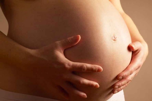
On eighth month of pregnancy Mom goes on maternity leave. She has more time to communicate with her baby, who will soon be born. It is during this period that the expectant mother will have to pass the third, final prenatal screening. And at the 31st week of pregnancy, she may receive a referral for an ultrasound scan as part of a screening study of the third trimester. About what is waiting for her in the ultrasound room and how her baby has become, we will tell in this article.
Objectives of the survey
At week 31, a third prenatal screening is appointed, the task of which is to summarize the results of the first two and find out how the baby feels at this stage. The study is now very informative in terms of planning tactics for the upcoming delivery. Many babies already at this time adopt the final position of the body, in which they are to be born. This makes it possible to minimize the number and risk of birth injuries, because for pelvic or transverse presentation, a woman may be shown a cesarean section.
Ultrasonography at 31 weeks gestation allows you to clearly and thoroughly examine the structure of the internal organs of the baby, since all of them are already quite large and perfectly visualized. Ultrasound examination is planned, it is carried out free of charge in consultations at the place of residence of the expectant mother.
In addition to screening tasks, additional trials are imposed on the third trimester ultrasound. the scan should clarify the condition of the child, features of uterine placental blood flow, show the growth of the baby in dynamics, clarify the expected date of birth, which is not far off.
Out-of-plan at this time, pregnant women with twins or triplets who become pregnant through IVF, as well as women with severe obstetric history, including after a previous cesarean section, can count on ultrasound, as it is important to monitor the state of the postoperative uterine scar on such a long period.
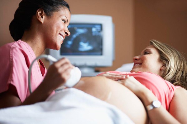
Carrying out the procedure
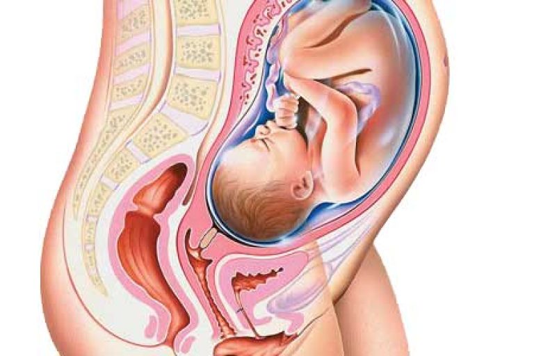
Ultrasound at 30-31 week make external, transabdominal way in which the sensor apparatus is located on the anterior abdominal wall of a woman. The uterus is so large that the build of a pregnant woman no longer plays any role, the extra weight and fatty folds on the abdomen can not complicate the visualization.
The doctor applies the vaginal sensor only if the woman has a threat of premature birth, and more precisely, the obstetrician-gynecologist is inclined to believe that there is an abnormal opening of the cervix that is abnormal for a given period.
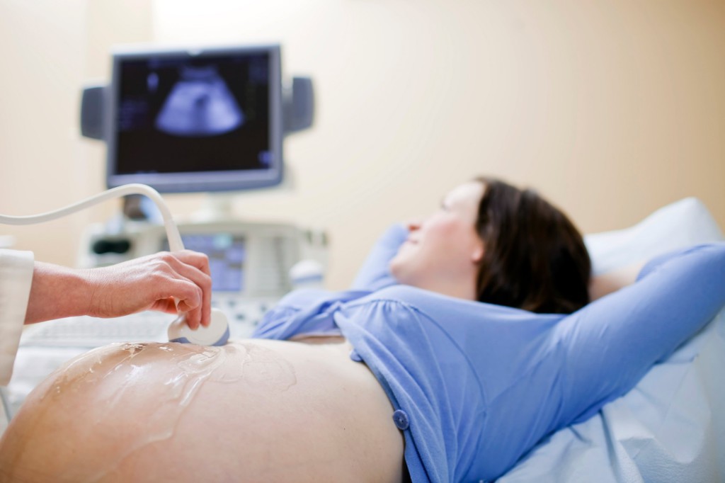
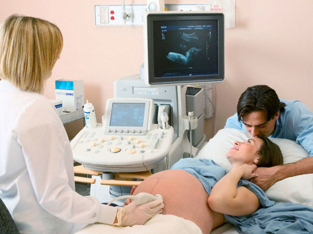
Preparation for an ultrasound is not required. If a woman wants to do a three-dimensional or four-dimensional ultrasound scan, then she should know that such an examination lasts longer than traditional ultrasound - from 40 to 55 minutes. With an ultrasound of any type, it makes sense to take an exchange card and passport, removable shoes and a clean diaper so that you can cover it with a couch, which you have to lie on during the scan.


What will the research show?
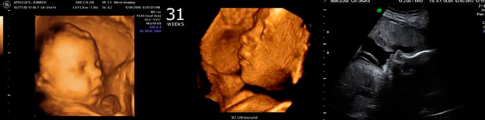
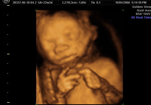
The size of the fetus at week 31 is quite large, so there is no possibility to look at the baby entirely, not a single sensor gives such a broad overview.The child will be examined in parts - the head and face, neck and chest, spine and stomach, limbs. The growth of the baby exceeded 40 centimeters, and the weight - more than one and a half kilograms. In some babies, the weight may be close to 2 kg.
The baby is stored with subcutaneous fat, which allows him to gain weight quickly, and after giving birth, he will protect the baby from freezing, because the thermoregulation functions will not be debugged yet. Due to the fat, the body of the baby gets a more attractive appearance - on the handles and under the knees there are cute folds, on the cheeks - dimples, wrinkles are smoothed. The baby at week 31 looks almost like it will look immediately after birth.
The child has fully formed all the senses. He hears and even partially sees, distinguishes the voices of his own people from unfamiliar and frightening sounds, distinguishes darkness from light. If you turn on the flashlight on your stomach, the baby will close his eyes reflexively. He already opens his eyes when he is awake, and, if lucky, he can demonstrate it during an ultrasound.
The lungs are preparing for independent breathing. So far the baby does not know how to do this, but a special substance begins to be produced in its lungs - a surfactant that will not allow the alveoli to stick together during expiration after the baby is born.
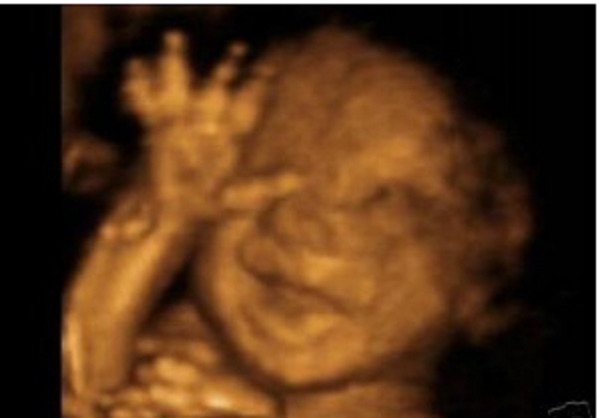

At week 31, the formation of furrows or convolutions in the child’s brain continues, it becomes “smarter”, and the connection between the brain and muscles becomes more efficient. The movements of the crumbs are now less active than before, but they are filled with meaning - he can purposefully pull his fists into his mouth, grab the umbilical cord, touch his little face.
Motor activity is not as intense as in the second trimester, because the baby already becomes significantly tighter in the mother's womb.
The crumb takes a compact position, because of which it becomes difficult to see the genitals, so the floor in the third trimester is more difficult to determine than in earlier periods.
Actively work the stomach and intestines, bladder and kidneys. The baby swallows amniotic fluid, hiccups, pisses, and in the intestine, the original feces, which is called meconium, is deposited. Scarce is sleeping and awake. In periods of wakefulness, he can play with his fingers or the cord cable, which can also be seen on the monitor of the ultrasound scanner.
Decryption and norms
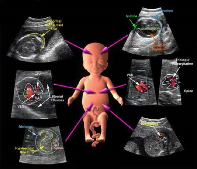
Understanding what is written in the conclusion of an ultrasound is not as difficult as it seems at first glance. The doctor necessarily measures the basic parameters, which are called fetometric. These are the dimensions of the head — the frontal-occipital and bipariented, as well as the length of the paired bones of the legs — the femur and tibia, and the hands — the shoulder and forearm bones. Also measured is the diameter of the chest, and two circles - the abdomen and head. Then the results are compared with a special table.

Table Fetometry 30-31 weeks of pregnancy (average values only):
BPR, mm | LZR, mm | DBK (thigh), mm | DKG (calf), mm | WPC (shoulder), mm | DKP (forearm), mm | The diameter of the chest, mm | Head circumference, mm | Abdominal circumference, mm |
78-80 | 97-101 | 57-59 | 53-56 | 53-55 | 46-48 | 79-81 | 285-294 | 264-274 |
In conclusion, ultrasound necessarily describes the anatomical features of the child's development. At this time, the doctor has all the possibilities to examine the internal organs of the baby, concluding that there are or no birth defects.
The placenta at week 31 has the first degree of maturity, in some cases it qualifies as a transition from the first to the second, although normally it is normal for the placenta to normal to the second degree after 35 weeks. The amount of water is gradually becoming less, because the baby is growing, and it needs more and more space in the uterus. This week is considered normal. the amniotic fluid index is 82-88 mm (on average).

Possible problems
Deviations of indicators of a particular child from the average values indicated in the table can be both physiological and pathological.If an ultrasound scan at 31 weeks reveals a developmental delay, then the lag is quite significant - about 2 weeks from the fixed obstetric period (at 31 weeks the size of the child corresponds to the 29th week). Excess of indicators on the same difference can also talk about pathology - edema, genetic abnormalities, malformations, intrauterine infection.
Minor deviations should not disturb the future mother and her loved ones. The physiological difference may be due to the hereditary features of the child’s appearance and physique - some are born larger, others are thin, some have “long in mother’s” long legs, and others are “long” long. The noses are snub-nosed and with a crook, the heads are also different. At week 31, the baby is already developing according to the individual, inherent nature of the program, it is what it is.
The turbidity of the waters, as well as the suspension detected in them according to the results of an ultrasound scan, may indicate a child’s distress — hypoxia, or a prenatal infection. With a history of anamnesis, a woman will definitely have an ultrasound scan with a doppler in order to understand how well the placenta is supplied with blood, if the child has enough oxygen and nutrients.
The threat of premature birth can be indicated by shortening and softening of the cervixas well as the uterine muscles and incomplete closure of the cervical canal A child who is born at week 31 can survive, for this he has everything he needs, but will require more thorough care. The task of the doctors is to preserve the pregnancy for at least one and a half months.
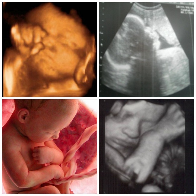
Snapshots
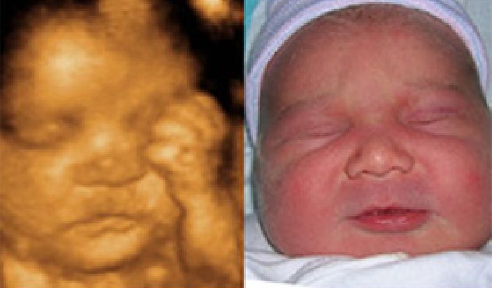
Ultrasound images at week 31 give an idea of the future mom and dad not only about what their child looks like, but also about what it does in the womb. If you want to get funny and touching pictures, it is better to visit the three-dimensional ultrasound and the "photo" of the baby with his first greeting to your family, if you are lucky, it will be yours.

You can find out what happens to mother and baby during the 31st week of pregnancy in the following video.


