Ultrasound in the 16th week of pregnancy: fetal size and other features
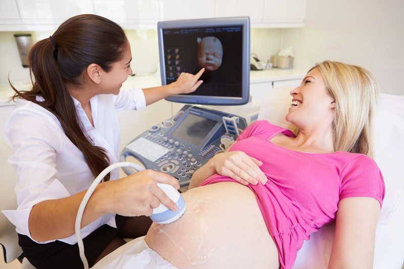
At the 16th week of pregnancy, the woman literally "flutters." Quite recently, the second trimester began, and as yet there is no excruciating fatigue and heaviness typical of the last weeks, but the tummy has already rounded and the expectant mother has replaced her usual wardrobe with more spacious and comfortable clothes. This week she can go for an ultrasound examination. What awaits her there, we will tell in this material.
Purpose of the study
Week 16 is the time that midwives use to calculate the date of birth. In fact, the child lives in the mother's womb for 14 weeks, and after the delay exactly 3 lunar months (12 weeks) passed. On this period, ultrasound is usually not appointed. The first screening is in the past, it was held from 11 to 13 weeks of pregnancy, inclusive, the second is still to be.
The directions for the blood test will be handed to the expectant mother this week, but Doctors try to prescribe an ultrasound procedure for a later period - from 18 to 21 weeks.
However, the reasons for the ultrasound diagnosis in a pregnant woman can be many. Screening studies directed to identify high risks the birth of children with chromosomal pathologies, developmental abnormalities, many of which are total and fatal. An ultrasound scan at week 16 will not pursue the goal of identifying markers of such pathologies, unless, of course, a doctor who is not satisfied with the results of the first prenatal screening sends a woman to such an expert ultrasound scan.
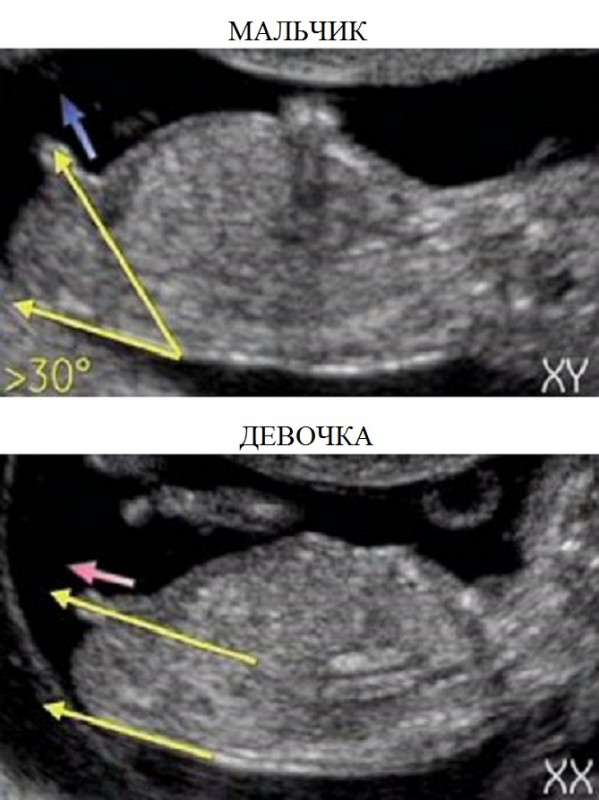
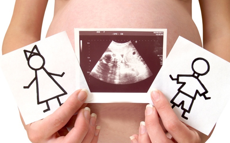
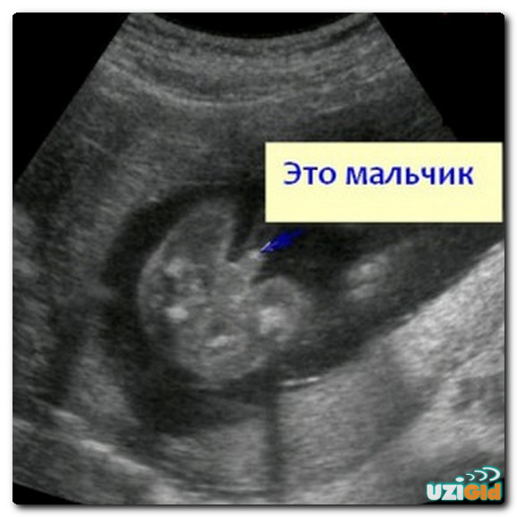
For the most part, this week women are recorded on ultrasound without the knowledge of the attending physician. to find out the sex of the baby. Determining gender from this week is not difficult. The external genitals of the baby are fairly formed, they have grown in size, and now it is much easier to distinguish the boy from the girl.
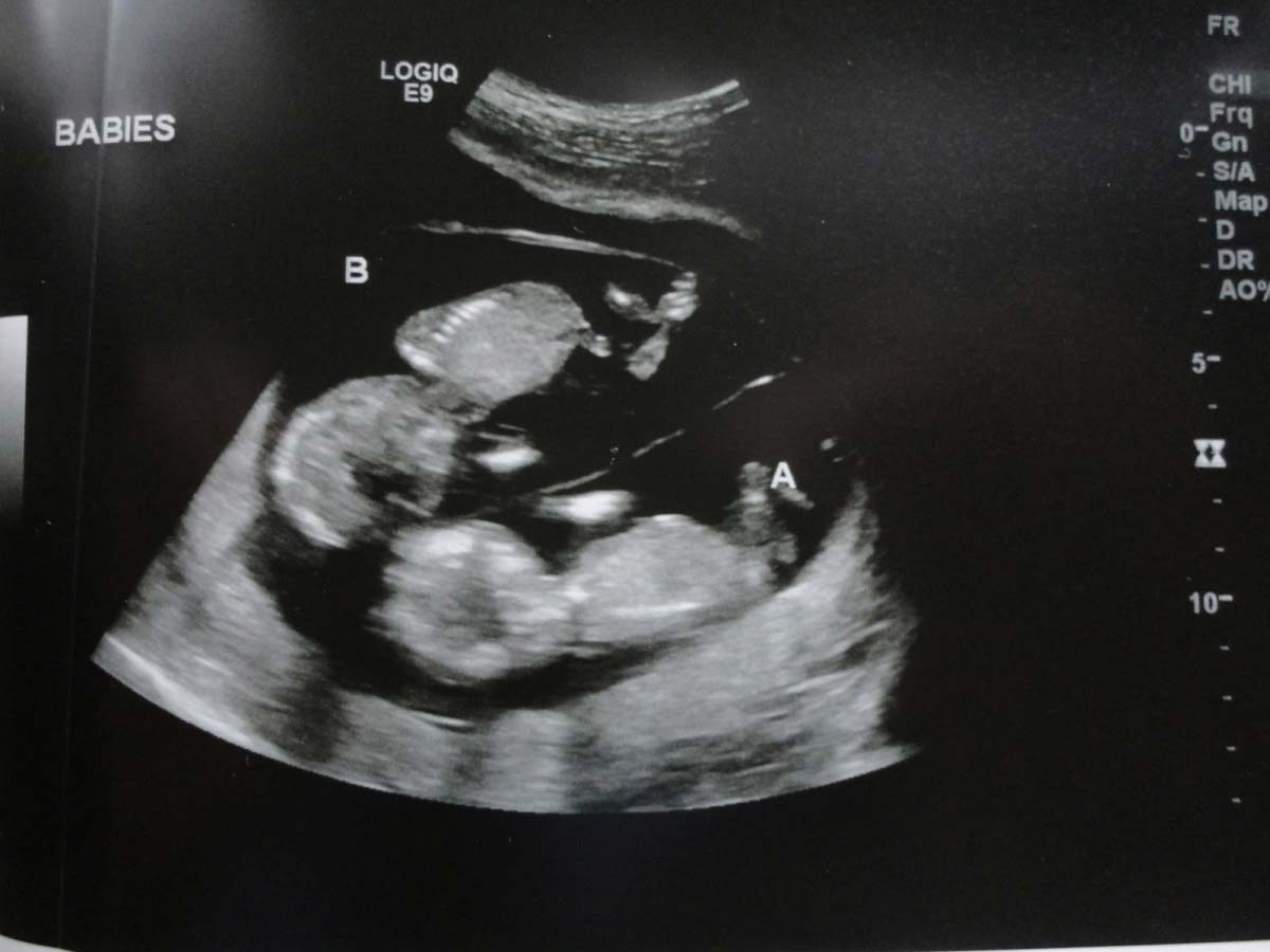
To the ultrasound room of a woman at 15-16 weeks of pregnancy may lead to complaints of well-being - there were pains in the abdomen, discharge, which normally should not be. Referral to an unscheduled examination can be obtained by a woman who has twins who become pregnant during the IVF process, since such pregnancies require an individual approach.
Women will also be sent to the ultrasound if the size of the uterus does not meet the deadline. In this case, it is important for the obstetrician to know how the child develops, if there is any intrauterine death of the fetus, or if there is a delay in development.

Method and preliminary preparation
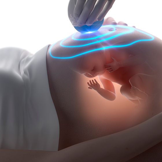
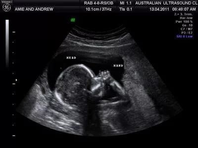
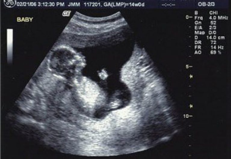
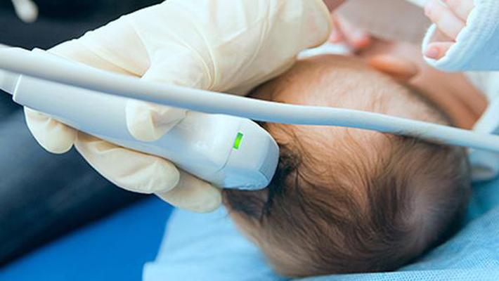
At week 16, it is most preferable to conduct an ultrasound scan using an external method, which is also called transabdominal. Through the front part of the abdominal wall, both the baby and the structural features of the placenta, the umbilical cord and the uterus are already well visualized. No training is required, there is no need to drink water and fill the bladder for this period.Because the amount of amniotic fluid at this time allows ultrasonic waves to pass easily and be reflected.
Vaginal ultrasound also does not lose relevance, but the doctor will examine the uterine cavity and everything in it through the vaginal wall only if the review through the stomach is greatly hampered by the curvaceous future mother.
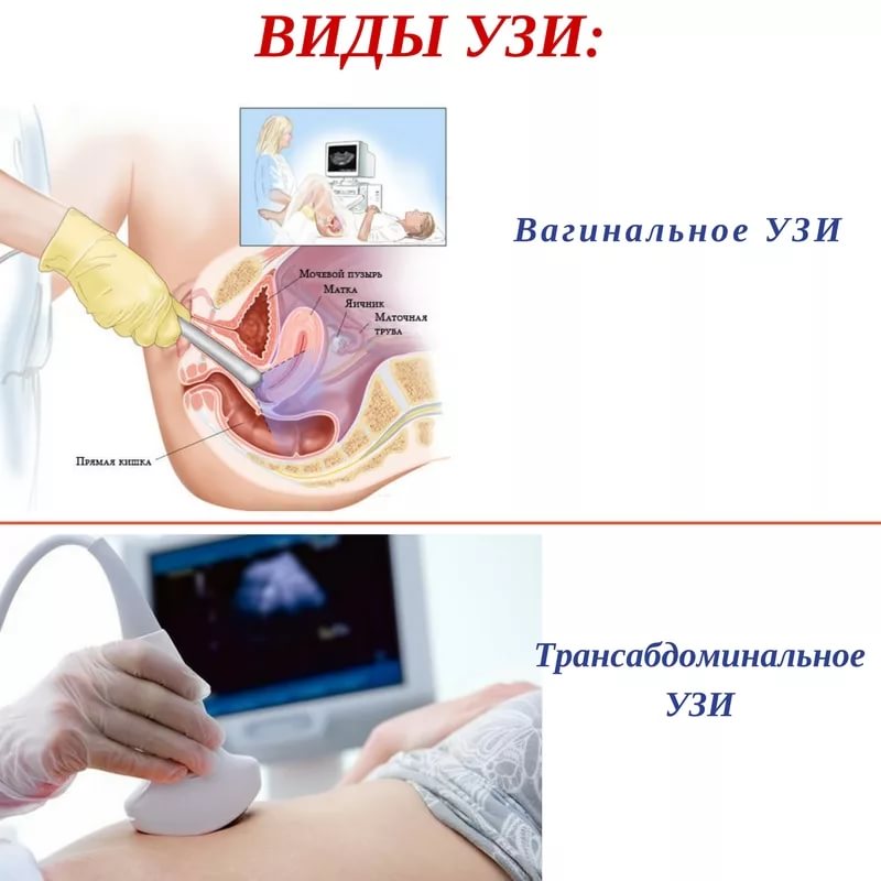
The doctor may use both methods if it is necessary to examine not only the child (through the abdomen), but also the cervical canal and uterine neck (through the vagina) with the possible threat of spontaneous abortion on this period.
In order to pass on the diagnosis, the future mother will only need to bring with her changeable shoes or shoe covers, as well as a clean diaper, which can be laid on the couch. Not hurt and a little chocolate. There is to eat it for 15-20 minutes before visiting the doctor, the child on the ultrasound will surely please the mother and the doctor with active movements, since the sweet children at this time are already very pleased.

What can be seen?
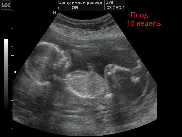
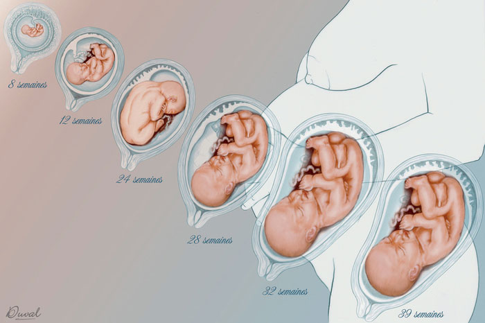
On the monitor of the scanner in the period from 15 to 16 weeks you can see a moving and fully formed baby. The size of the fruit, on average, ranges from 14 to 18 centimeters, and the weight of the crumbs is in the range from 70 to 115 grams. The child's neck stretched out, now the baby has got the opportunity to turn his head to the right and left, which he is actively beginning to use.
The formation of facial muscles continues, the crumb mastering all new grimaces. He winks, squints, opens his mouth, frowns. But he didn’t learn how to open his eyes. The girls this week get internal sexual signs - their ovaries sink into the small pelvis. Boys still have only external sexual characteristics - the scrotum and penis, their testicles remain in the abdominal cavity.
The kid has noticeably improved coordination of movements, which is associated with the intensive formation of the nervous system and muscle-neural connections. Now his movements, which are clearly visible on the monitor of the ultrasound scanner, does not resemble chaotic swing hands and feet. A kid can quite purposefully send a cam into his mouth or pull a cord on himself to play a little.

It is believed that at this time, the kids already have dreams, during the resting phase out of the eye, covered for centuries, quickly run back and forth.
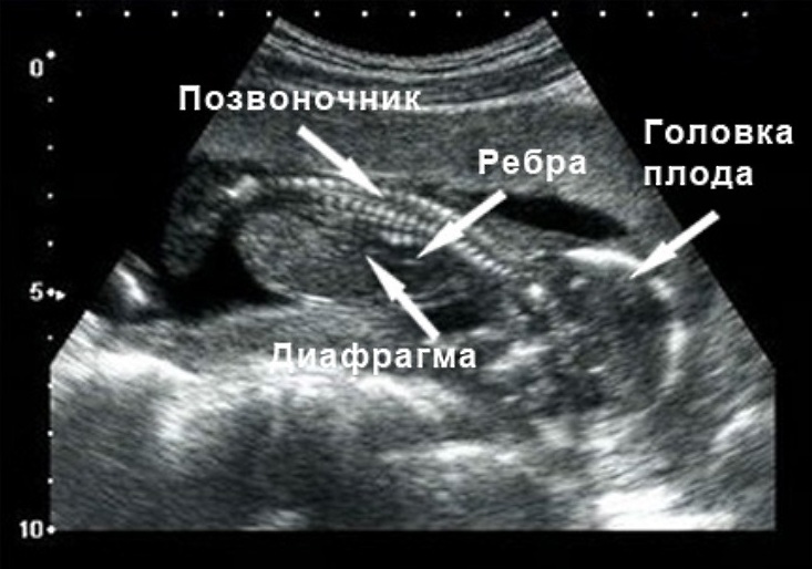
On the ultrasound at week 16 you can look at the fingers and toes. By the way, they already fully formed small nails. The spine is formed, practically all the internal organs of the crumbs work normally. He drinks, pees, trains the lungs, and original cal - meconium is formed in the intestines.
Decoding results
This week measures the main fetometric indicators of the child. It is their proportionality and timing that are the basis for judging whether a baby is developing normally. At week 16, the rate of fetometry looks like this:
BPR, mm | LZR, mm | Abdominal circumference, mm | Shoulder length, mm | Thigh length, mm | Fruit weight, gr |
31-37 | 41-49 mm | 93-102 | 15-18 | 15-18 | 77-118 |

Among the anatomical descriptions of the fetus are the main organs. The doctor must examine:
the ventricles of the baby’s brain are normally not dilated;
face profile - with normal development has no features;
eye sockets - are visualized, normally do not have features of development;
spine - if everything is in order, the doctor writes that he is normal;
lungs, spleen, gallbladder and intestines are normal without features;
the heart has a four-section cut;
liver, stomach and kidneys - are present, with normal development, they do not have features.
It describes in detail where and how the placenta is located. Its normal thickness for this period is from 18 to 18.5 mm. The umbilical cord should have 3 vessels. The cervix should not have pathological changes, the cervical canal is normally tightly closed.
Possible problems
The size of the child does not meet the standard
A child may lag behind or be ahead of the average norms for a given period of pregnancy for various reasons. Small fluctuations of a few millimeters are not considered pathological.However, if the dimensions of all parameters or one of them are lagging behind by more than 2 obstetric weeks or are ahead of the “schedule” for the same period, additional examination is required.
The reasons for deviations of the fetometric parameters can be malnutrition, developmental delays, oxygen starvation of the fetus, a tendency to heavy weight, and pathology of genetic origin.
Doctors will be able to answer this question in more detail after the second scheduled screening, in which the woman will go from 18 to 21 weeks.
Gender Mistakes
Such errors are not excluded. The accuracy of ultrasound as a diagnostic method is no more than 90%, the accuracy of determining the sex at this time is about 80%. If you want to get a higher percentage of accuracy, then with the question of determining the sex of the baby to the diagnostician better to contact after 20 weeks. Then the probability of correct "hit" will be within 90%.
The doctor may be mistaken due to the location of the child in the uterus. If the crumb is in pelvic presentation and at the same time sits with its back turned to the sensor, then it will be very difficult to examine the genitals.
Sometimes the error creeps in because the boy can hide his penis between the legs, and he will be mistaken for a girl. The girl can be mistaken as a boy because the umbilical cord loops fall between her legs and will be treated like a penis. The probability of such errors is about 1.5-3%.
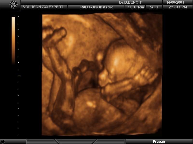
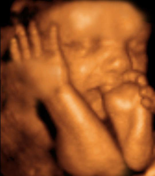
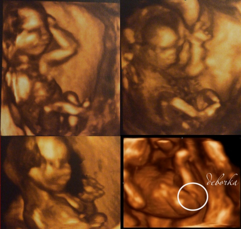
If you want to see the sexual characteristics of your child with your own eyes and not to worry about the strange stripes on the black and white ultrasound scan, better to do a 3D ultrasound. In a three-dimensional form, if the kid, of course, shows his “charms” to the doctor, the picture does not need explanations.
Fetal Choroid Plexus Cyst
Such a wording in the conclusion of an ultrasound at 16 weeks can lead a pregnant woman and her relatives to panic. In fact, worry is much more harmful. Cystic fluid formation in the plexus is called only for echographic signs. In fact, the accumulation of fluid in the choroid plexus is produced by the plexus itself and does not pose any danger to the life and health of the child.
Cyst of such a plan does not grow, does not degenerate into a tumor, therefore, there is no diagnosis of this, a disease with such a name is not described in medical reference books. Just an ultrasound doctor states a fact - there is an accumulation of fluid. Nobody talks about pathology.
After the baby is born, he will undergo an ultrasound of the brain, in 90% of cases such cysts dissolve on their own; in the remaining 10%, doctors choose observational tactics. Cysts can resolve a little later.

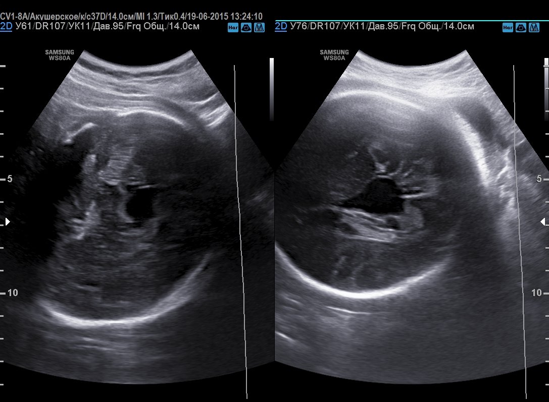
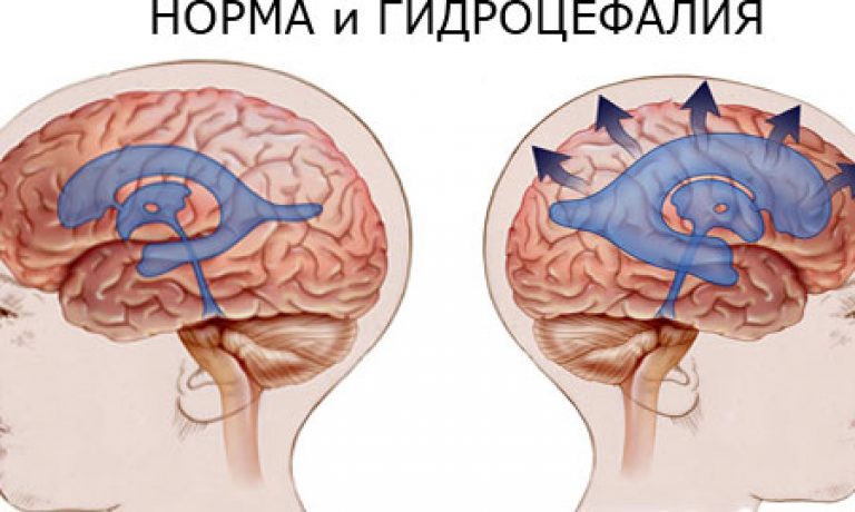
Brain asymmetry
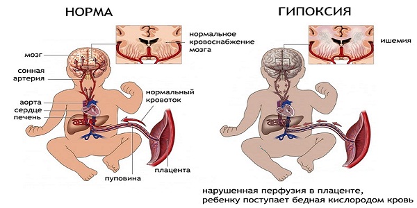
That is how future mothers formulate a medical verdict on the expansion of the lateral ventricles of the fetal brain. The condition requires additional examination, since the expanded and asymmetrical ventricles can become due to hypoxia experienced by the child, intrauterine infections, foreign bodies and organ tumors.
Much depends on the degree of asymmetry and other indicators of the development of the baby. Severe asymmetry may indicate the risk of hydrocephalus. (along with an increase in the size of the head).
In this case, the mother will have to visit the pediatric neurosurgeon while still carrying the baby, in order to assess the risks together with the specialist and make a decision to provide the child with help after birth.
Snapshots
In the pictures in three-dimensional format, the child at the gestational age of 16 weeks is seen in more detail than in the usual two-dimensional “photo”. Twins looked slightly worse. The sex of a child on a 3D ultrasound at 16 weeks looks like this.


To save pictures of the baby in the womb for a long memory, you should ask the diagnostician to give you electronic files on any carrier. Printed on special paper pictures lose their image clarity and color too quickly.


