Ultrasound in the 17th week of pregnancy: fetal size and other features
17 week of pregnancy does not provide for mandatory ultrasound diagnosis. This is a calm and measured period in which the mother feels good. Toxicosis, if it was, almost ended, and the tummy is still small and does not give the woman any unpleasant sensations, she does not feel tired. However, the need to do an ultrasound may still arise. How this procedure takes place at this time and what can be seen on the scanner monitor will be described in this article.
Purpose of the survey
The first screening was left behind, until the second is still about one and a half weeks.
In some consultations, the first tests of the second screening, a triple or quadruple blood test, may be prescribed at 17 full weeks.
In the 17th week, there may be several reasons for a woman to go to the ultrasound diagnostics office, except for curiosity (there are such restless women who go to ultrasound in paid clinics almost every two weeks to see how the baby is developing) :
the appearance of pain, bloody, smear, blood-like discharge;
severe toxicosis, edema, increased pressure in the future mother;
expert ultrasound (if during the screening of 11-13 weeks dubious or disturbing signs were identified);
if the obstetrician-gynecologist had any doubts (the manual examination indicates the obstetric and the actual term, there is a suspicion of missed abortion).
Preparing and conducting research
The woman's uterus is already quite large, so there is no need for any special preparation for the study. The doctor chooses a way to examine the situation. If a woman is thin, then this week it is possible to carry out the procedure transabdominal, examining the uterus and baby through the anterior abdominal wall. If a pregnant woman does not have thinness, it is sometimes easier to see the fetus through the vaginal wall, and then the doctor uses a transvaginal method of research.
If a woman turned to an ultrasound because of discharge and pain, then the study will be carried out only by a vaginal sensor, since there is an urgent need to carefully examine the cervical canal and cervix to eliminate the possibility of spontaneous abortion.
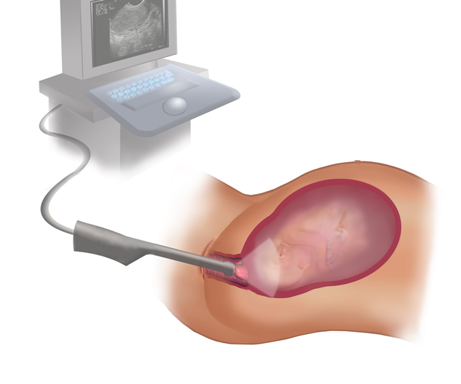
The duration of the procedure at this time can vary from 5 to 10 minutes. For a child and a future mother, such a diagnosis is considered harmless and painless.
What will show?
At week 17, the baby begins a stormy and important process of bone mineralization, in the gums there is a laying of future milk teeth. The formation of the hearing aid has already been completed, and on the high-resolution ultrasound machine you can already try to make out the small ears. In the female fetus at this time, the formation of the main reproductive organ, the uterus, begins. The system of blood vessels is actively developing and forking, but there is no possibility to consider this in ultrasound diagnosis.
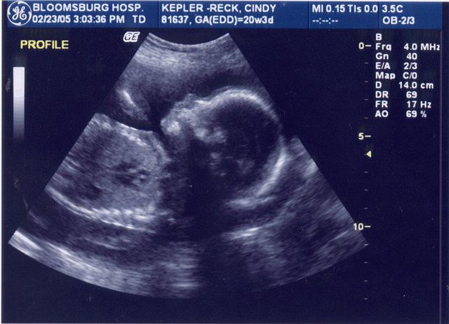

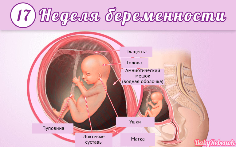
A pretty grown-up baby will be clearly visible to the doctor and future mother during the scan at week 17. The size of the fruit is now about 11-12 centimeters, and its weight is more than 100 grams. The fact that the child was able to hear will become noticeable during the procedure, because noises from the outside already cause certain reactions to the crumbs. Thus, the noise of the sensor can cause the baby to activate movements, he will quickly move the arms and legs, which are also noticeably lengthened.
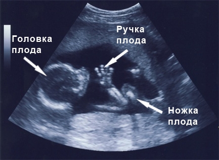
The diagnostician will carefully examine the condition of the uterus, the young placenta, the formation of which has already ended, the amniotic fluid. It will measure the basic parameters of the baby himself, which will allow to verify the timing, as well as to judge the pace of development of the child.
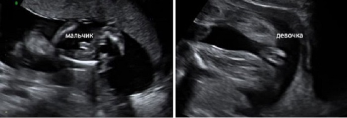
If the crumb is conveniently located for the review by the sensor, then the diagnostician will be able to recognize the child's sex with a high degree of accuracy. Now the fetus is in the very stage of development, when it is no longer small, but not so large that intimate places are covered due to the uncomfortable pose of the toddler. Therefore, now is the time to ask the doctor, the child of what sex will soon become a full member of the family.
Rules and interpretation of results
At week 17, the doctor defines a completely standard set of data that are important in order to understand how a little person feels.
Presentation of the fetus
At this time it can be any - head, pelvic or transverse. A child is not fixed for a long time in any position and almost constantly turns over, taking advantage of the fact that he is still very free in the womb. Therefore, you should not worry about pelvic or transverse presentation and possible problems in childbirth associated with this position of the baby. Karapuz again and again change the position.

Fetometric indicators
Such measurements include the growth indicators of the baby - the longitudinal and transverse size of his head, abdominal circumference, and the length of paired bones. Based on these values, the ultrasound scanner program calculates the estimated weight of the baby, and the doctor will use the tables to determine whether fetal fetometry values are normal for his gestational age.
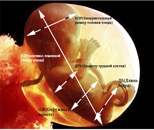
Fetal head and belly sizes
Gestational age | BPR (bipartial size), mm | LZR (frontal-occipital size), mm | Head circumference, mm | Abdominal circumference, mm |
16-17 week | 34-38 | 45-50 | 124-135 | 102-112 |
Paired bone length
Thigh length (DBK), mm | Shin Length (DKG), mm | The length of the humerus (WPC), mm | The length of the bones of the forearm (DCT), mm |
20-24 | 18-21 | 18-21 | 15-18 |
Nose bone length
Gestational age | Nasal bones - normal, mm | Allowable vibrations, mm |
16-17 weeks | 5,4 | 3,6-7,2 |
Lagging behind the norms for 2 weeks or more can be regarded by doctors as intrauterine growth retardation, a sign of genetic pathologies or infection of the fetus. Excess rates can be regarded as an error in calculating the duration of pregnancy, which may be in women with an irregular cycle or late ovulation.

A slight lag or advance of the norms cannot be regarded as a pathology; most likely, it is a matter of heredity, because the baby is already developing according to an individual program laid down in it by parents and nature. Long legs, a nose "button", a small head may simply be features of appearance, inherited from mom and dad.
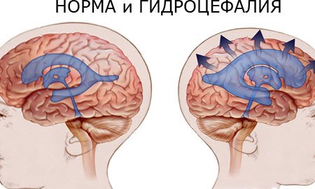
The size of the baby’s head is important.. If BDP and LZR are 2 weeks or more behind the norm, doctors can prescribe a detailed study including invasive methods, because such a decrease may be a sign of chromosomal abnormalities or microcephaly. Excess of 2 weeks or more is also an alarming sign, which may indirectly indicate the likelihood of hydrocephalus. Minor excess of average norms are regarded as an individual trait of appearance.
Fetus anatomy
At the 17th week of pregnancy, the doctor can evaluate the profile of the baby, examine its facial bones. The structures of the brain, heart, kidneys, stomach and intestines, lungs and bladder will also be examined.When pathologies are detected, genetics will be engaged in detailed research, as often congenital malformations of internal organs “coexist” with various syndromes and disorders of the central nervous system.
If all the organs are in order, then the doctor will indicate that they "do not have features," "are normal," or simply "examined", each will not be described in detail.


Placenta and umbilical cord
The nutrition of the baby, providing it with oxygen-rich maternal blood, depends on the state of the “children's place” and the umbilical cord. Normally, the umbilical cord has 3 vessels, about which the doctor ultrasound necessarily make an entry in the protocol of ultrasound.
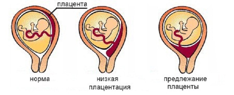
The normal thickness of the placenta for the 17th week of pregnancy is from 15 to 25 mm, the degree of maturity is zero. If the placenta is located close to the internal pharynx, the doctor will place a low placentation or placenta previa. Both of these conditions are dangerous for the child, and therefore treatment and supportive therapy should be started immediately. If you follow all the recommendations of the obstetrician, there is a fairly large chance that with the subsequent growth of the uterus, stretching its walls, the placenta will also rise.
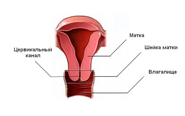
Uterus, cervix, appendages
On ultrasound at this time, not only the size of the female reproductive organ is assessed, but also the state of the uterine walls. If thickening is detected, then doctors may suspect hypertonia, which accompanies the threat of abortion. Normally, the cervical canal should be closed, the appendages and cervix should not have diagnostic features.
Accuracy
Whatever the convenient ultrasound diagnosis, it is impossible to call it an exact method. The accuracy of research on this period is estimated at about 85-90%. In matters of sex determination accuracy is even lower - about 80%. The doctor may be mistaken because of poor review, outdated equipment. When determining the sex of girls, umbilical loops can be confused with the penis, and the boy can be “recorded” as a girl because he has his legs “manhood” and the doctor could not see it.

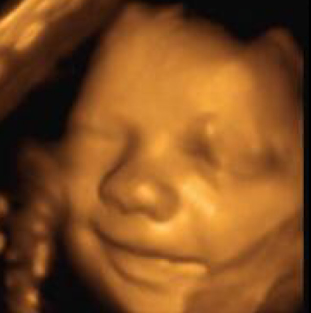
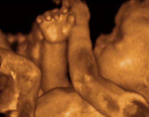
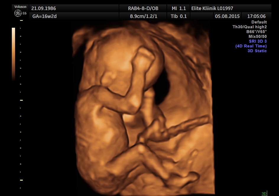
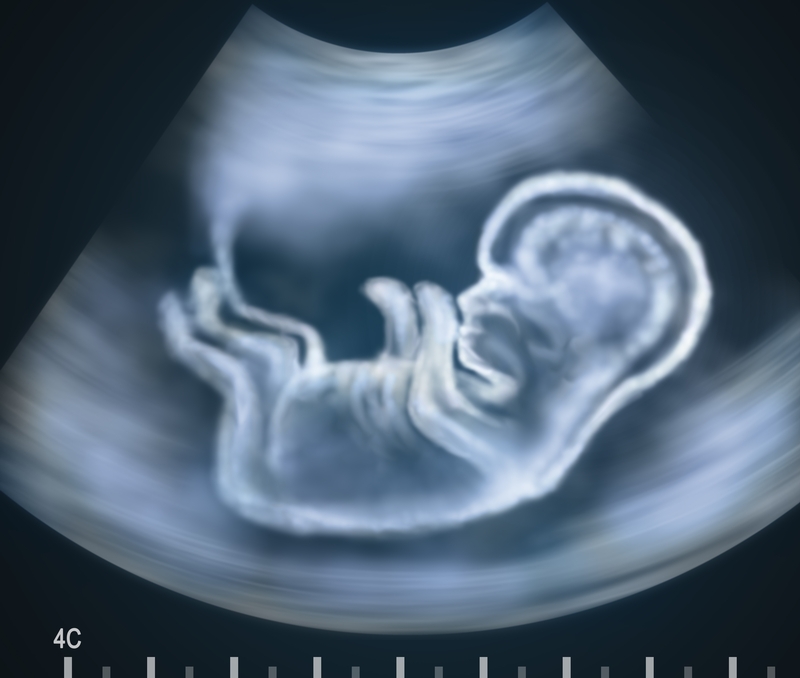
3D ultrasound gives the best overview and images, and although the procedure is recommended from 20–22 weeks of pregnancy, and at week 17, according to the mothers ’reviews, it was possible to obtain accurate data on the child’s sex, and the doctors and future parents were able to examine intimate places on beautiful volumetric shots.