Ultrasound in the 15th week of pregnancy: fetal size and other features

The intrauterine development of a baby is a rather complicated process. Detect any pathology in this period is possible during the ultrasound. This simple study helps to identify even the most hidden diseases that have formed in the fetus.
Purpose of
15 obstetric week is a close time to planned screening for 2 trimesters. To perform such a study or not during this period, decides only a gynecologist, who observes a pregnant woman. Examination performed at the 15th week of gestation, mainly for certain medical conditions.
At this stage of intrauterine development, a specialist in ultrasound diagnostics can identify only some pathologies. Also using this method are determined by certain chromosomal and genetic diseases. The appearance of technical errors at this time occurs much less frequently than in the initial weeks of pregnancy.

The beginning of the second trimester is the period when the child has fine audible heartbeat. The baby's heart beats at a speed of 135-170 beats per minute. This heart rate is normal. After birth, the heart rate will gradually decrease. This indicator is physiological and does not require any correction.
During this period, the baby, who is still in the womb, begins to work the thyroid gland. It is still difficult to call such functioning fully. The fetal thyroid captures iodine, but cannot yet synthesize hormones.

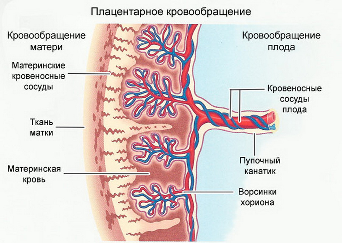
At this time, an experienced ultrasound specialist can determine the functioning of the baby’s respiratory system. The child continues to develop the development of the lungs, bronchi and alveoli. Blood fetus at this stage through the placental vessels. He has no own independent blood supply yet. It is important to note that By the end of week 15, the “baby seat” or placenta begins to function fully.
Some future moms already feel the active movements of babies. The fruit develops and begins to actively push. Some pregnant women have similar symptoms 2-3 weeks later.
Many pregnant women note that they noticed active movements of babies only by the end of the 2nd trimester.

On the fifteenth week, doctors evaluate the work of all these internal organs. All fetal membranes are also evaluated. Conducting a study, an ultrasound specialist determines the pathology not only of the fetus, but also of the mother. To do this, he carefully examines with the help of an ultrasound transducer all the organs of her reproductive system.
Sex determination can also be carried out at this stage. However, for this purpose, research is carried out somewhat earlier. Qualified doctors can determine the sex of the future baby already at 11-12 weeks of pregnancy.
The composition of the amniotic fluid that surrounds the fetus is sterile at this stage. Their number is a very important indicator.
An excessive amount of amniotic fluid indicates that the child is forming dangerous pathologies.
Survey methods
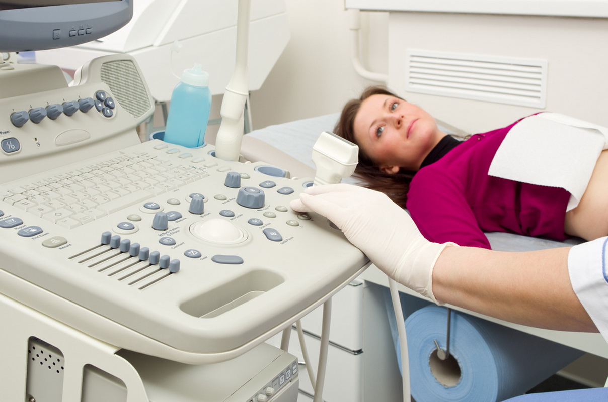
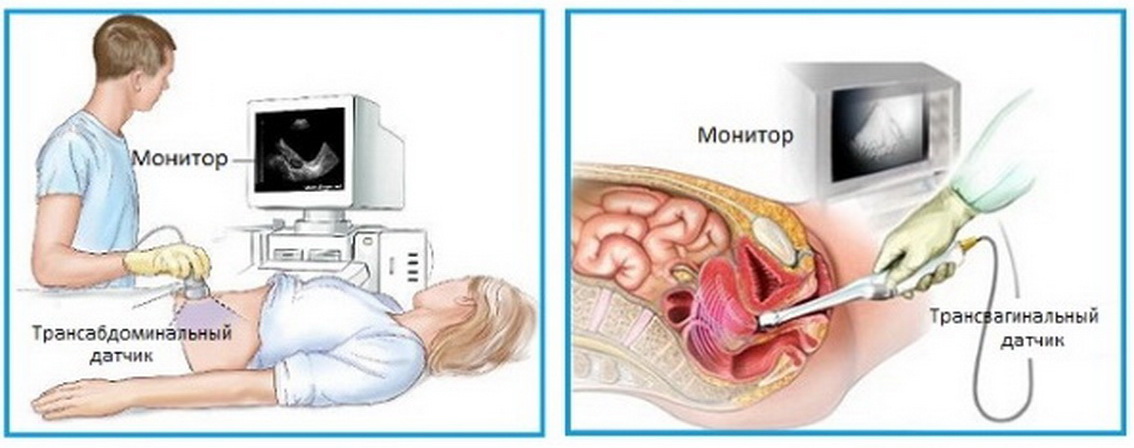
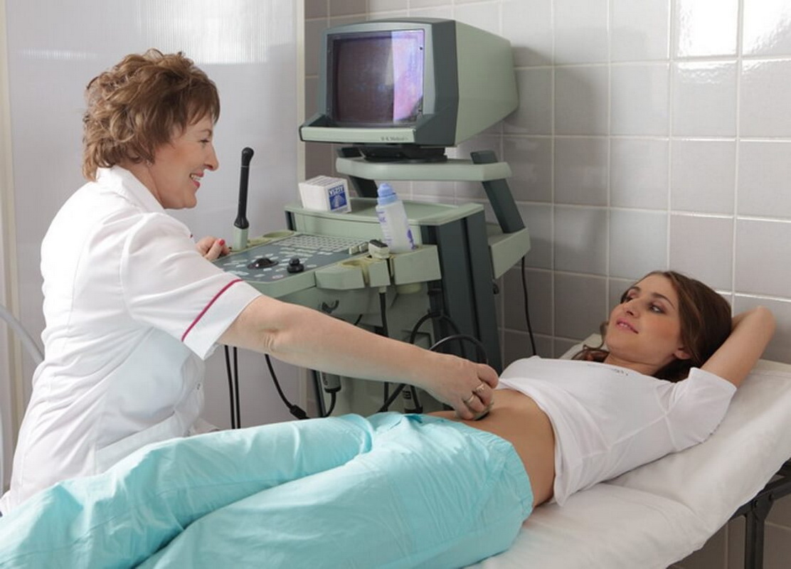
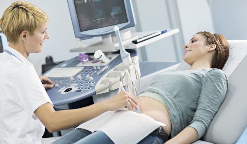
During this period of prenatal development, it is possible to conduct research in different ways. The most commonly used method is transabdominal examination.To do this, the doctor examines the belly of the future mom with a special sensor.
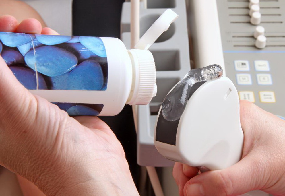
For the purpose of better refraction and reflection of ultrasonic waves, a special diagnostic gel. It is applied to the skin of the abdomen of a pregnant woman. The hypoallergenic chemical composition of the gel is not capable of causing any allergic reactions in women.
If the obstetrician-gynecologist recommends transvaginal examination, then the doctor inserts an ultrasound probe into the vagina. This method of ultrasound is usually chosen in the early weeks of pregnancy.
A study is conducted in a conventional ultrasound room. Before the procedure, a pregnant woman is laid on a special couch. In some cases, an ultrasound specialist may ask the expectant mother to turn on her left side. In this case, the uterus lends itself to a better visualization.
If the expectant mother goes for an examination at a regular clinic, then she should take with her paper napkins and towel. With the help of a handkerchief, it will be possible to remove the remaining gel from the skin Towel spreads on the couch before the procedure. If the study is conducted in a private clinic, then in this case, all consumables are provided and included in the cost of the procedure.
The duration of the study may be different. This is largely determined by the experience and qualifications of the specialist who conducts the survey. On average, the procedure lasts 30-40 minutes. If additional dopplerography is required, the duration of the study in this case may be about an hour.
What structures can be defined?
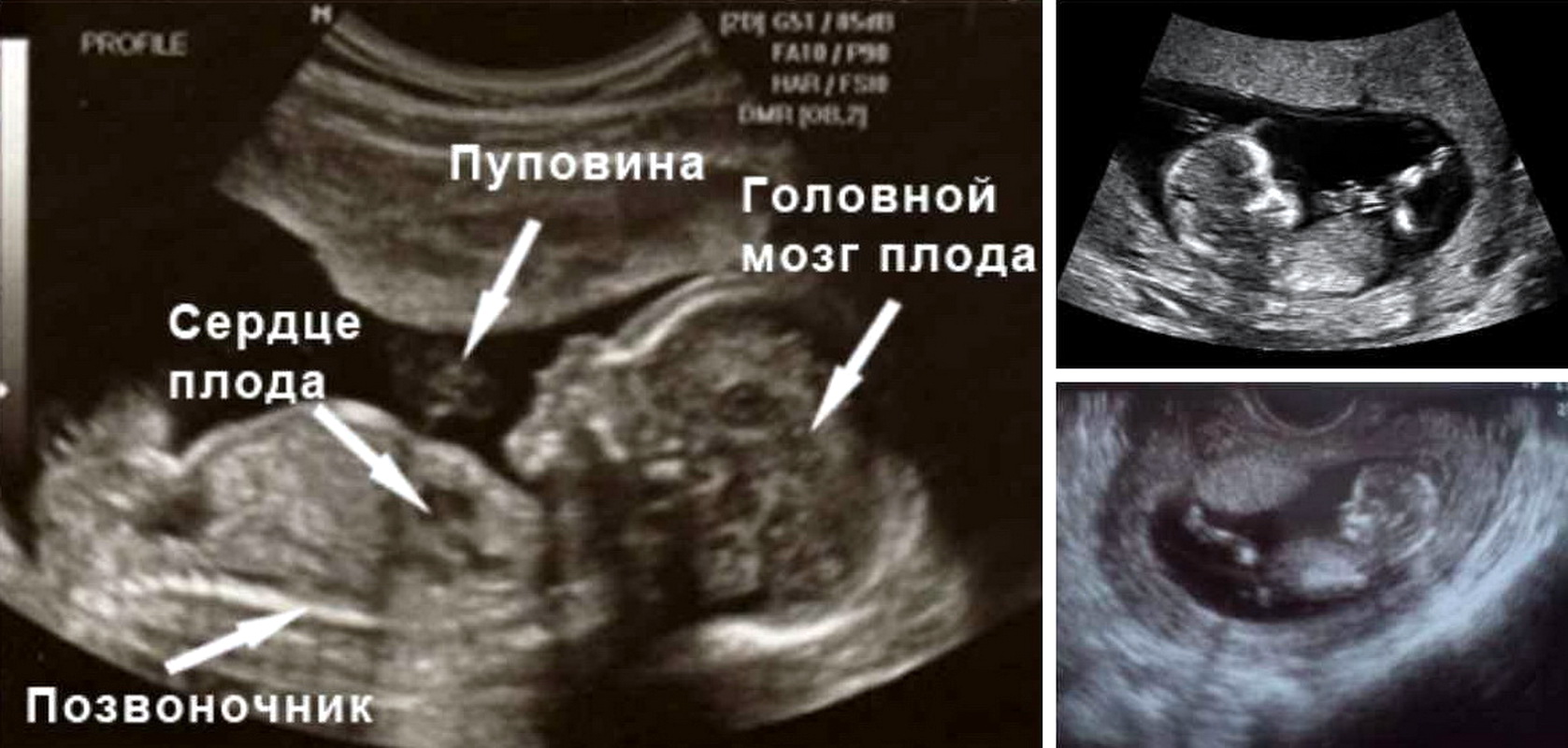
At the 15th week of pregnancy in the uterus, the fetus is already perfectly visualized. Modern devices allow you to get a fairly clear image. It is displayed on a special screen. With the help of such a monitor, the future mother can examine her future baby with the doctor.
If during the study the doctor identifies any serious pathology, he can immediately tell the pregnant woman about it. Also, these violations are made in a special form of medical opinion, which is issued to the patient in her arms. It is permissible to take pictures while conducting such a study. They are also handed out and attached to a medical card.

Modern 3D and 4D techniques allow to obtain three-dimensional images in which even the smallest structures of a baby’s body are visualized. The future mommy can see how her baby moves or flexes fingers. Also during the examination, spontaneous movements of the fetal chest are often seen.
A doctor at this stage of intrauterine development may identify some heart defects in the fetus. Normally, a baby's heart consists of 4 cameras. Between them there are special openings that are closed by valves. The presence of anomalies of the valvular apparatus of the heart and leads to the formation of heart defects.

Week 15 is the time for mandatory evaluation of the cervix of a pregnant woman. Gynecologists identify several types of pathologies that may develop in the future mother in the presence of defects in this reproductive organ. One of these dangerous pathological conditions is the development of cervical insufficiency.

Also during the study necessarily conducted a study of the ovaries. Such a diagnosis is especially important for women in whom, before the onset of pregnancy, various cysts or neoplasms in these reproductive organs were identified. The ovaries play a very important role during pregnancy. They provide optimal hormonal balance, which is necessary for the proper development of the fetus.
Standards of performance
Week 15 is a very close time to conduct a second screening. This leads to the fact that not many of the studied parameters are used to assess this period of fetal development.
The size of the fetus at this stage of pregnancy is usually 10 cm. The weight of the baby is 60-70 grams. Immediately it is worth noting that for multiple pregnancies, these parameters may be different. Quite often at this stage of pregnancy, future twins or triplets weigh less than 50 grams.
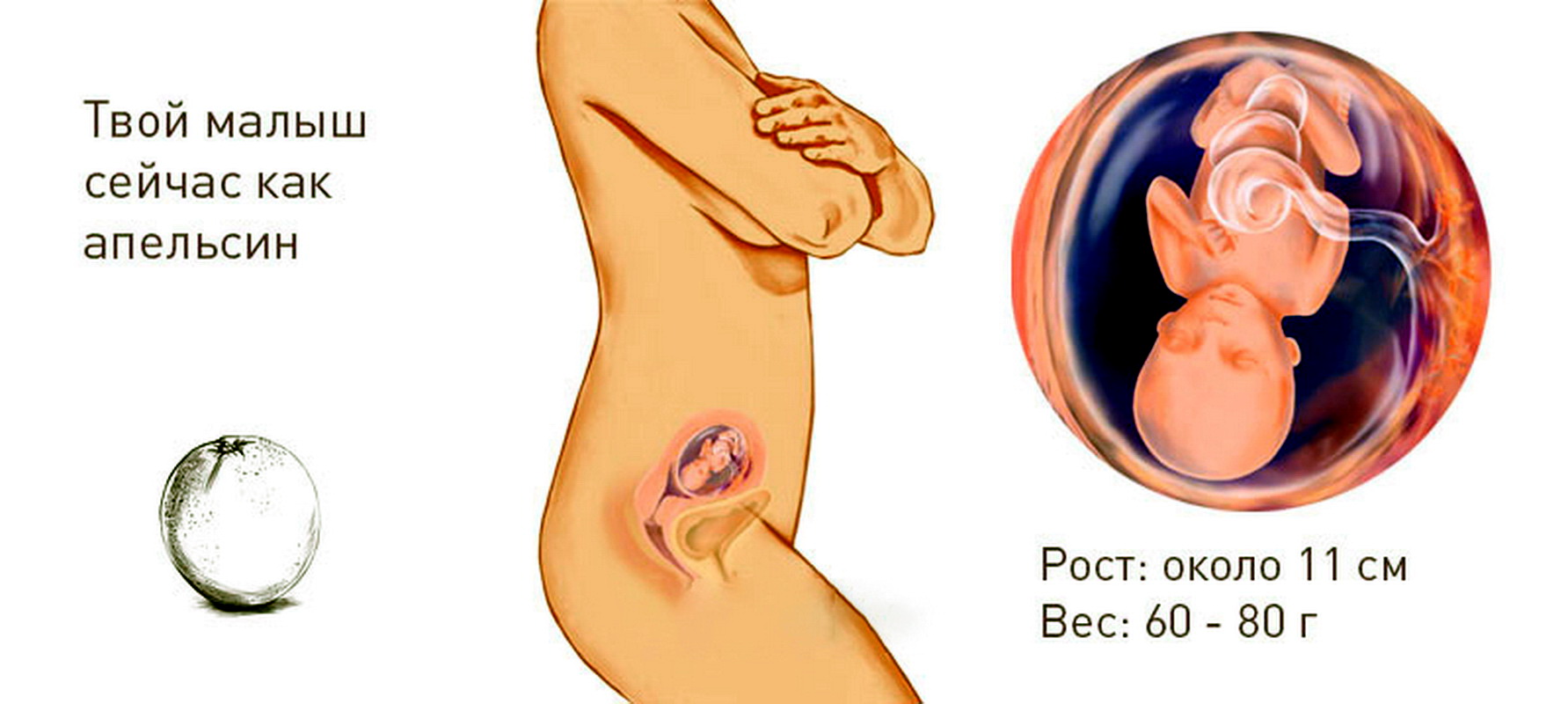
To visualize what the baby looks like at this time, you should just remember an orange. By its weight, the fruit at this stage, as a rule, corresponds to the size of this fruit.
The baby in the 15th week of its intrauterine development already resembles a real person. He has fingers, hairs and even marigolds.
To determine the uteroplacental blood flow is determined by the blood flow in the blood vessels of the placenta. An experienced specialist conducting research can identify pathological constrictions or even torsion of such vessels feeding the fetus. If such a situation occurs during pregnancy, then This requires a mandatory emergency consultation with a gynecologist.

The inner layer of the uterus is also being examined. It is important that there are no myomatous nodules or tumors on the endometrium. The thickness of the muscle layer of the uterus at this stage is usually 2-3 cm.
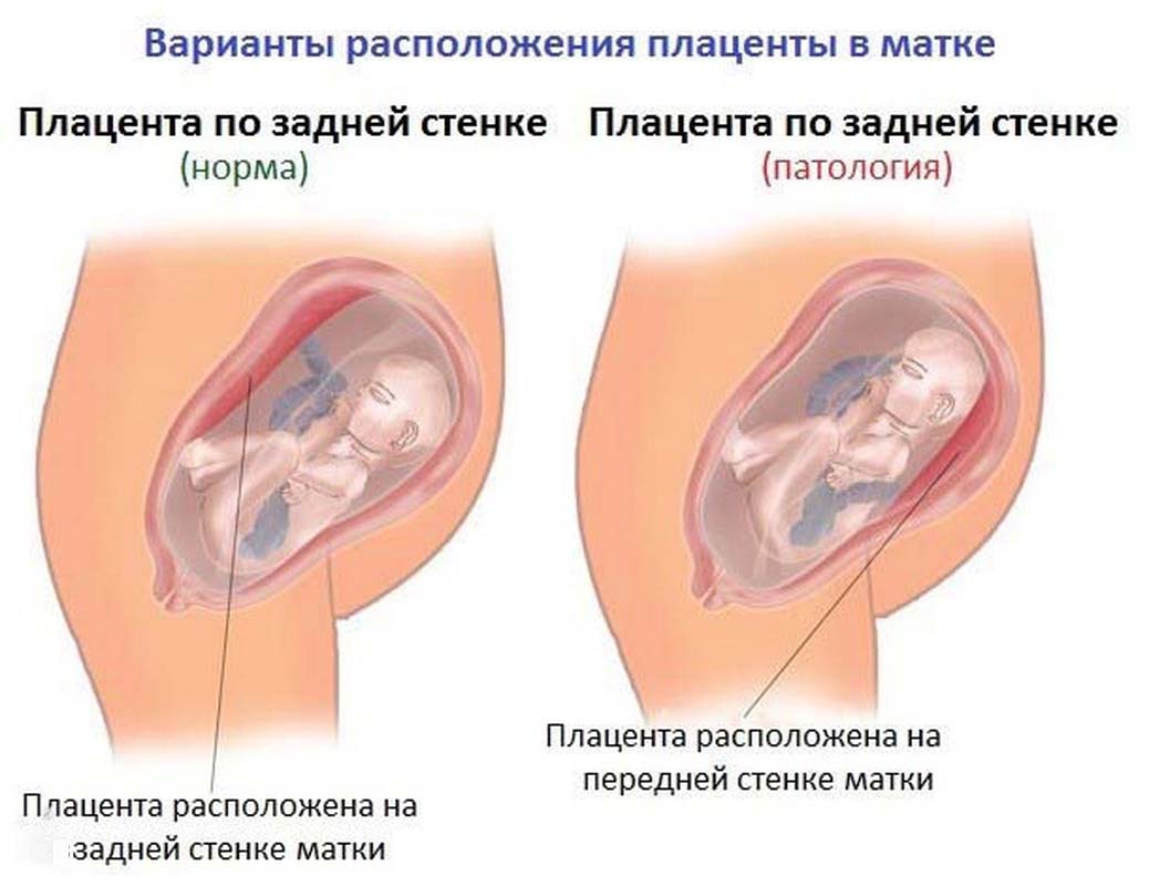
During the study, the position of the placenta is also determined. It is usually located either on the front or on the back of the uterus. Excessive growth of the placenta should not be. If it grows to the lower corner of the uterus at this time, then in the future, careful monitoring of its growth is required.
The results obtained are necessarily interpreted by an obstetrician-gynecologist. If some pathologies are suspected, determination of hCG. This biochemical marker allows you to identify various violations in various periods of pregnancy. The conclusion of one of the ultrasound in any case can not be diagnosed.
Is it harmful?
The second trimester is the time when conducting many studies is not dangerous at all. However, future moms should be aware of the possible risk of adverse effects. at abusing carrying out ultrasonography. Frequent use of this method without special medical indications can lead to various neurological disorders in the future baby.
During the normal course of pregnancy there are no strict indications for an ultrasound examination at this stage. In this case, it is reasonable to wait for the second screening. It is usually carried out from 16 to 20 weeks of pregnancy. In this case, identify any pathology is much easier.
About what changes occur in the 15th week of pregnancy, see the following video.


