Ultrasound in the 11th week of pregnancy: fetal size and other features

Until the end of the first trimester remains quite a bit. At week 11, the expectant mother is usually already registered at the antenatal clinic. This is where she can receive referral to the first prenatal screening this week, although most often this examination is prescribed from 11 full weeks, 12-12.5 weeks of gestation. That can be seen on ultrasound at week 11 and what are the norms of child development, we will tell in this article.

Objectives of the survey
Week 11 by obstetric standards is about 9 weeks from the date of conception of the baby. And about 7 weeks from the delay of the next menstruation. Pregnancy is already obvious, no doubt. Ultrasound during this period are assigned important medical tasks - diagnosis of probable pathologies and abnormal development of the child both genetic and not related to genetics.

The first screening, to which ultrasound can already be attributed this week, provides information for calculating the probability of having children with total chromosomal pathologies that are not subject to any correction or treatment. These are Down and Edwards, Patau, Turner, Cornelia de Lange syndromes and other anomalies that are not only incurable, but in most cases 100% lethal.
In addition to ultrasound scanning, the screening includes blood chemistrywhich determines the concentration in the blood of a pregnant woman of certain hormones and proteins, peculiar to the period of carrying the baby. In addition to this main task, an ultrasound scan at week 11 helps to find out how the baby develops, whether this pregnancy has any complications, identify possible risks of miscarriage of the child and other adverse factors, and also helps to “check the clock” - establish exact dates of pregnancy according to obstetric standards and determine the estimated day of birth.
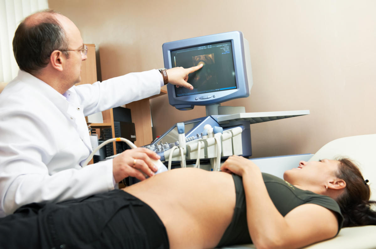
Method and preparation
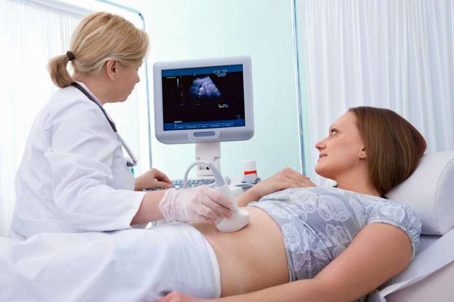
At this time, ultrasound diagnostics themselves can be performed in two ways - through the vagina (by the tranvaginal or intravaginal method) or through the peritoneum (by the transabdominal method). The choice of a particular method is the prerogative of the doctor, which is based not only on the duration of pregnancy, but also on the physique of a pregnant woman.

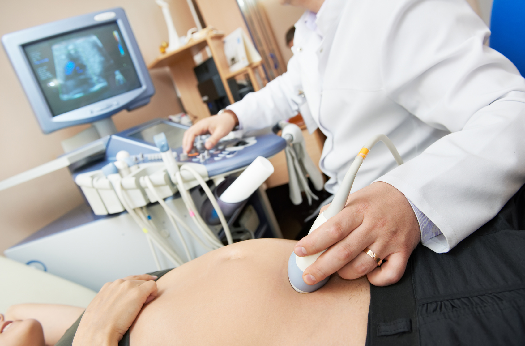
Fat and plump women often undergo transvaginal examination, because through the fatty layer on the anterior abdominal wall with a relatively small amount of amniotic fluid, which is observed at this time, the review of the baby is very, very difficult.
Only very slender and thin women can count on external ultrasound, which is carried out by the abdomen sensor, at week 11.
Prepare for transvaginal ultrasound need 2-3 days before passing the diagnosis. A woman needs to limit herself in sweets and sparkling water, in legumes and cabbage, as well as in cottage cheese and fatty yogurt. These foods cause fermentation in the intestines, and accumulated gases can put pressure on the pelvic organs. Therefore, such a diet, intestinal cleansing is desirable, and before going to a consultation for a few hours you should drink a dose "Simethicone" or "Espumizana». The bladder with intravaginal examination does not need to be filled.


But it is desirable to do this before visiting transabdominal ultrasound diagnostics. Although much depends on the size of the uterus and the amount of amniotic fluid. It may happen that the doctor sends a prudent future mom to empty a bladder so diligently filled.
On the ultrasound, you should take a diaper so that you can lay it on the couch, as well as napkins to remove excess special gel that is used during transabdominal diagnostics. The study will take no more than 10 minutes and will not cause the slightest harm to the woman or her baby.
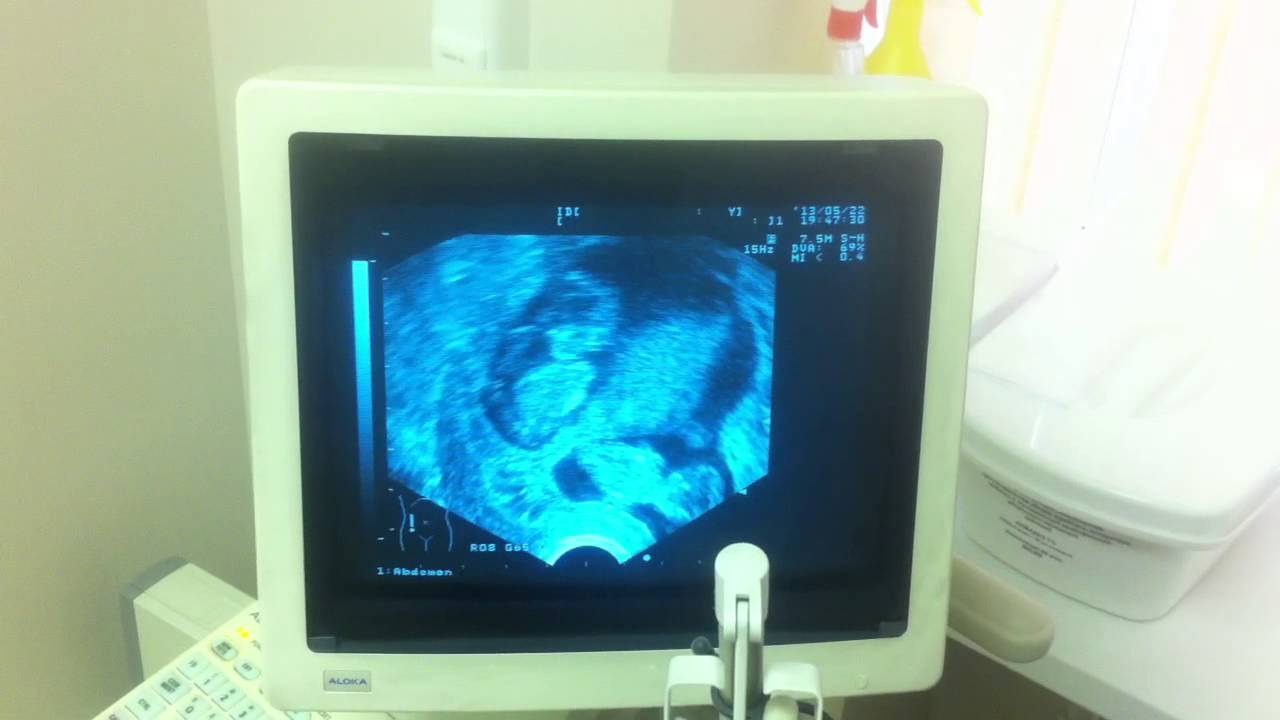
What can be seen on ultrasound?
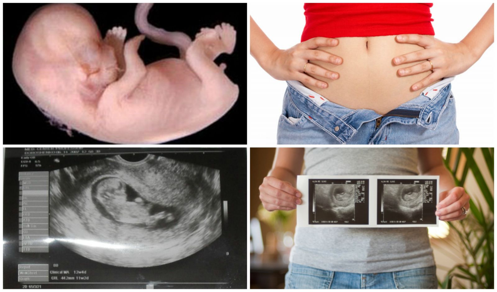
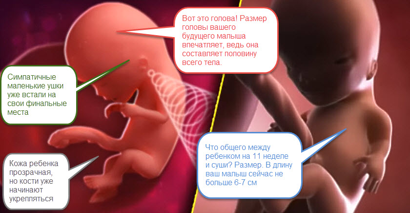
The baby at week 11 already has a lot and is capable of much. He has formed all the organs, including the spinal cord, there are basic structures of the brain. The baby's liver has already begun to synthesize proteins that are needed as a building material for a growing body. This organism grows not by leaps and bounds, and at this time the size of the fetus already inspire respect - its height is about 50 mm, and its weight is about 8-9 grams.
This week, the baby can show the diagnostician, and at the same time the future mother, how smartly he learned to move his arms and legs, his movements become more energetic, because the formation of muscle and bone tissue is in full swing. The skin of the baby on the entire area of the body and head becomes sensitive, the baby can touch. There are ears and very soon he will be able to hear. This week is the formation of taste buds, and also formed vocal folds - the only powerful weapon of the baby immediately after birth.
On ultrasound at this time it is impossible to see all the incredible transformations that yesterday's embryo is experiencing, which has already become a fetus with all the external signs of a human baby.
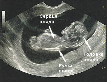
But parents can appear in all their glory a tiny creature with a big head and movable handles, with legs tucked to the stomach. When making a movement, the baby has already learned to help himself with his feet, raking in the right direction. If the period of activity coincides with the time of the study, then on a good device with high resolution one can already see such activity.

The sex of the child is not yet visible, only the genital tubercle is defined, which so far is exactly the same in both boys and girls. The doctor at week 11 is clearly visible all the organs of the baby, his position in the uterine cavity.
Rules and interpretation of results
Having found a fetus in the uterus, the doctor necessarily assesses its position - in the upper, middle, or lower part of the uterus, a fertilized egg is fixed. The definition of the vital activity of the child is also considered primary - his heart should be beating and signs of motor activity should be observed.

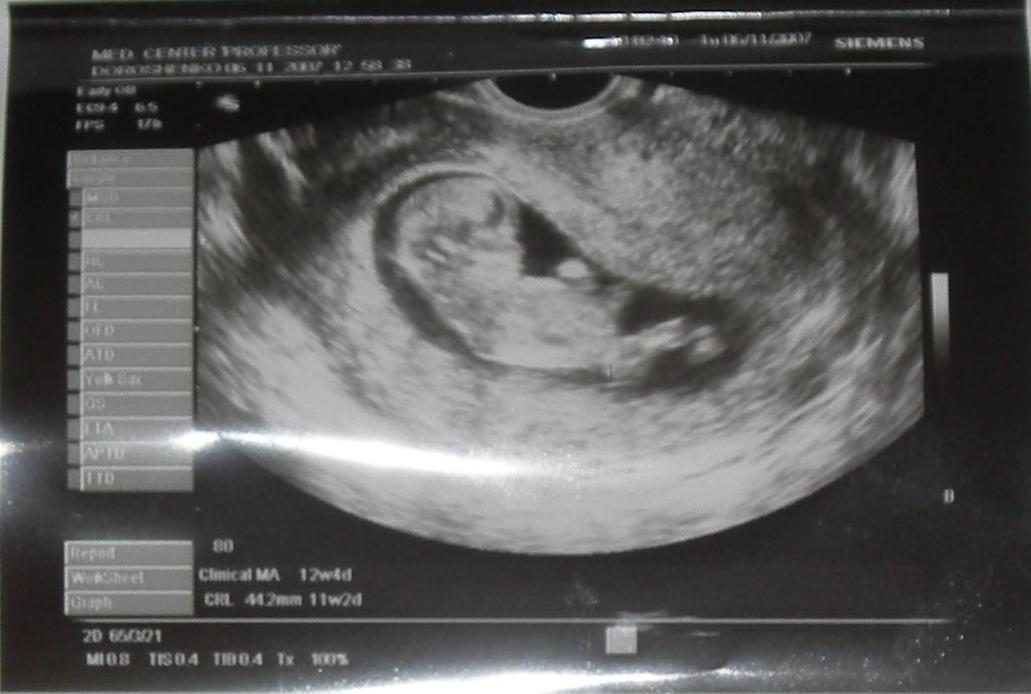
The main parameter that speaks about the pace of development, about the state of the child, about the compliance of his gestation with the date of the last menstrual period is the size of the fetus from the tailbone to the crown. It is called so - coccyx parietal size or simply KTR.
Kopchiko-parietal size at week 11:
The average rate of KTR, mm | Possible fluctuations, mm | Matched gestation |
33 | 28.0-39.2 | 10 weeks + 1 day |
35.2 | 29.5-40.5 | 10 weeks + 2 days |
36.7 | 31.0-42.5 | 10 weeks + 3 days |
38.3 | 32.5-44.0 | 10 weeks + 4 days |
39.9 | 34.0-45.6 | 10 weeks + 5 days |
41.5 | 35.5-47.2 | 10 weeks + 6 days |
43 | 37.0-49.1 | 11 weeks exactly |
According to the same table, which is always at hand with the doctor of ultrasound diagnostics, compliance with the date is checked. If the size of the fetus is normal, the doctor indicates that the KTR corresponds to the gestational period.
The heart of the baby knocks rhythmically and loud enough. Doctors necessarily give future moms listen to him. In conclusion, however, indicate the frequency of contractions of the heart per minute. Heart rate is an important indicator of the child’s condition. Normally, at week 11, a small four-chamber heart knocks at an average speed of 153-170 beats per minute. The rhythm is clear, without interruption, the tone is clear.
One of the important parameters, which are measured at week 11, are the so-called markers of genetic disorders.Knowing that some defects tend to shorten and thicken the neck, as well as swelling in the back of it, measure the thickness of the collar space.
The NTP rate for this week is 1.5 mm, increases or decreases are allowed from 0.8 to 2.2.
Another marker - nasal bones. In children with chromosomal abnormalities, the face is often flattened, and signs of such flattening make it possible to see measurements of the bones of the nose. At this time it is very difficult to see them, even harder to measure. Their average size is only about 1 mm, so when they are examined at week 11, doctors simply indicate whether the bones of the nose are visualized in the fetus without measuring them.
Possible problems
Let us examine them in more detail:
- TVP more than normal. With a significant excess of this indicator, a control ultrasound is appointed after 1-2 weeks, and blood results taken as part of the screening are also evaluated. If there are fears that the risk of pathology is high, a woman is sent for a consultation to genetics, which may offer an expectant mother an invasive procedure, for example, a chorion biopsy. She answers with high precision the question whether the child has anomalies.
- KTR does not meet the deadline. The most common cause of inconsistencies is an error in calculating the duration of pregnancy, as well as in late ovulation. If the embryo is implanted later, then the discrepancy may be from a week to one and a half weeks. If the difference is more than 14 days, then the woman needs additional examination, since the slow growth of the baby can be a sign of abnormalities at the gene level, and a symptom of malnutrition, developmental delay, intrauterine infection.
A small lag (in a few days) should not cause concern in pregnant women. All children are now individual, and they have different rates of development.


- The bones of the nose are not visible. If the bones of the nose could not be considered at 10-11 weeks, then this is not a sentence, but a reason to visit the doctor again, after a week, when the fetus grows, and its nasal bones will also increase and be available for inspection and measurement. An alarming sign is the absence of nasal bones at 12-13 weeks of pregnancy, but not at all at 11 weeks. If all other parameters and indicators for ultrasound are normal, then the decrease or absence of the nasal bones this week can also be regarded as a hereditary feature of appearance - parents with neat little noses, obviously, presented the same little nose to their child. In a week, the bones will be visualized.
- Fading pregnancy. Fetal death of the fetus can be the result of a variety of reasons - from gross malformations to the effects on the maternal organism of radiation, infectious diseases, poisons, medicines, hormones. If the baby does not develop, ultrasound will show not only a decrease in all sizes, but also an absence heartbeat and movements. There is only one way out - to surgically clean the uterus in order to avoid inflammation and sepsis, and then perform a biopsy of the chorion and fetal tissues, in order to determine in laboratory conditions which cause led to the tragic finale. This will help when planning the next pregnancy.

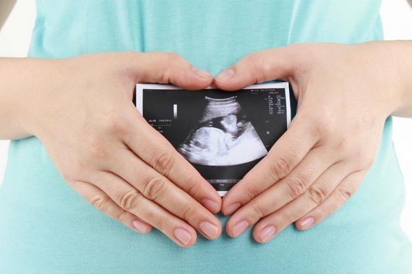
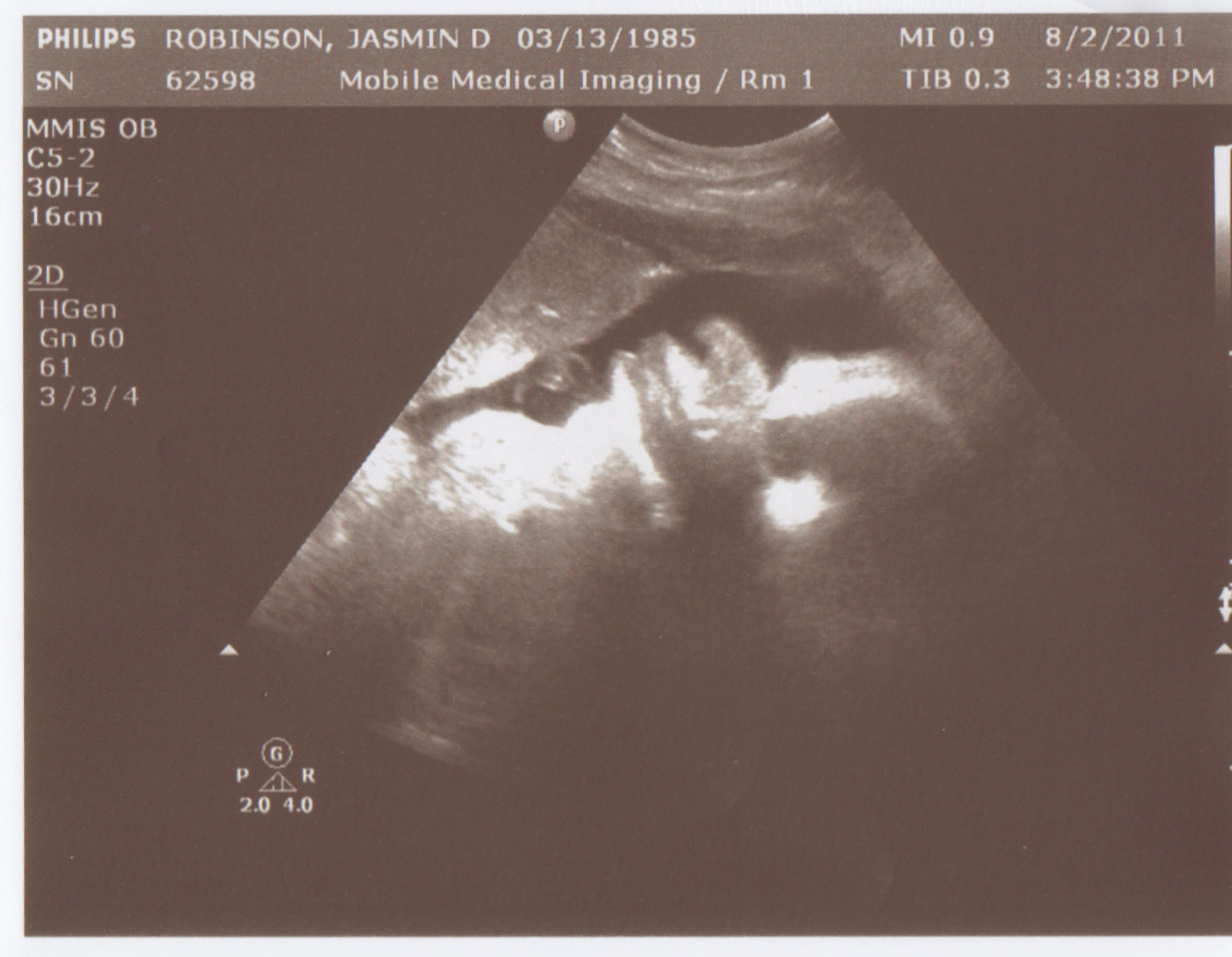
Snapshots
The ultrasound images taken at this time show the first outlines of the profile of the baby’s face, his tummy and limbs. If you are wearing twins, you can take pictures on a three-dimensional device - 3D-ultrasound. Children are well visible, even if they are identical twins. The main thing is to remember that ultrasound paper shots are short-lived, and if you want to keep the beautiful moments of the 11th week of pregnancy forever, it is better to ask the doctor for a photo or video on electronic media.