Ultrasound on the 6th week of pregnancy: fetal size and other features
For many women, an ultrasound scan at week 6 is the first meeting with a little man who was conceived only about a month ago. This is a special diagnosis, filled with a lot of emotions, because it confirms what has already been suspected and what home tests to determine pregnancy have indicated. What is the size of the fetus at this time and what are the average rates, we will tell in this material.
Objectives of the survey
Usually, at the sixth obstetric week, a woman goes on an ultrasound examination on her own to make sure that a delay of 2 weeks is not a disease of the reproductive system and not a hormonal failure, but pregnancy. With a ready conclusion, most often women come to the antenatal clinic in order to get registered.
Sometimes at this time the woman is referred to an ultrasound scan by an obstetrician-gynecologist, to which she appealed for the lack of regular menstruation. Such research is required to make sure that the pregnancy is there, it develops.

Women who have had miscarriages, missed pregnancies and abortions earlier in this period or so may also be referred to ultrasound. This is especially true of women who have had two or more miscarriages early in a row. Ultrasound diagnosis in the sixth week can be recommended for women who become pregnant through IVF to find out how many fruits have taken root, and how they develop.
Such an earlier examination was also shown to women who had previously had ectopic pregnancies, as well as women with pathologies of the pelvic organs - myoma, ovarian cyst, and myomatous nodes.
If a woman has previously been treated for such diseases, the doctor must make sure that the absence of menstruation is a sign of pregnancy, and not the appearance of a tumor or inflammatory process.

Ultrasound this week will make and women who have complaints of pain in the lower back and lower abdomen, bloody or blood-like discharge, "daub." It is important to understand whether the baby is alive, whether there has been a detachment of the ovum, or if a miscarriage has begun. Strong toxicosis, rise and fall in blood pressure, fainting from the expectant mother is also a reason to visit the ultrasound room.
Ultrasound scanning at this time is not mandatory and ubiquitous. The first general examination awaits the expectant mother from 11 to 13 weeks, when she will undergo the first prenatal screening.

Procedure and preparation for it
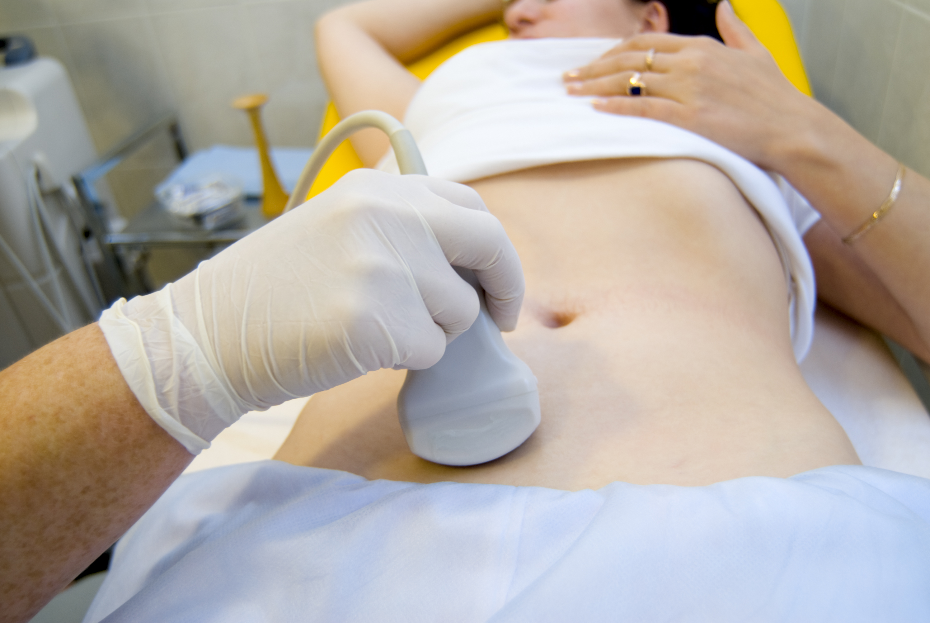
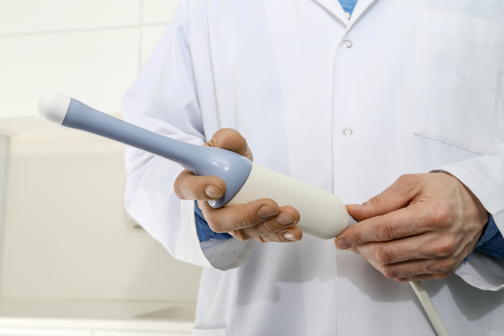
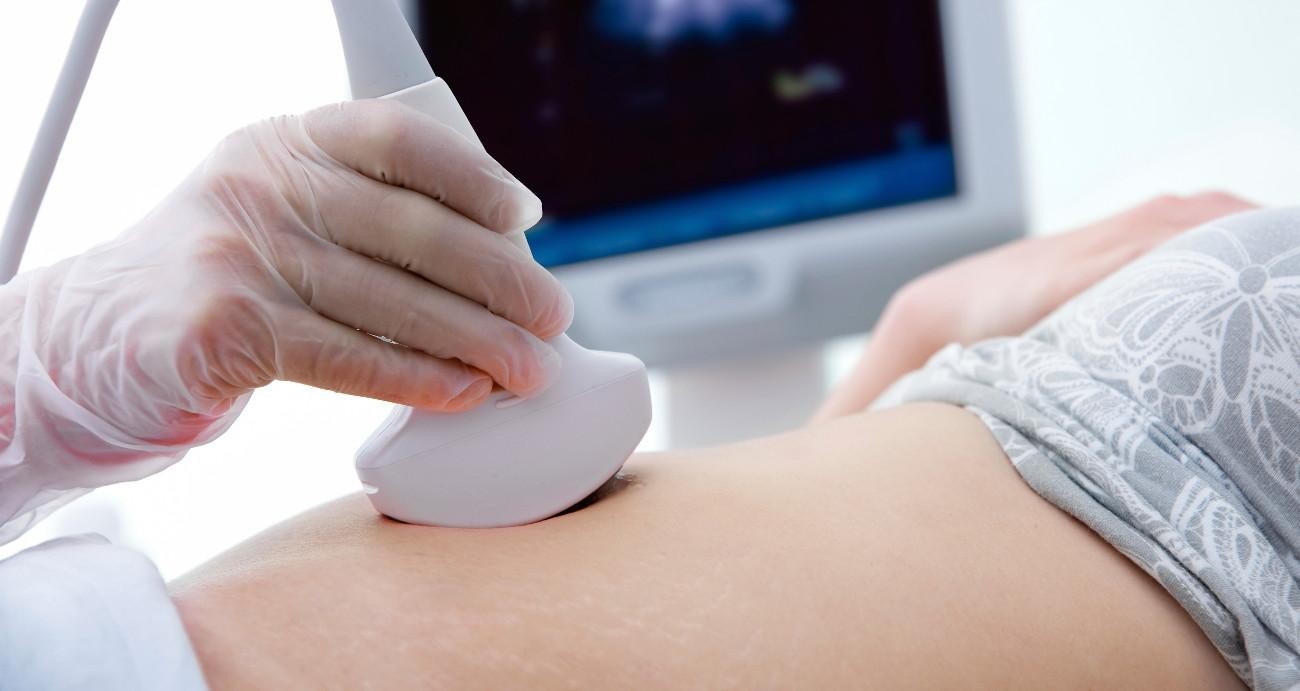
Inspect the pelvic organs and the baby, who probably already lives in the uterus of a woman, the diagnostician can in two ways. Transabdominal and intravaginal. Most often, in the early stages, preference is given to so-called internal ultrasound. The diagnostician puts a condom on the sensor and examines the uterine cavity through the vaginal wall.
When viewed transabdominal, the sensor is located on the abdomen, richly lubricated with a special gel, the review is carried out through the anterior abdominal wall.This method is not considered to be the most successful for the sixth week of pregnancy, since the embryo is still so small that it will be problematic to examine it through a sufficiently dense layer of muscles, subcutaneous fat.
For women "in the body," such a method of examination in the early period is generally not suitable.

Preparing for the study in a transvaginal way includes emptying the bladder, not filling it, as many women mistakenly believe. It is also desirable to empty the intestines and eliminate the possible accumulation of intestinal gases, taking a couple of hours before visiting the doctor "Espumizan"Or" Simethicone. " Crowded intestines can squeeze the pelvic organs, distorting the real picture on the monitor of the ultrasound scanner.
If the ultrasound is planned, then for a couple of days before going to the doctor it is advisable not to eat foods that contribute to gas formation - beans, kefir, cabbage, as well as sweets and pastries from yeast dough. If the study is to be outside the method, and a half hours before the appointed time should drink a few glasses of waterso that by the time of the examination the bladder is filled, it will facilitate the passage of the signal from the sensor and give the doctor the opportunity to see the uterus and its contents more clearly.

On ultrasound, regardless of its type, you should take a diaper or cloth napkin, a small towel. The diaper is needed in order to lay it on a couch or viewing chair, and a towel will help wipe the remnants of the gel from the stomach, if the diagnosis is transabdominal.
The procedure does not harm either the woman or the child. It is painless and lasts only about five minutes.
What does ultrasound show?
No matter how hard the specialist, and no matter how expert the apparatus on which the diagnostics is carried out at this time, would be, it would not be possible to see something intelligible to the expectant mother. The baby is still too small. The length of the embryo between 5 and 6 weeks of gestation is from one and a half to 4 mm. Weight - less than a gram.
Such an "tiny" on the scanner monitor can only see an experienced doctor. If he is in a good mood, he can show this dark point on the screen to a woman.
6th week is considered one of the most important. It is now that the nervous system of the crumbs is actively being formed, the immune system is beginning to form, the thymus gland appears, a small heart begins to beat, and this is what you can already try to hear on the ultrasound. Atria is not yet, the heart looks like a small pulsating tubercle. At this time, the formation of the baby’s handles begins, the face is laid, the mouth and the places where the eyes will be located are already determined, there are dark spots at the place of the future ears.
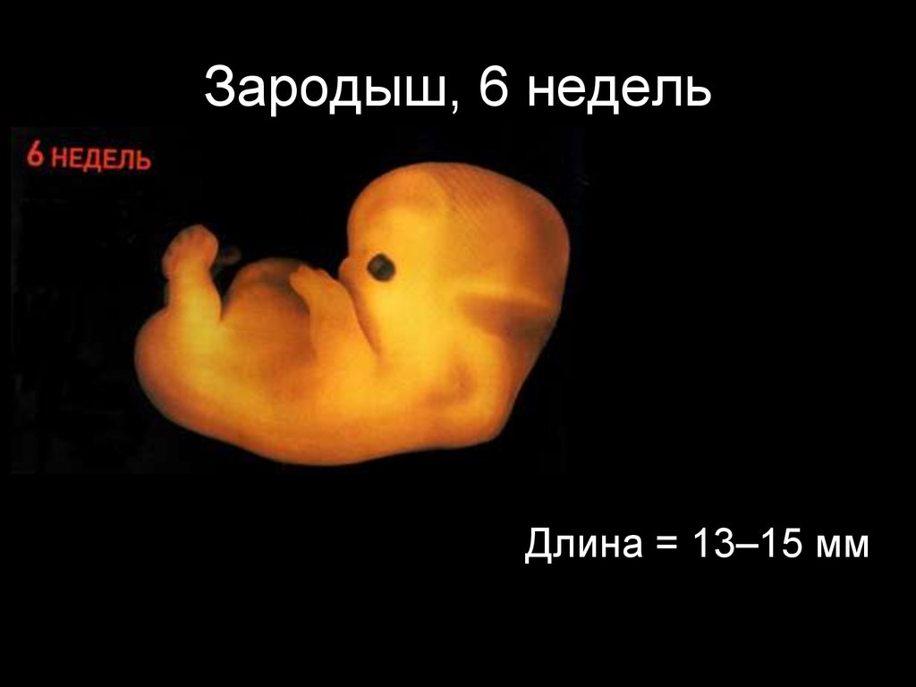
The skin of the embryo barely begins to acquire sensitivity, the neural tube on this period begins to branch, creating a “platform” for the subsequent development of the central nervous system. The baby has not yet begun to move, at least in the general sense of the word. After a couple of weeks on the ultrasound will see how he moves the arms, but now the movements are more like nerve impulses, they are involuntary.
The umbilical cord is formed, the baby gets the opportunity to swim in the surrounding amniotic waters. Food he gets from the yolk sac. While the placenta is being formed, this temporary organ is a children's “food store”.
The placenta itself still looks like a slight thickening, there is still no “child's seat” as such.
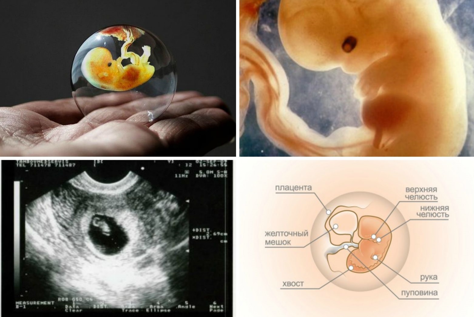
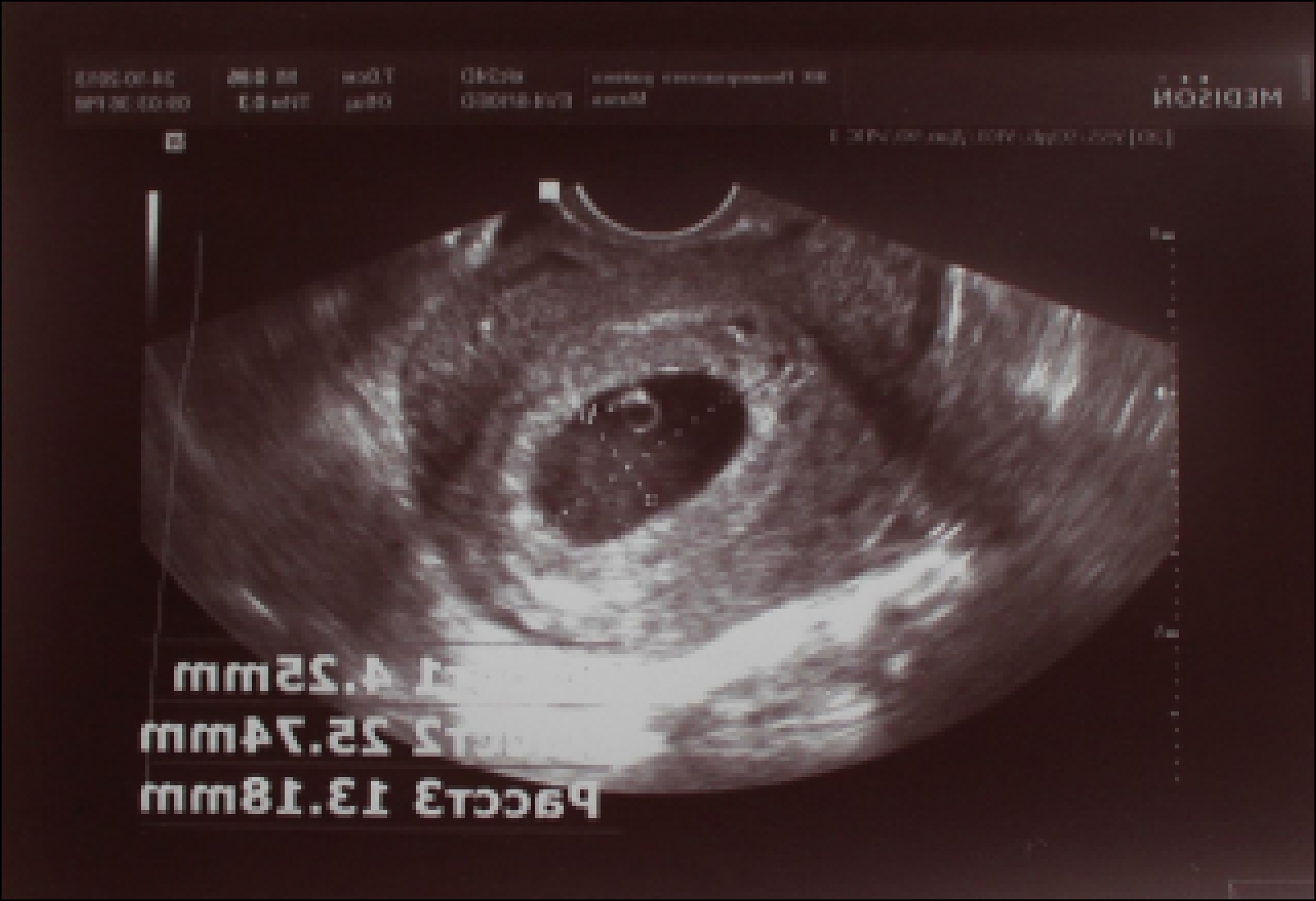
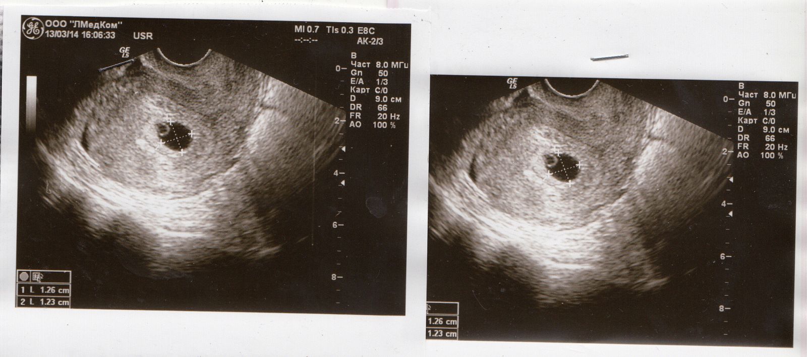
On ultrasound at this time, the doctor will be able to see the fetal egg with the embryo inside, the place of attachment of the ovum in the uterus, by the end of the sixth week it is theoretically possible to measure the baby from the coccyx to the crown, but usually doctors do not. To determine the size is still too early.
The sex of the child is predetermined from the moment of conception, but the genitals are not yet formed, and therefore it is premature to ask the doctor whether the boy or girl settled in the stomach.The doctor will not be able to answer it for at least 5-6 weeks, and even that is not a fact.
Sometimes sex can be established only after 16 weeks.
Interpretation of results and norms
The human embryo at this stage resembles the embryo of any mammal. It is human traits that will appear and be measured and described by a doctor later. In the meantime, the well-being or distress of the fetus can be judged only by the growth rate of the ovum.
To assess development at 5-6 weeks measure the average internal diameter of the ovum (from wall to wall along the inner surface, a segment is laid on the ultrasound machine). SVD of the ovum is still the only diagnostically important indicator, based on which the doctor can determine the exact duration of pregnancy, as well as notice some alarming symptoms indicating problems with gestation or fetus formation.
SVD cannot be called the exact parameter, the size and outline of the ovum varies, and the error can be as long as a week or more. To clarify the timing is best suited to another size - coccyx parietal. But in most diagnostic centers it is only started to be measured from 7-8 weeks of pregnancy.
For the sixth week, the average internal diameter of the ovum is usually normal - from 6-7 to 12 mm. Such a large "scatter" is associated with the rapid growth of the ovum. If at the beginning of the sixth week it is only 6 mm in diameter, then by the end it can be doubled.
In more detail, the size of the SVD at the sixth week looks like this:
The diameter of the ovum (SVD), mm | Matching Date |
6 | 5 weeks + 1 day |
6-7 | 5 weeks + 2 days |
7 | 5 weeks + 3 days |
8 | 5 weeks + 4 days |
9 | 5 weeks + 5 days |
10 | 5 weeks + 6 days |
11-12 | 6 weeks exactly |
Ultrasound in 5-6 weeks and measured yolk sac. Its normal size by the end of the sixth week is about 3 mm. The number of fruits, the place of attachment of the ovum in the uterus is estimated. In addition, the doctor pays attention to the very egg of the fetus - its contours, shape. It is important that it does not look squeezed, deformed.
Heart rate at this time is not always measured. Much depends on the quality and capabilities of the apparatus on which the study is conducted. If the equipment is “weak”, the doctor can simply put a “+” in the column “Fetal heartbeat».
On good modern equipment It is possible to measure the heart rate indicator. Between the fifth and sixth weeks this value is usually at the level of 110-120 beats per minute. And at the beginning of the week, the heart can beat with the rhythm of 80-85 beats per minute, but by the end of the full sixth obstetric week heartbeat becomes more confident and fast and reaches 102-126 beats per minute.
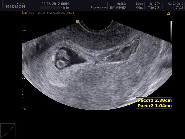
Separate attention on this period deserve the organs of the small pelvis of a woman and the assessment of the general reproductive health of the future mother. The diagnostician carefully examines the uterus, appendages, fallopian tubes and ovaries, assesses the condition of the cervix, cervical canal.
On ultrasound, signs of compaction of the uterine wall are clearly visible, which indicates an increased tone, which is a true symptom of the threat of miscarriage.
Possible problems
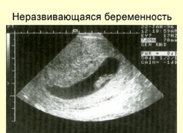
Undeveloped pregnancy
On ultrasound, the embryo's development in this period is characterized by a lag in the rate of increase in the ovum. Also, the doctor cannot diagnose palpitations or pulsations. Fetal death can occur due to various reasons - severe genetic malformations of the fetus, poisoning, taking medicines, especially antibiotics, autoimmune processes in the body of a woman, which lead to the rejection of the ovum.
To confirm or refute suspicions should control ultrasound in a week, as well as a blood test for hCG, in the case of the death of the embryo, the level of this chorionic hormone in the blood of a woman begins to fall.
Ectopic pregnancy
If the test conducted at home, gives an undoubted positive result, and the fetal egg at the sixth week in the uterus was not detected by ultrasound, then we are talking about ectopic pregnancy. The reason for it lies in the fact that the fetus was fixed somewhere outside the uterus, the “mistake” happened during the implantation period, 7-8 days after conception.
It is not always possible to detect a fertilized egg with the help of ultrasound, sometimes urgent laparoscopic diagnosis is required. A woman is necessarily hospitalized because pregnancy outside the uterus threatens her life.

Retrochorial hematoma
Complaints of the woman to the aching aching pain in the lower abdomen, to the bleeding of different intensity may be signs of detachment of the ovum. Retrochorial hematoma (place of detachment) looks like a dark spot between the ovum and the uterine wall.
The prognosis depends on the size of the hematoma.. In most cases, with a small or medium detachment, it is possible to save the pregnancy, if you comply with all doctor's prescriptions, take prescribed medications and follow bed rest.
Small fertilized egg
Lagging in size from the average norms of the ovum is not a reason for panic and worries. It is possible that the woman had late ovulation, respectively, the implantation occurred later, and the actual duration of the pregnancy is less than the woman and her obstetrician-gynecologist thought. There is nothing pathological.

However, slowed growth in the future, with dynamic observation, can be interpreted as a delay in the development of the fetus. In any case, should undergo additional tests.
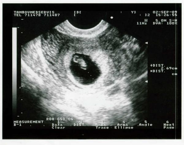
Snapshots
Pictures taken at this time may well indicate the presence of twins or triplets. In the sixth week, it is already possible to distinguish twins from identical twins.
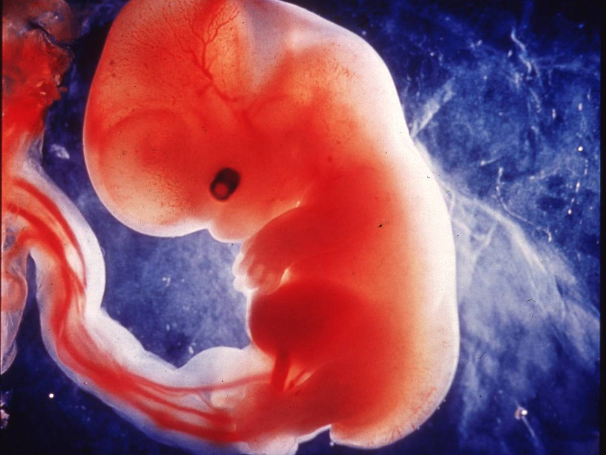
The picture taken at week 6, although it does not give a clear idea of what the baby actually looks like, may well be the first in the children's photo album. If you want this, you should ask the diagnostician an electronic copy of the image. Ultrasound paper photographs tend to fade quickly and lose their appearance.
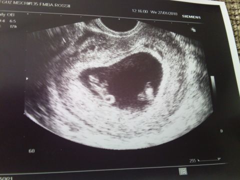

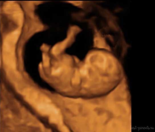
Three-dimensional, popular among future mothers Ultrasound (3D) is not usually performed on this period.. It does not make much sense, because there is nothing to consider in the volume - the embryo is still too small. The photo shows the embryo at the 8th week of pregnancy. A kid two weeks younger looks almost the same, you can get such a photo only on high-precision equipment.