Ultrasound at 4 weeks gestation

Ultrasound examination in early pregnancy is carried out only under strict medical indications. At week 4, the fetus is actively growing and developing. With the help of ultrasound it is possible to identify not only pregnancy, but also to determine the primary anomalies of its course and development.

What does the study show?
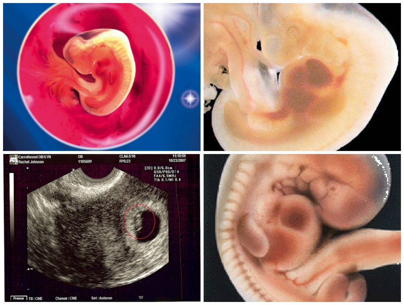
3-4 weeks of pregnancy is a very important stage of organogenesis. At this time, the laying of vital organs. In this period, doctors call the future baby embryo. In its size, it reaches only a few millimeters.
It is important to note that to assess the intrauterine development of the fetus, doctors determine the obstetric duration of pregnancy. With this medical term, the future mom will subsequently face throughout the life of the baby.
Some doctors also use the concept "Embryonic term". It should be noted that embryonic development lags behind the obstetric for a couple of weeks. This is because the obstetric period is calculated from the estimated date of the last ovulation and conception for 2 weeks.
In order not to get confused with the terms, doctors recommend future moms to use those that they use in their work. Especially in the future obstetric gestational age will be found in the medical records of a pregnant woman much more often.
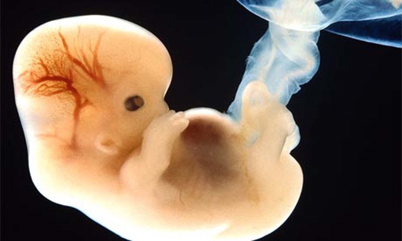
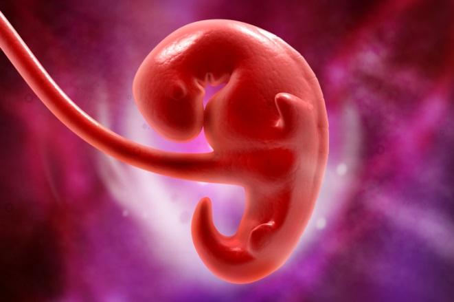
At this time there is an active development of the embryo. At this time period of pregnancy, a small embryo represents only a group of actively dividing cells. Usually it has a three-layer structure. In the future, these layers will be transformed into the development of fetal structures. At this time, the rudiments of the brain, spine, gastrointestinal tract, future skin and other internal organs are actively formed.
Chorion is an actively developing anatomical structure. It is necessary for the blood supply to the fetus. The blood vessels of the chorion in this case strongly adhere to the inner lining of the uterus.
Through such a message, the fetus receives all the necessary nutrients and oxygen dissolved in the blood. Also from the chorion will be formed in the future and the placenta.


By the end of the fourth and the beginning of the fifth week of pregnancy begins to form amnion. This is the formation within which the embryo itself and the fluid component are located. In the future, fetal membranes and water will be formed from the amniotic sac.
Yolk sac - Another important anatomical structure of the embryo. It is the germ of the formation in the future of some internal organs. At this time, the size of this formation does not exceed a few millimeters. It is important to note that only experienced ultrasound diagnostics specialists can determine this anatomical structure.

What is it for?
Carrying out an ultrasound at the earliest stages of pregnancy is the exception rather than the rule. Absolutely all women do not do such research. At this time, the size of the embryo is so small that the doctor can accurately and not install them.
4 week gestation is the time when it is determined fact of pregnancy, as well as associated diseases of the internal genital organs of the future mother. It is important to note that such an examination plays a very important role in the compilation of further patient management tactics.
It is possible to carry out ultrasound and multiple pregnancy, when the expectant mother is expecting the birth twins or triplets. A reproductologist may also assign this study to establish the result after in vitro fertilization. In this case, in the uterus, the specialist performing the ultrasound will see several gestational (fetal) eggs, which will indicate a favorable outcome of implantation.
It is also necessary to conduct research at such early stages. to establish signs of ectopic pregnancy. In this case, the ovum in the uterus is not visualized. High blood levels of hCG in this condition only confirms the presence of an ectopic pregnancy.
Doctors may prescribe such a study also in cases of suspected trophoblastic disease. This disease can lead to spontaneous miscarriage or the formation of intrauterine malformations.
Also, ultrasound diagnostics helps to identify various cysts of the corpus luteum, which also often develop during pregnancy due to altered hormonal imbalance.
Usually screening in the first trimester of pregnancy is carried out much later - at 10-12 weeks. In this case, you can already have a better look at all the anatomical structures of the fetus. Also at this time, doctors can determine various pathologies of intrauterine development.
The norms of the studied parameters
The small size of the embryo does not yet allow ultrasound diagnostics specialists to determine all its internal elements in this period of pregnancy. A future baby looks like at the 4th week of its intrauterine development, usually like a poppy seed. Its longitudinal size at this time is 1-1.5 mm. By weight, it is even less - about 0.6 grams.
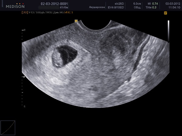
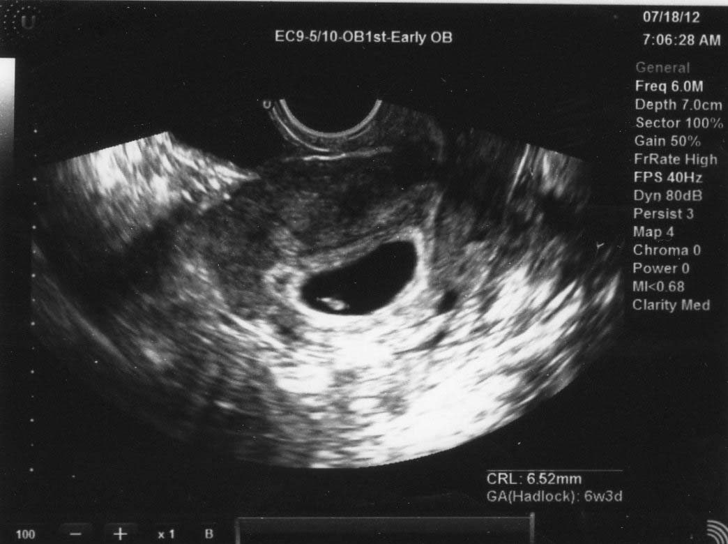
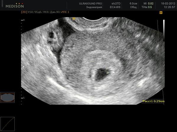
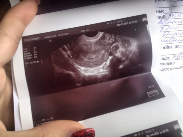
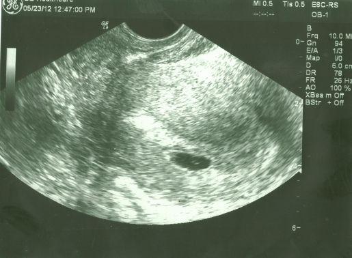
The main anatomical element, which is determined by doctors of ultrasound diagnostics at this stage, is the fetal sac. It is a formation located on the inner wall of the uterus. In size, the sacculate sac is usually 3-5 mm.
Week 4 is also time when all the indirect signs of pregnancy are excellent. During this period, doctors determine the expansion of the uterine blood vessels. This phenomenon is due to the fact that the growing embryo needs more nutrients and oxygen for its active growth and development.
At this period also determines the size and tone of the uterus. The growing embryo and the effect of pregnancy hormones lead to the fact that the size of the reproductive organ gradually begins to increase. The size of the cervical canal is also determined. Normally, it should not be less than 3 cm. If its shortening is determined, then in this case the patient must turn to a gynecologist for a second consultation in order to exclude ICN.

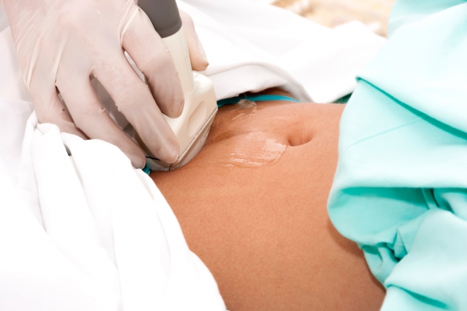
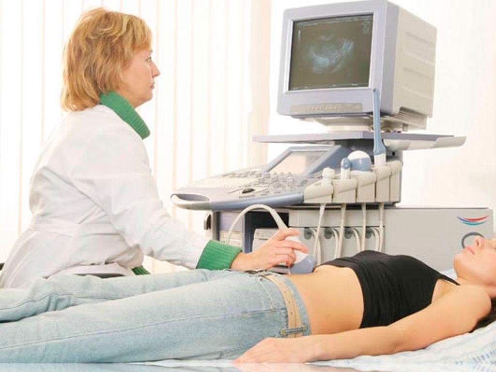
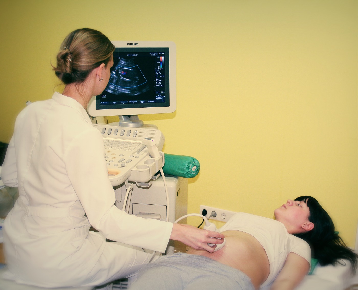
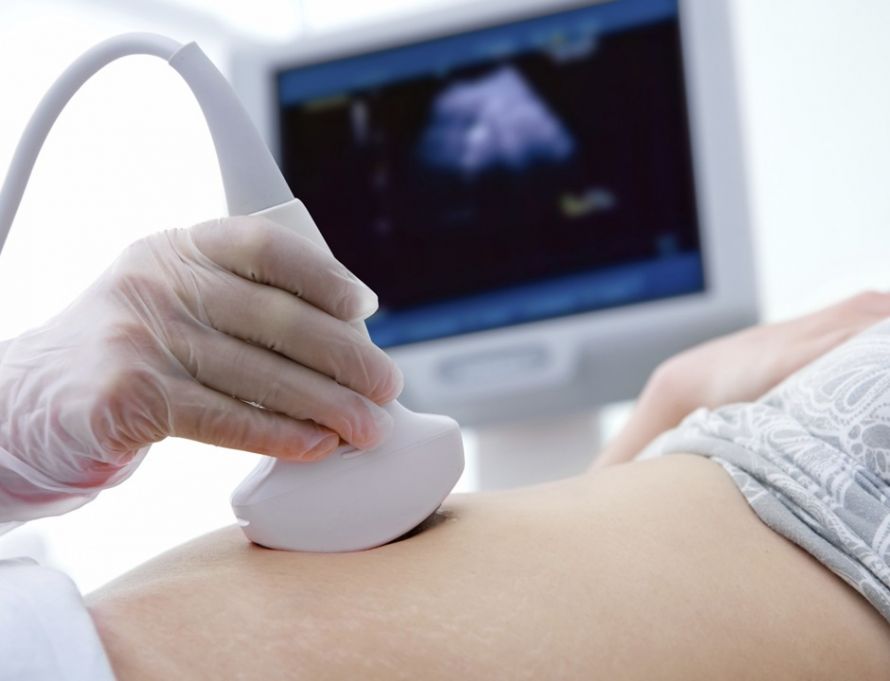
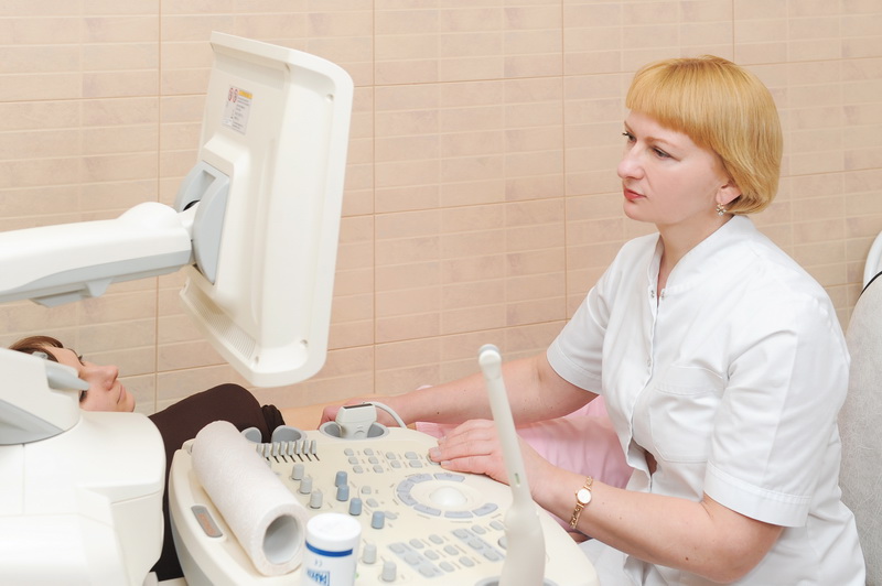
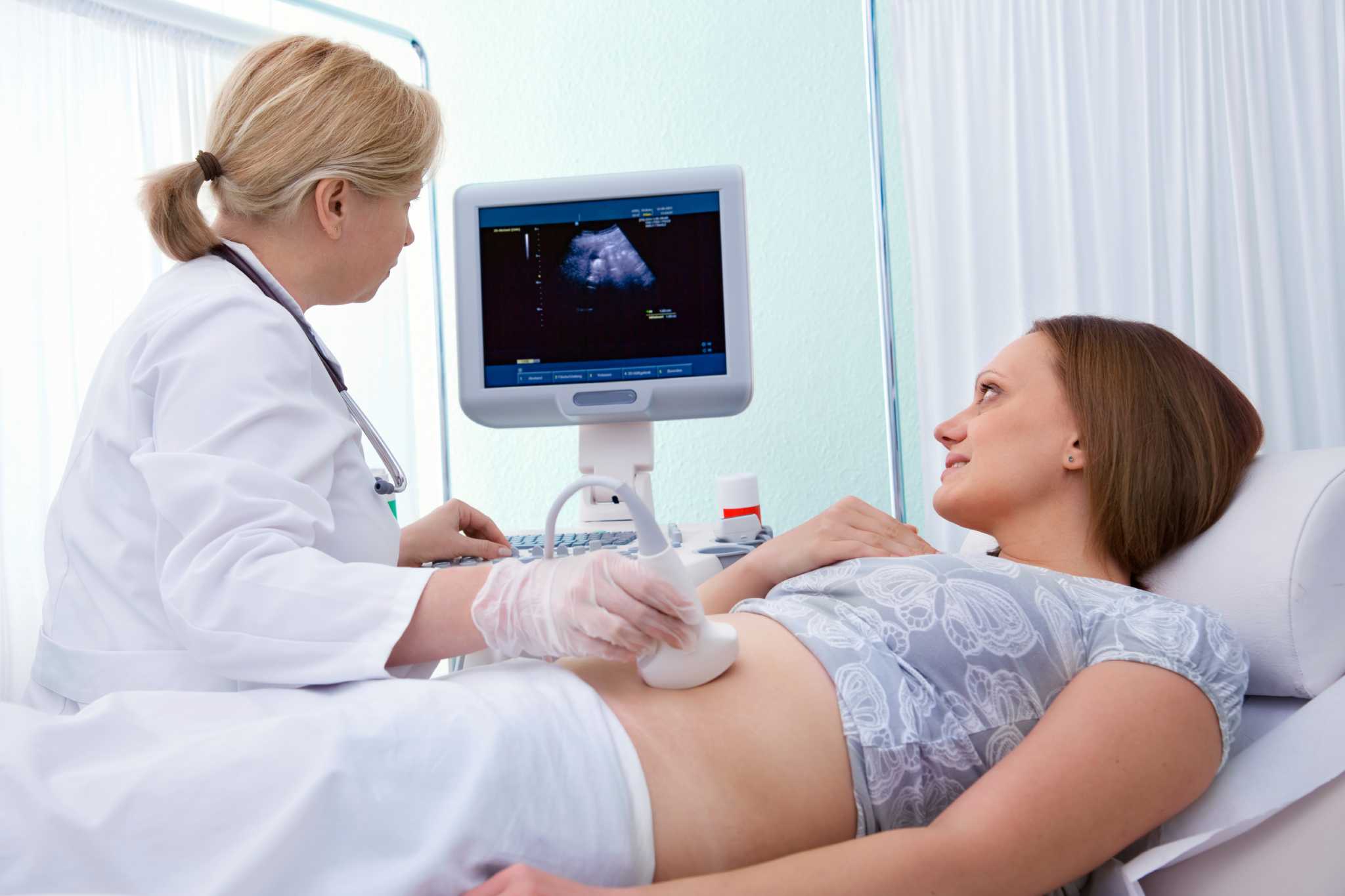
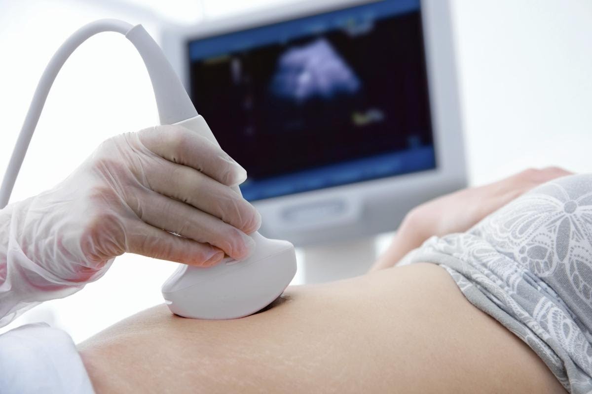
To conduct research in the early term in several ways. The most common is transabdominal. In this case, the study is conducted through the anterior abdominal wall. The doctor touches the skin with a special ultrasound sensor, and an image appears on a special screen.
To obtain the best picture uses a special transparent substance. This gel is pre-processed skin of the abdomen. It is necessary to improve the penetration of ultrasonic waves into the body.
Many pregnant women believe that such a gel can be dangerous. However, this is not the case. Its hypoallergenic composition completely eliminates the occurrence of any allergic reactions in expectant mothers. After the examination, the gel is removed using a normal paper towel.

Another way to get the image on the screen - use of transvaginal ultrasound. In this case, the doctor conducts research using a special sensor that is inserted into the vagina. Most doctors consider this method to be the most accurate and informative, especially in such early terms.

It is important to note that there are several contraindications to the doctor for transvaginal ultrasound. If a pregnant woman has an exacerbation of colpitis or vaginitis, then most likely, this examination will be postponed. In such cases, doctors replace transvaginal ultrasound with transabdominal examination.
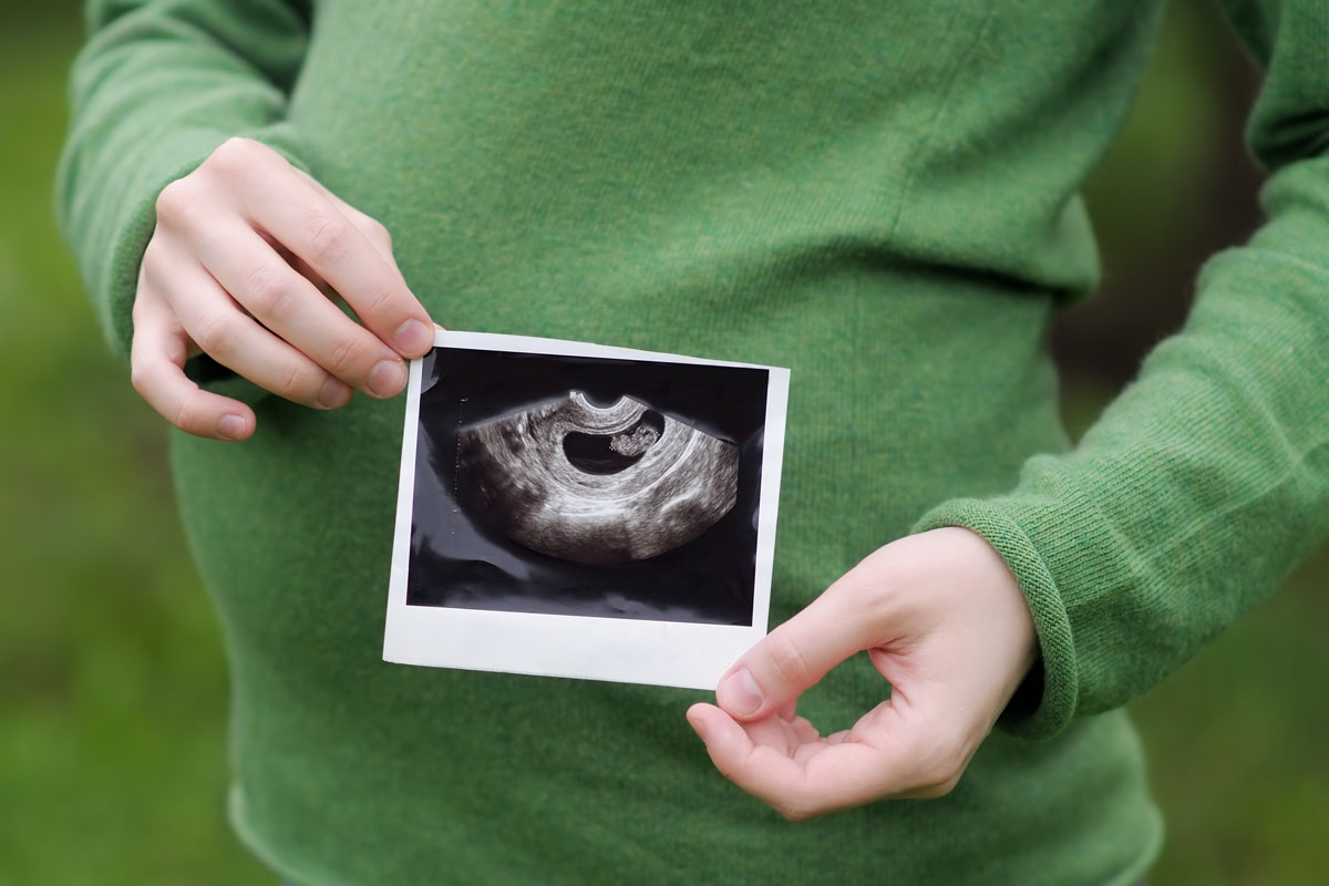
During such an examination, the doctor can take the first pictures of the baby. In such an image it is still impossible to distinguish the contours of the body of the unborn child. This picture shows only a rounded or elongated formation. In some cases, the first “photo” of the child is made at the request of the future mom.


If, after the ultrasound examination, the doctor did not detect any signs of pregnancy, but the woman still has quite a high level of pregnancy hormones in her blood, then she will be re-examined after 1-2 weeks. This situation quite often occurs when it is difficult to diagnose ectopic pregnancy. Also, such a case may be due to a "frozen" or "frozen" pregnancy.
Is it possible to do this?
Early ultrasound is an exception, not a mandatory practice. The first regulated period of the survey is 10-12 weeks of gestation. At such a time, an ultrasound scan can reveal all the specific pathologies of gestation and evaluate its intrauterine growth.
Frequent ultrasound, especially in the early stages of fetal development, will inevitably lead to adverse effects. Scientists from many countries annually conduct hundreds of scientific experiments that determine the negative effect of ultrasound on the developing fetus after abusing ultrasound in pregnant women.
Week 4 is the time for the most intensive growth and fetal organogenesis. The impact of ultrasonic waves has a mechanical external effect. This contributes to the fact that the embryo can form various abnormalities in its intrauterine development.
Ultrasound is performed at this time only under strict medical conditions. Doctors in such a situation mainly conduct this research in order to exclude a spontaneous miscarriage or an ectopic pregnancy. Especially unfavorable to conduct research in focus mode. Doctors say that 3D and 4D ultrasound on this period is also not worth carrying out.
Future mom should remember that The suspicion of pregnancy at this time is not a 100% reason for an ultrasound. In order to confirm the fact of pregnancy, in this case, the doctors use some laboratory tests and perform a clinical examination. In the absence of contraindications, ultrasound should not be done at the 4th week of pregnancy, so as not to harm your unborn baby.


