How long can a pregnancy be determined by ultrasound?

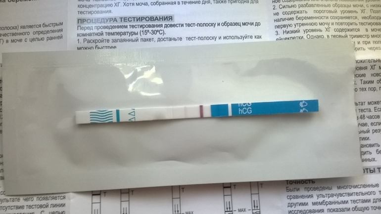
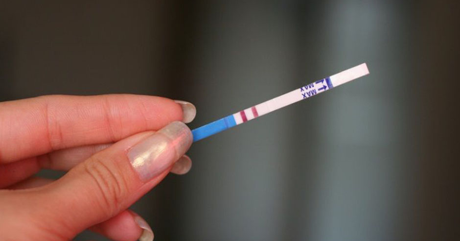
Suspicions about a possible “interesting situation” may appear in a woman long before the next menstruation is delayed. Modern test strips can determine the content of the specific hormone HCG in the urine already on the first day of the delay, and some even a few days before it. Whatever the test result, a woman wants to make sure she has a pregnancy as soon as possible. About when the baby can be seen for the first time on the ultrasound will be discussed in this article.
Minimum terms of determination
After conception took place, inside the future mother begins intensive processes, which she most often does not realize. On the very first day, the fertilized egg cell divides and progresses along the fallopian tube, where conception took place, into the uterine cavity. The journey lasts about four days. In the uterus, it is no longer a set of individual cells that is being lowered, but a blastocyte — a formation in the shape of a sphere. It is embedded in the shell of the uterus. This is an implantation. This happens 6–7 days after fertilization, and sometimes the woman feels implantation due to slight pulling sensations in the lower abdomen.

The earliest symptom of pregnancy is sometimes the so-called implant bleeding - a few drops of bleeding or blood secretions at the time of the introduction of blastocytes into the endometrium. This does not mean that it is time to run for the test or register for an ultrasound.
Test strips react to the formation of the so-called pregnancy hormone - hCG, and it is just beginning, the hormone level is below the control level of sensitivity of the test strips. And on the ultrasound blastocyte can not be seen - its size is only 0.2 mm.
There is no placenta yet, nutrition for the embryo is “supplied” by the uterine lining. But from the very first day after the attachment, the crumb begins to produce hCG, this hormone gives the entire female body a massive "mobilization" team. The restructuring of all systems of the female body begins to create the most comfortable conditions for the future growth of the child.
In two weeks after conception, the child grows up to 1 mm, the menstruation begins, and during this period the pregnancy can already be determined with high probability by the level of hCG in the blood (if the woman gives a blood test from a vein), the test strips begin to "strip". However, on the ultrasound, the woman is still not happy with anything, the pregnancy is not yet visible.

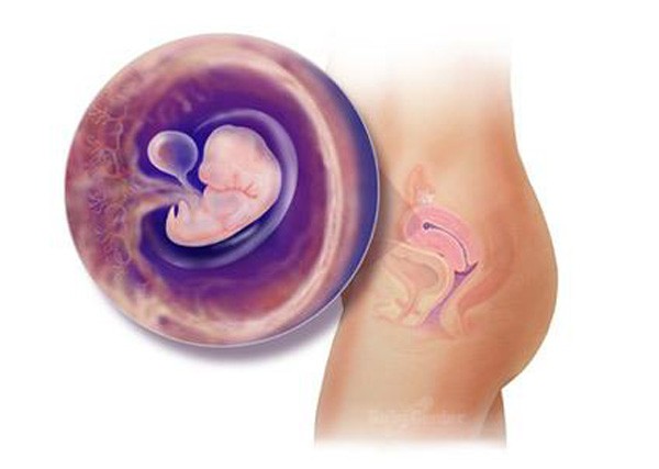
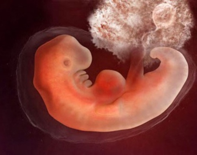
In 3 weeks after conception (this is the fifth obstetric week, which is measured from the first day of the last menstruation), the size of the baby reaches 4 mm. He laid the neural tube, and begins the formation of the placenta. The embryo takes on an oval appearance - a fertilized egg appears. The formation of the brain and spinal cord begins exactly at 3 weeks after conception, and the heart begins to beat in the embryo.
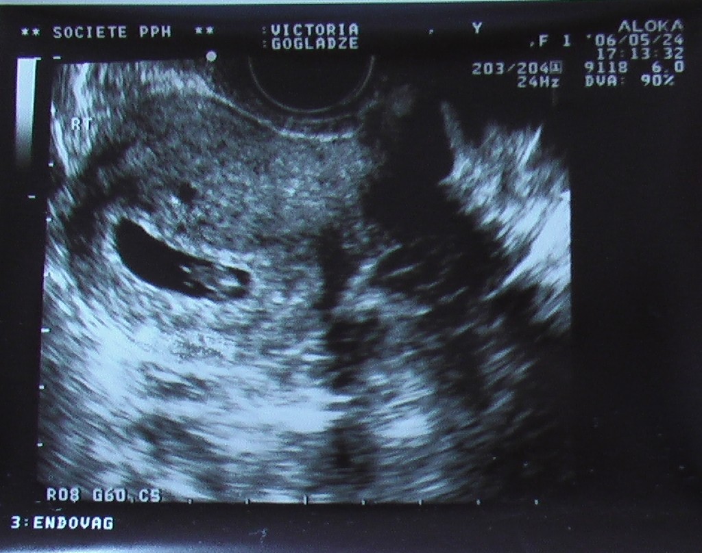
It is a week after the start of the delay (this approximately corresponds to the 21st day of embryo development or the full 5 obstetric weeks) that the embryo can be seen for the first time using ultrasound diagnostics.True, this possibility depends on many factors.
- A woman should not have polyps and diseases of the uterine lining. If there are such pathologies, the doctor may confuse the ovum with a fragment of a polyp, and it will not be possible to confirm the pregnancy.
- The scanner should have a good resolution, it is possible to determine pregnancy at such a minimum period only with modern good equipment and, naturally, with the help of an experienced and qualified doctor.

Indications for examination
If there are no periods, the test is “striped” or it does not show an interesting situation, then 10 days after the start of the delay, in any case, you should contact the antenatal clinic. A small increase in the uterus by an obstetrician-gynecologist at this time can be determined manually when examined by a woman.

Ultrasound examination after 10 days from the day of delay provides sufficiently accurate indicators of the presence, absence, and characteristics of the embryo. This does not mean that all pregnant women without exception in such early terms should walk around the ultrasound diagnostic rooms and do an ultrasound, as much as they want. The effect of ultrasound on the embryo is not considered harmful, but it is impossible to call it useful, it is not well understood.

There are certain indications, according to which the doctor will recommend a woman to undergo an ultrasound procedure on such a short period:
- The delay is accompanied by unpleasant, painful sensations, there are secretions that are not menstrual;
- Previously, women had ectopic pregnancies, early miscarriages;
- If there is a delay, the test shows a positive result, and the size of the uterus and features of the organ during palpation do not tell the obstetrician about the occurrence of pregnancy;
- If earlier the woman had surgery on the uterus, including cesarean section;
- If a woman does not remember the date of the last menstruation.

Diagnostics with an ultrasound scanner in these cases will allow to establish whether implantation has occurred in the uterus, if a woman develops a tubal (ectopic) pregnancy, and also allows to establish whether there is a detachment of the ovum if abnormal discharge occurs. It is in the early stages to establish gestational age can be accurate to the day, because in the embryonic period all embryos grow at approximately the same rate.
For women who have undergone surgery on the uterus, an ultrasound will help to find out the condition of the postoperative scar, whether the fertilized egg has fixed in the area of the scar. If a woman doesn’t have any concerns and complicated history, then there is also an urgent need for ultrasound, and for the first time, the expectant mother will be able to look at her baby at 11-13 weeks, when the doctor will refer you to the first prenatal screening.
How is the ultrasound?
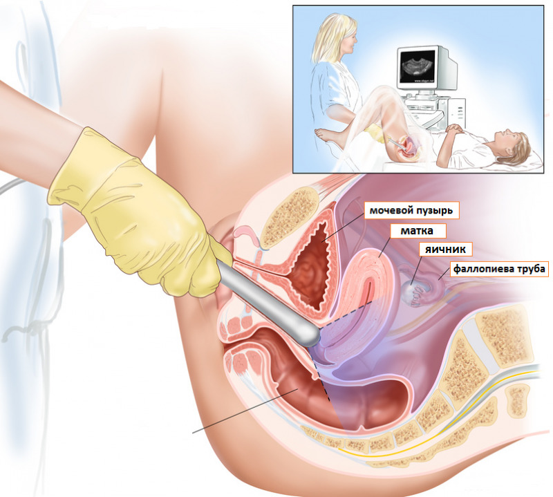
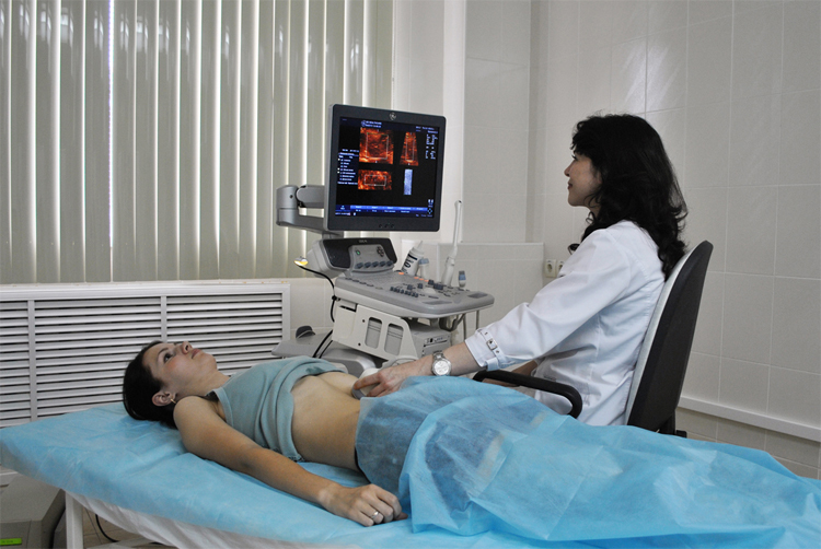
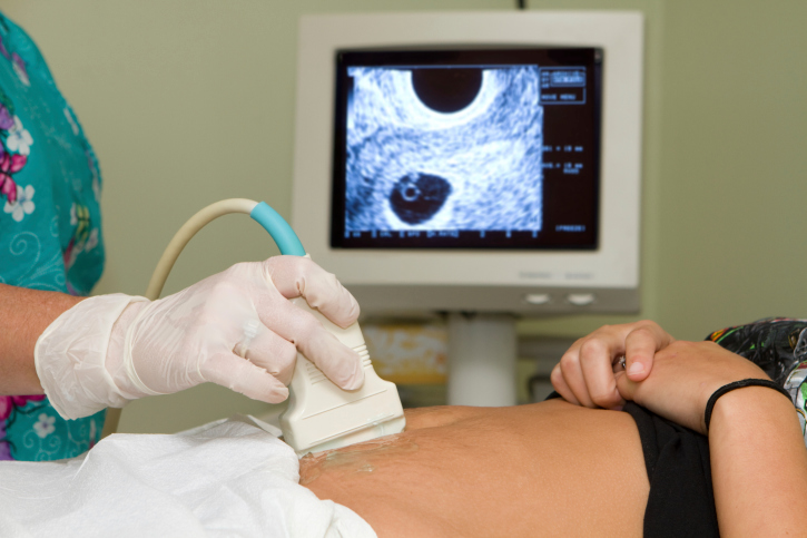
Two types of ultrasound examinations are used to determine pregnancy - tranvaginal and transabdominal. In the first case, the doctor examines the uterus and its contents with a vaginal sensor. In the second case, the inspection is carried out by the sensor through the abdominal wall. Most doctors prefer the first method when it comes to early pregnancy. Through the vagina it is much easier to see the embryo and its structure.
Ultrasound examination of the pelvic organs by the abdominal method is recommended with a full bladder, transvaginal - with an empty one, and it is better to make sure in advance that the intestine is not inflated with gases. To do this, a few hours before going to the doctor to the woman, it is advisable to take "Espumizan" or "Smektu».


It should be noted that the transvaginal way of pregnancy can be seen earlier than the transabdominal, for several days. So, a vaginal sensor and a good specialist can tell a woman about her “interesting position” as early as 5-6 days from the day of the delay, and scanning through the abdomen may not show pregnancy and 8-10 days. The procedure is painless, not dangerous for women and children, it lasts no more than 5-7 minutes.
Interpretation of the first ultrasound
At the very first ultrasound study on the definition of pregnancy, the diagnostician will be able to detect an echogenic formation. This is the fertilized egg. Its size will indicate the exact duration of pregnancy.The doctor will also determine the size of the yolk sac, the position of the ovum, the thickness of the endometrium, eliminate the inflammatory processes in it, as well as the presence of cysts, polyps and other unwanted formations. The sizes of the ovum and the timing table are presented below.
Obstetric term (from the date of the last month) | The diameter of the ovum (in mm) | KTR (distance from coccyx to crown), mm | BPR (bipartial size), mm | The diameter of the yolk sac, mm |
5 weeks | 18 | 2 | Not determined | Not determined |
6 weeks | 22 | 5 | Not determined | 3 |
7 weeks | 24 | 9 | Not determined | 4 |
8 weeks | 30 | 16 | 6 | 4,5 |
9 weeks | 33 | 23 | 8,5 | 5 |
10 weeks | 39 | 31 | 11 | 5,1 |

Are mistakes possible?
The method of ultrasound diagnosis is considered to be one of the most accurate for determining early pregnancy, however, one should not assume that its accuracy is 100%. In gynecology, the accuracy of this study is estimated at about 90%. In early pregnancy, accuracy decreases to 75%.. A doctor is primarily a person, not a machine with a program embedded in it. He has the right to make mistakes, especially if the woman has problems with the health of the reproductive system. So, the doctor can confuse uterine fibroids with pregnancy in the initial periods, if a woman has not been diagnosed with fibroids before, and she only found out about her presence on ultrasound. A cyst or polyp can be confused with the ovum, since the cyst is also an echogenic formation.

If the woman had late ovulation, then the pregnancy a week after the delay may not be detected at all by the ultrasound specialist, since the fertilized egg later descended into the uterus and has not yet been visualized. Naturally, the doctor will write in the conclusion that no signs of pregnancy have been found, but after 7-10 days on a re-examination he will be able to determine both the fertilized egg and its structure. Only sizes will help to understand that ovulation was really late.
Common Questions
On the Internet, inexperienced pregnant women and those who are still dreaming about an “interesting situation” ask a lot of questions concerning the very early diagnosis. On the most common situations is to talk in more detail.
Pregnancy test gave a positive result, and ultrasound - no
There may be several reasons for this. First of all, we should not exclude that the test turned out to be defective, it happens, and quite often, especially if we are talking about inexpensive test strips that are sold at almost every corner. In the desire to see the two cherished stripes, some ladies go too far, starting to look for the ghost stripes on the strips of dough. If they do, they automatically begin to consider their test positive, although in reality there may be no pregnancy.
If the test is still not deceived, then the reason for the negative conclusion of the doctor of ultrasound diagnostics may be that the woman turned to the doctor too early and the fertilized egg was not yet visible. The device itself may be outdated, with low sensitivity and poor resolution. The reason for the absence of signs of pregnancy on ultrasound may be late ovulation, and the presence of an inflammatory process in the uterus, and, of course, insufficient qualifications of the doctor.
Pregnancy test gave a negative result, and ultrasound - positive
The reasons for this situation may also be sufficient. Firstly, the woman herself could have conducted the test at home with an error, the test could be defective or expired, and it is also possible that she was conducted too early when the level of the hormone hCG in the urine was still insufficient for the test to respond to it brightly. second stripe.
Ultrasound diagnosis in this case is rarely premature, since a woman after a negative home test is not in a hurry to see a doctor, patiently waiting for the onset of delayed periods. After one and a half to two weeks delaywhen the lady still goes to the doctor, the pregnancy on the ultrasound is clearly visible. Therefore, the results of ultrasound should be considered more reliable than the results of a home test. In case of doubt, you can donate blood for hCG to get even more accurate data.
How to calculate the pregnancy by ultrasound?
To do this, you can use the table above. If more detailed term is required, a term correspondence table is used with an accuracy of one day to the mean internal diameter of the ovum (SVD). The table of the terms of pregnancy in accordance with the SVD is given below.
The value of the average internal diameter of the ovum | Gestational age | Allowable fluctuations |
6 mm | 5 weeks +3 days | 3 weeks +6 day - 6 weeks +6 day |
7 mm | 5 weeks + 3 days | 4 weeks - 7 weeks. |
8 mm | 5 weeks + 4 days | 4 weeks + 1 day - 7 weeks. + 1 day |
9 mm | 5 weeks + 5 days | 4 weeks +2 days - 7 weeks. +1 day |
10 mm | 5 weeks +6 day | 4 weeks +3 days - 7 weeks. +2 days |
11 mm | 6 weeks | 4 weeks + 3 days - 7 weeks. + 3 days |
12 mm | 6 weeks + 1 day | 4 weeks + 4 days - 7 weeks. + 4 days |
13 mm | 6 weeks + 2 days | 4 weeks + 5 days - 7 weeks. +5 days |
14 mm | 6 weeks + 3 days | 4 weeks + 6 days - 7 weeks. +6 day |
15 mm | 6 weeks + 4 days | 5 weeks - 8 weeks |
16 mm | 6 weeks + 5 days | 5 weeks + 1 day - 8 weeks. + 1 day |
17 mm | 6 weeks + 5 days | 5 weeks 2 days - 8 weeks. + 2 days |
18 mm | 6 weeks + 6 days | 5 weeks + 3 days - 8 weeks. + 3 days |
19 mm | 7 weeks | 5 weeks + 4 days - 8 weeks + 4 days |
20 mm | 7 weeks + 1 day | 5 weeks + 5 days - 8 weeks + 5 days |
21 mm | 7 weeks + 2 days | 5 weeks + 6 days - 8 weeks + 6 days |
22 mm | 7 weeks + 3 days | 6 weeks - 9 weeks |
23 mm | 7 weeks + 4 days | 6 weeks - 9 weeks |
24 mm | 7 weeks + 5 days | 6 weeks + 1 day - 9 weeks. + 1 day |
25 mm | 7 weeks + 6 days | 6 weeks + 2 days - 9 weeks + 2 days |
26 mm | 7 weeks + 6 days | 6 weeks + 3 days - 9 weeks + 3 days |
27 mm | 8 weeks | 6 weeks + 4 days - 9 weeks + 4 days |
28 mm | 8 weeks + 1 day | 6 weeks +5 days - 9 weeks +5 days |
29 mm | 8 weeks + 2 days | 6 weeks + 6 days - 9 weeks + 6 days. |
30 mm | 8 weeks + 3 days | 6 weeks + 6 days - 9 weeks + 6 days |
31 mm | 8 weeks + 4 days | 7 weeks - 10 weeks. |
32 mm | 8 weeks + 4 days | 7 weeks + 1 day - 10 weeks. + 1 day |
33 mm | 8 weeks + 5 days | 7 weeks + 2 days - 10 weeks. + 2 days |
34 mm | 8 weeks + 6 days | 7 weeks + 3 days - 10 weeks. + 3 days |
35 mm | 9 weeks | 7 weeks + 4 days - 10 weeks. + 4 days |

What if 1 week is more obstetric?
This may indicate late ovulation, as well as the fact that there are prerequisites for the fruit to be large enough, although first trimester not considered diagnostically important in predicting the estimated weight of the fetus. A woman in such a situation should be alerted by the fact that the home test, if it was done, showed pregnancy with a week's delay, since the tests begin to indicate two strips a couple of weeks after conception. If ovulation was late, then both implantation and hormone production was also late.

For information on how to determine early pregnancy, see the following video.


