Ultrasound in the 13th week of pregnancy: fetal size and other features

The first trimester of pregnancy ends, and with it the discomfort associated with toxicosis and hormonal changes take place. Future mother becomes more calm and reasonable. At week 13, most pregnant women are waiting for a long-awaited meeting with a baby on an ultrasound. This week, referrals to the first prenatal screening receive up to 90% of all expectant mothers, since this week is considered the most informative.

Objectives of the survey
An ultrasound scan, conducted between the 11th and 13th week of pregnancy, is part of the prenatal diagnosis complex, which is administered to all women who are registered at a antenatal clinic by order of the Russian Ministry of Health. Ultrasound at 12-13 weeks is considered the most important, because the fetus is not so small that the diagnostician could miss possible deviations in its development.
The purpose of the study is in a comprehensive assessment of the condition of the baby, in search of markerswhich may indirectly indicate possible genetic abnormalities, defects and anomalies. The obtained data are compared with the results of the blood test, which is designed to establish the concentration of certain hormones and proteins inherent in a woman during the period of carrying a child.
Both studies allow to calculate the potential risks of congenital abnormalities in a child. If it turns out that the risk of Down syndrome or Edwards disease is high, a woman is recommended to undergo a more accurate invasive examination, during which chorion particles with baby DNA are taken for analysis.
In addition to the early diagnosis of defects, ultrasound at week 13 allows you to establish the exact timing of pregnancy, the growth rate of the baby, see the problems of gestation, threats and risks of spontaneous abortion. The procedure this week is considered to be very informative, and the screening conducted during this period is the most important during the entire pregnancy.
13 week of pregnancy - these are obstetric values, which on average are 2 weeks ahead of a woman’s own calculations. Thus, 11 weeks have passed from conception, and almost 9 weeks have passed since the day of the delay and the first two cherished stripes.
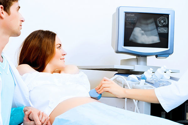
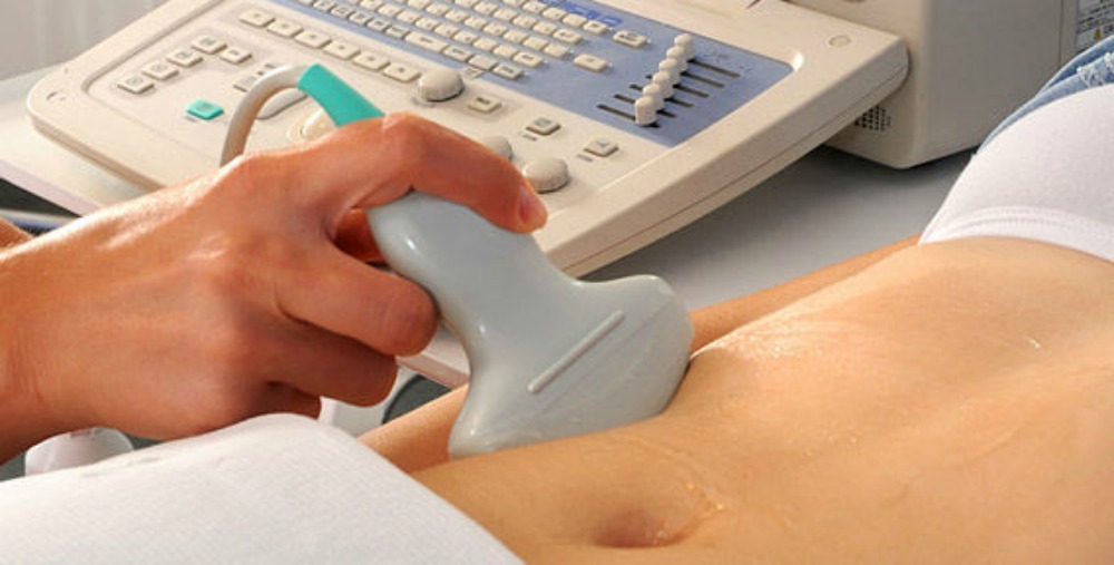
How is the ultrasound, do you need training
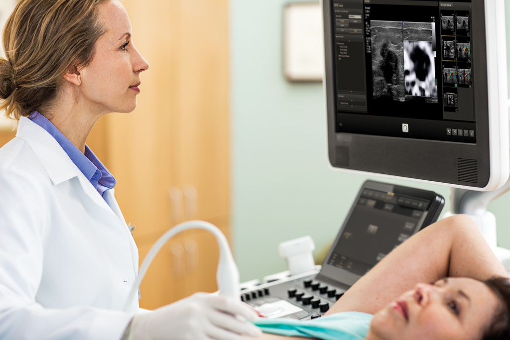
An ultrasound scan is done at week 13, most often in a transabdominal way, in which the sensor touches the skin of the woman’s abdomen, and the doctor gets an overview through the anterior abdominal wall. However, if the expectant mother is overweight, fat wrinkles in the abdomen, the doctor may decide in favor of another method of examination - transvaginal.
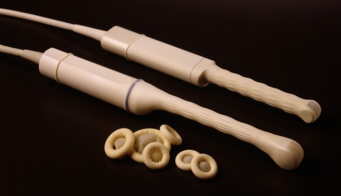
Internal ultrasound is performed by a sensor in a condom. A view through the vaginal wall is considered more accurate.therefore, it is used in all cases when there is a suspicion of threatened abortion, in order to examine not only the child, but also the characteristics of the cervix, the uterine wall.
Special preparation for the passage of ultrasound at week 13 is not required. The uterus is already quite large, and the amount of water allows you to conduct an examination without a filled bladder. Before the passage of vaginal ultrasound, it is generally desirable to empty the bladder and intestines from the contents, and 2-3 hours before the examination to take a single dose of the drug that reduces the amount of intestinal gases.
You can drink "Smektu", "Espumizan"Or" Simethicone. " For pregnant women, they are harmless.


What can be seen on this date?
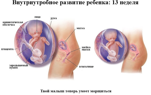
At week 13, the size of the fetus is 10-12 centimeters, its weight is 20-30 grams. The child still looks disproportionate - the head is still the largest part of his body, but the features of his small face are already beginning to acquire individuality, the rudiments of milk teeth have formed. this week the nervous system of the crumbs continues to develop, its movements are now controlled by the brain, and new muscle-neural connections are formed almost every hour.
The baby's pancreas began to synthesize insulin. From this week, the head of the baby begins to grow more slowly, but the rest of the body "accelerates." The formation of external genital organs continues, they are already distinguishable and, under favorable circumstances, the doctor will be able to examine the sex of the child by ultrasound.
The placenta has formed, and this week it takes over the “control” of the life processes and nutrition of the baby. The yolk bag is thus no longer needed; it ceases to exist. On ultrasound at this time, the child shows everything he has learned over the past few weeks - he moves his arms and legs energetically.
In response to loud sounds and unfamiliar voices, it can react either by an increase in motor activity, or, conversely, by silence. He hears everything, is able to distinguish the usual "safe" sounds - mother's bloodstream, heartbeat, breathing and the voice of strangers, and therefore potentially dangerous - other people's voices, equipment buzz, door slamming.
The ultrasound is clearly audible heartbeat baby, and mom must give it to listen. Baby every day more and more like a person in appearance. An ultrasonic sensor can detect and examine all the internal organs of a baby.

Decoding results
Decoding the results should be handled by specialists, but future mothers are very curious and inquisitive people, and therefore questions are often asked what the ultrasound protocol issued on hands can mean and everything is fine with the child. Unfortunately, the doctor does not always have the time to explain to each pregnant woman that he uses the abbreviations and numerical values to describe the state of the child, because the screening takes place in a queue. That is why the following is a detailed transcript.
First of all, the doctor marks the date of the last menstrual period, so that obstetricians have the opportunity to calculate the expected date of birth. He then indicates the way in which the survey was conducted — transvaginal, transabdominal, or both, if the survey required the use of two methods.

Following this is the number of fruits. If a woman expects twins, the doctor notes whether there are signs of life in each child. These include heartbeat and motor activity.
The position of the child in the uterine space is also described - it can be head, transverse and pelvic, at this time it is not significant, since the crumbs are still free and spacious in the mother's womb, and he constantly turns, tumbles and changes his position.
From week 13, doctors no longer measure the size of the coccyx-parietal, it is no longer considered informative. More useful data is now called fetometric. These are the BPR (bipariental size of the head is the segment between the temporal bones), LZR (the fronto-occipital size is the segment from the frontal to the occipital bone), as well as the length of the paired bones - femur, tibia, forearm and humerus.
Not all of these measurements can practically be made at week 13. The table of normal fetometry values at week 13 (12-13 weeks) is as follows:
BPR, mm | LZR, mm | Thigh length, mm | Shin length, mm | Shoulder length, mm | Forearm length, mm |
21-24 | Not determined | 7,3 -9,4 | Not determined | Not determined | Not determined |
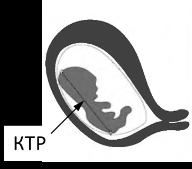
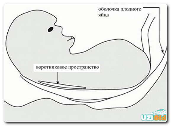
The abdominal circumference of the baby on this period is on average from 61 to 69 mm.The yolk sac, like the fetal CTE, is no longer measured this week. The rates of heart rate this week on average range from 147 to 171 beats per minute. Important for the diagnosis of possible genetic abnormalities are such markers as the thickness of the collar space and the length of the bones of the nose. These parameters are evaluated once during the first screening ultrasound. The table of TVP and the length of the nasal bones at week 13 is as follows:
Gestational age | TVP, mm | The length of the bones of the nose, mm |
12-13 weeks | 0.8 - 2.5 (on average, 1.6) | 2.0 - 4.2 (average, 3.1) |
The amount of amniotic fluid at this time is not measured. In addition to the development of the child, the doctor describes the features of women's health - the size of the uterus, ovaries, the condition of the fallopian tubes and other indicators.
Sex determination is not included in the screening, and therefore this service will be paid. It should be noted that the probability of an error in determining the sex at this time is quite high - boys are confused with girls if they cover their genitals with their legs, and girls can easily be mistaken for boys if there is a umbilical cord between the legs.
Therefore, at week 13, doctors do not speak with confidence about the field of babies; more accurate answers to an important question for parents can be obtained at the next examination, which is conducted from 18 to 21 weeks.

Possible problems
Ultrasound problems can reveal a wide variety. At this time, considering the specifics of the survey, the most frequent mothers are at a loss and panic because of the disparity between the size of the baby’s head and the obstetric gestational age, as well as the dubious results of identifying markers of chromosomal abnormalities.
If the BDP and LZR of the fetus have significantly less than the norm, doctors can talk about intrauterine growth retardation, and that the child may also have genetic abnormalities. Excess head size - also needs a separate medical "investigation". It is possible that an error occurred in the establishment of the timing of pregnancy - This often happens in women with an irregular menstrual cycle, with late ovulation.

The increase in the thickness of the collar space, as well as the absence of the bones of the nose, which shows ultrasound - reason for turning to genetics, as well as conducting a control ultrasound study in about a week.
At week 13, it is believed that the period, which is dangerous in terms of the probability of an early miscarriage, is already behind us. However, the diagnostician can still see the increased tone of the uterine walls, and then the obstetrician will prescribe a woman a treatment to save the pregnancy.
The revealed low placentation is not a serious problem at this time, because the uterus has yet to grow, and together with its walls, the placenta is more likely to "migrate" higher.


Snapshots
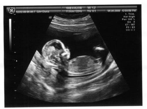
Ultrasound images at week 13 will allow a mother to examine her baby in more detail in a calm home environment. She will be able to see the profile of the baby, his chest and tummy, the structure of the brain.
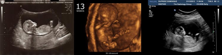
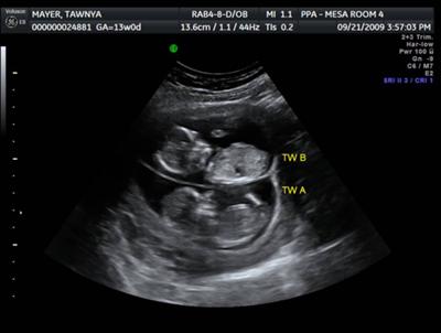
On a three-dimensional ultrasound, you can already see the tiny man more holistically, imagine what he looks like. Twins in this period looks like this.
In order to preserve the image of the baby at this time for a long family memory, it is better to ask the doctor to give an electronic copy, on any digital medium, since the printed images quickly fade.
About what happens to the mother and baby in the 13th week of pregnancy, you can see in the next video.


