Ultrasound in the 33rd week of pregnancy: fetal size and other features
There is only one and a half months left until the birth, and the woman is very worried. The 33 week gestation is often marked by the passage of the third, final prenatal screening. Having received a referral for an ultrasound at this time, the woman is waiting for a new meeting with her baby.

Purpose of the survey
An ultrasound scan at the 33rd week of pregnancy completes a prenatal diagnostic campaign. Exciting screenings of the first and second trimesters have already been left behind; quite a lot is already known about her baby to the future mother. The third ultrasound examination is intended to put an end to the determination of not only the risks of congenital abnormalities of the fetus (this issue is revealed with addiction in the first two screenings), but also to assess the possible risks that await the mother and baby during the birth process. Usually, the final screening examination is scheduled from 30 to 36 weeks, most often from 32-34 weeks.
Ultrasound during this period allows you to find out how pretty grown-up baby feels in the womb, how preparations are underway for his birth, whether his health is in order with his mother.
A referral to the diagnostician this week can be given not only for screening:
- It is more likely to get women who are carrying twins or triplets;
- Women whose pregnancy has become possible through in vitro fertilization (IVF);
- Women who have previously had surgery on the uterus, including cesarean section (one or more), which need to assess the viability of the postoperative scar.
- On the ultrasound this week, women will go to the obstetrician with complaints about stopping or increasing fetal movements, for backache pain, for blood pressure jumps;
- Diagnosis is also shown to those who have an obstetrician-gynecologist suspect intrauterine growth retardation due to the discrepancy between the height of the uterus standing and the gestational age;
- Rhesus-negative mothers who are carrying a Rh-positive baby can be sent to the ultrasound room to determine the severity and manifestations of Rh-conflict.
Sometimes ultrasound diagnosis at this time is required in order to correct the expected date of birth, as well as to assess the baby’s ability to survive outside the womb if urgent delivery is required at this time.
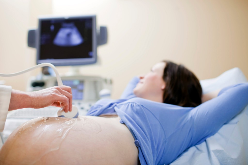
How is the research going?
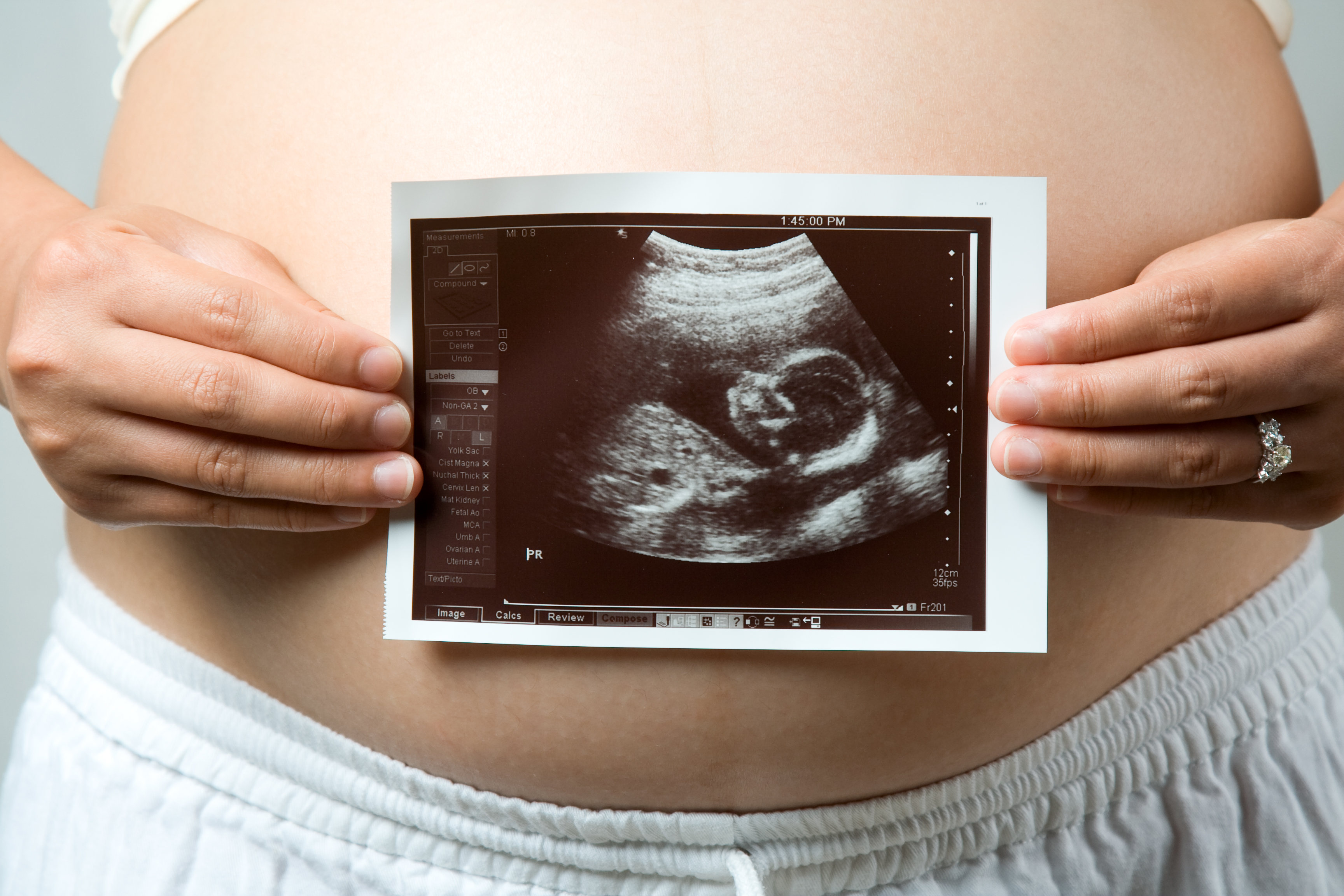
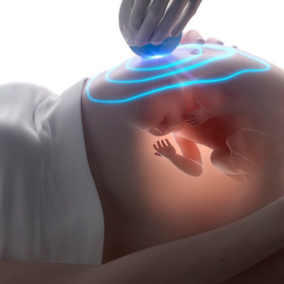
On such an impressive period, ultrasound diagnostics is performed only in a transabdominal manner. The sensor apparatus is placed on the anterior abdominal wall of the woman. The generator of ultrasonic waves provides waves of a certain length and intensity. When penetrating through tissues and body fluids, the waves repel or are absorbed, and a signal about this is sent back to the sensor. This information will be visible on the monitor of the device.
Preparations for this method of ultrasound diagnosis at the 33rd week of pregnancy are not required: the amount of amniotic fluid is large, ultrasonic waves can penetrate through them with ease. The child is already big, it can be clearly seen

The so-called internal (intravaginal) method of examination on ultrasound at this time can be applied only in cases when the doctor should exclude or confirm the readiness of the female body for premature birth.Through the vaginal wall, it is easier to examine the state of the cervix, which shortens and becomes more looseif a slight disclosure is noticed, then the woman will be sent to the maternity hospital, where they will decide on the delivery or preservation of pregnancy in the hospital.

What does ultrasound show?
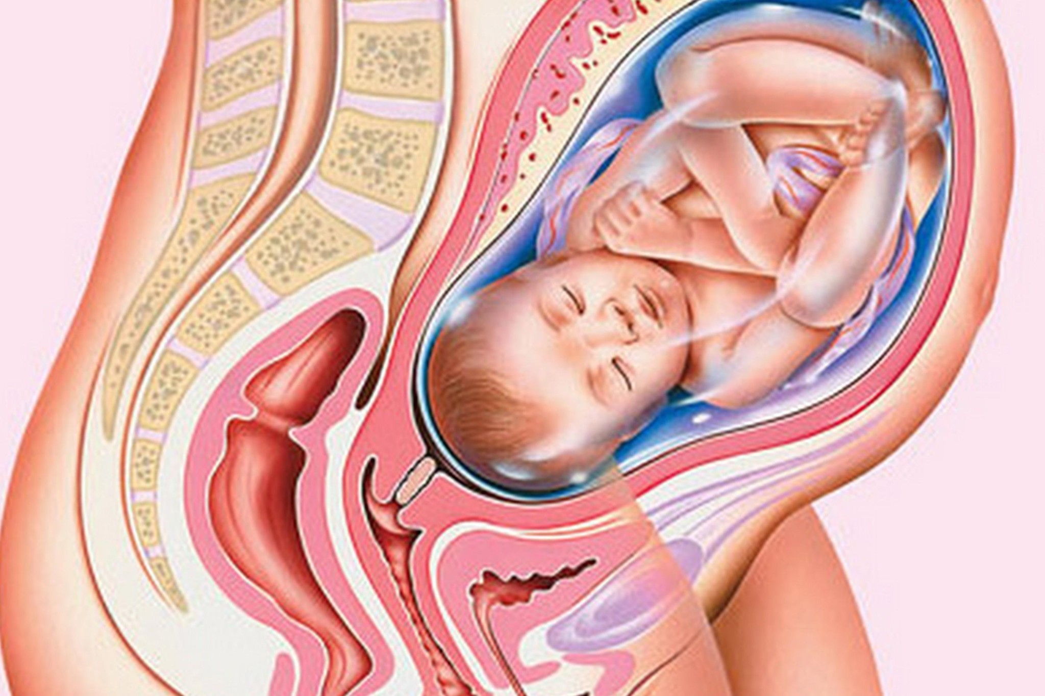
The baby is already quite big, almost the same as the mother sees him immediately after birth. The size of the fetus at this time - about 43 centimeters, and the average weight - about 2 kilograms. A month and a half before birth, the weight of babies varies greatly. It depends from hereditary features. In large parents, the expected weight of the children exceeds the mark of 2 kilograms, and in lean mothers and fathers the child may not yet reach this mark.
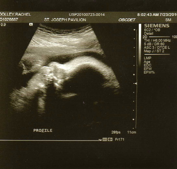
It is no longer possible to see the child on the monitor of the ultrasound device; only parts of the body — arms, legs, head and face, tummy, spine — can be examined. The baby is too big for the doctor to show his mother to his full height. He has fully formed all organs and systems, the crumb actively accumulates subcutaneous fat, increases its weight, acquires puffy cheeks, which will cause tenderness in adults as soon as the baby is born.
In the 33rd week of pregnancy, neither the mother nor the doctor of ultrasound diagnostics will please the child with active movements - the baby becomes cramped, he can no longer, as before, make somersaults and coups, swing his arms, swim. But it becomes obvious that the body of the little man has become more proportionate: the difference between the big head and the rest of the body has decreased.

The baby has a fairly well developed nervous system, although its formation is still ongoing and will continue even after birth. Scarce well coordinates his movements. During the sleep phase, his eyeballs move quickly, neonatologists suggest that the child is already dreaming.
The kid studied the voices of close people, first of all, of his own mother. Other people's voices and unfamiliar sounds can cause fear in him, so there is nothing surprising in the fact that during the passage of ultrasound diagnostics, the child will calm down cautiously, will not lurk and will not want to be shown to strangers.
At 32-33 weeks of pregnancy is already quite difficult to determine the sex of the child. The big baby tries to take the most compact position in the cramped uterus, while it presses the legs to the stomach, sometimes sits in the pelvic presentation, completely closing the view of intimate places. If before the floor was not recognized or there was no such possibility, then the accuracy of sex determination at this time will be even lowerthan in the first trimester - only slightly more than 65-70%.
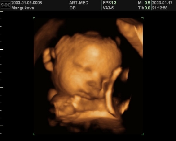
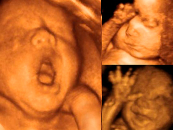
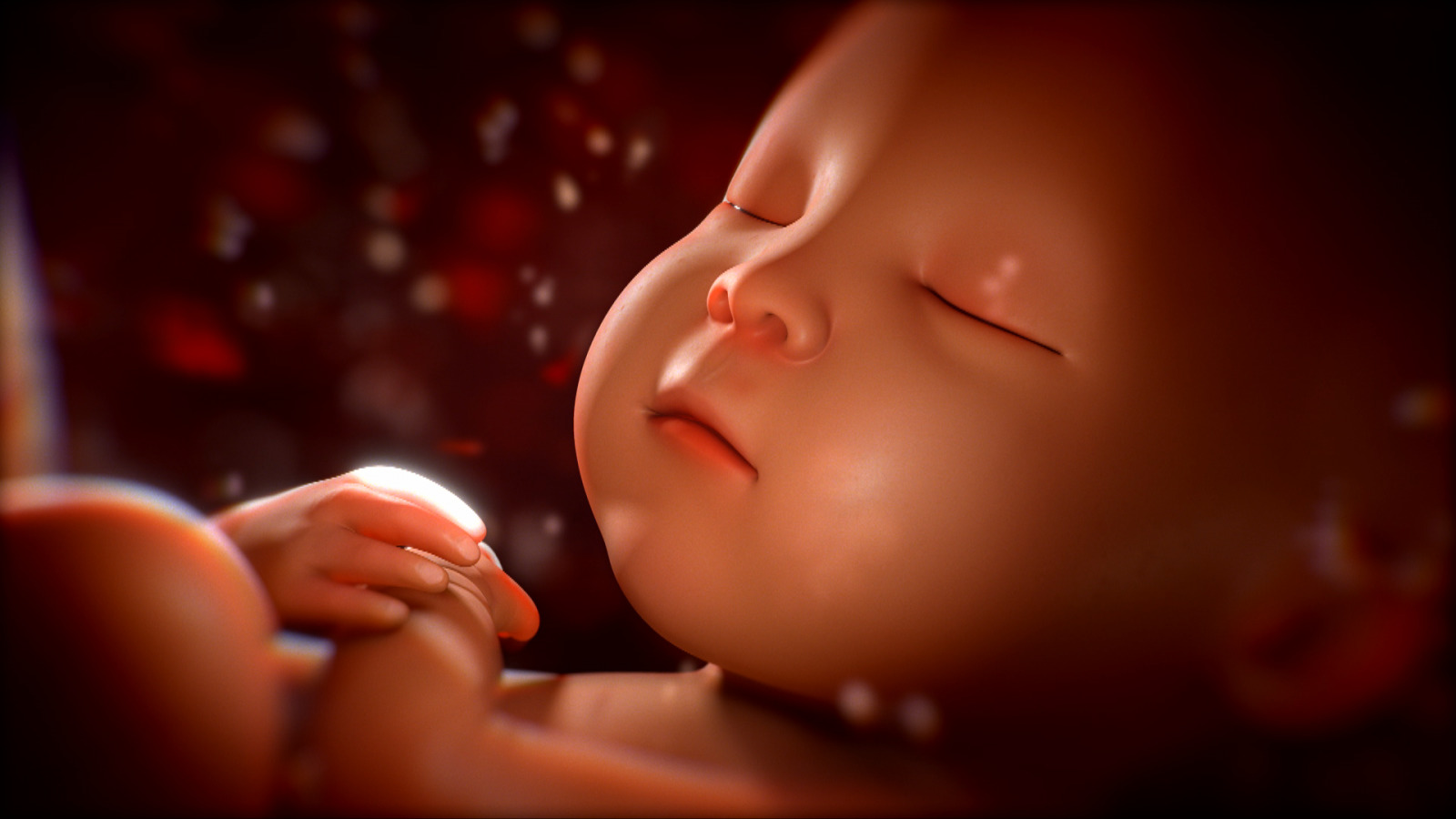
If a woman has decided to make a three-dimensional or four-dimensional ultrasound at this time, then an unforgettable sight is waiting for her. On the 3D ultrasound, you can see the baby, understand who he is more like, enjoy his rich mimicry, because the expression on the face of the children changes very often.
4D ultrasound will show all the same, but in motion, in real time. Future parents admit that they seemed to have watched a small film about the life of their crumbs in the womb.

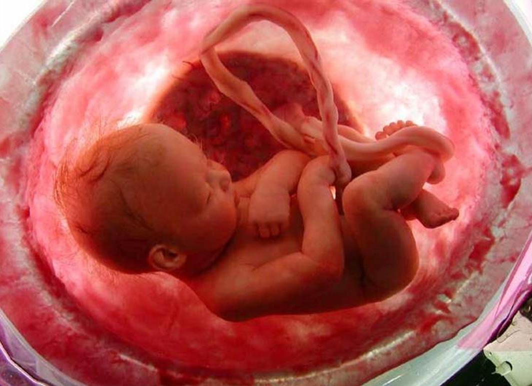
The kid leads an active life - drinks amniotic fluid, pissing, sleeping and awake, examines himself, touching his face, umbilical cord, legs, pulls in his mouth not only fists, but also his toes. He is almost ready for birth. If childbirth occurs this week, the baby will be considered viable., it does not threaten anything, just for a premature baby will require more thorough and time-consuming care.
Decryption and norms
The conclusion about the passage of the study, the pregnant woman receives immediately after the procedure. It reflects the main information: the size of the fetus, the characteristics of its anatomical features, the position in space of the uterus, as well as a description of the supporting structures - the placenta, umbilical cord, water.Fetometry is the main dimensions expressed in numbers, which can be measured with a sensor and which give a complete picture of the proportions of the child and its compliance with the gestation period.
These values include two head sizes - transverse (BPR) and longitudinal (LZR). On the structure of other parts of the body speak such dimensions as the diameter of the chest, the length of the paired bones - the femur, the bones of the lower leg, shoulder and forearm. The doctor compares the obtained numbers with the table of average values, after which he makes a conclusion about the compliance of the development of the baby with the term.

Table fetometry at 32-33 week of pregnancy is as follows:
BPR, mm | LZR, mm | The diameter of the chest, mm | Thigh length, mm | Shin length, mm | Shoulder length, mm | Forearm length, mm |
82-84 (admissible fluctuations from 75 to 91) | 104-107 (admissible fluctuations from 95 to 116) | 83-85 | 63-65 | 83-85 | 56-58 | 48-50 |
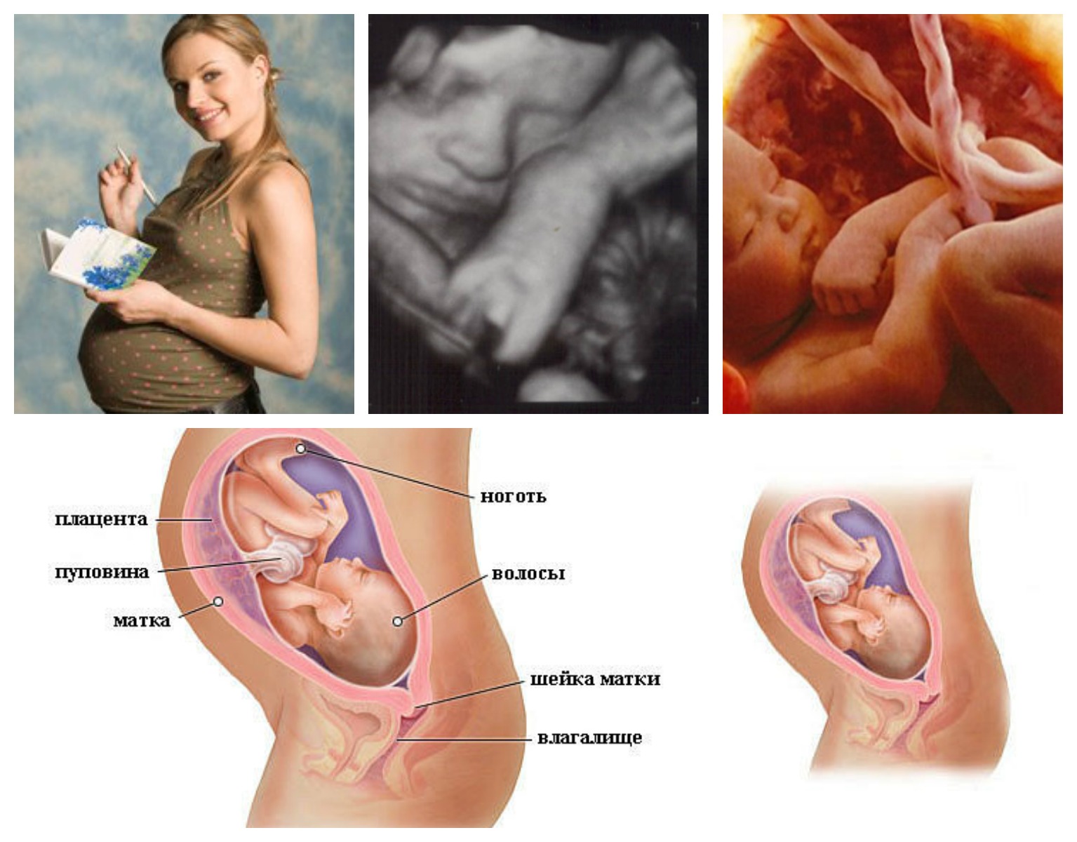
Anatomy of the fetus includes an assessment of the internal organs - the heart, lungs, liver, stomach and intestines, kidneys, brain structures. If the doctor does not find out during the examination of gross malformations of these organs, then in conclusion he indicates at his discretion either “norm”, or “examined”, or “without pathologies” or “+”. If an organ defect is detected, the doctor details, as far as the quality of the equipment allows, describes the violation found. At this time, an important indicator is considered the position of the fetus in the uterus.
The normal previa is considered normal: the baby is located head down, he is ready to "start" in the small pelvis in the generic process. However, sometimes the doctor can determine the position of the fetus, as pelvic (if the child sits head up) or transverse previa.
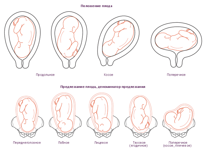
Usually at this time the baby assumes the position which will be until birthAfter all, it becomes more difficult for him to roll over. But pelvic or transverse presentation is not a sentence yet, there is a chance that the baby will change the situation; if this does not happen, doctors will consider options for the method of delivery. The amount of amniotic fluid gradually decreases by the end of pregnancy. Normally at week 32-33, the amniotic index is 144-245 mm. Moderate low water in this period can be diagnosed at index 77-83 mm.
Exceeding normal values is considered high water, a slight deviation qualifies as moderate high water.

The placenta is the main nutrient organ, without which the baby can not survive. This week she is in the transitional stage of her maturity - from the first to the second, and this is completely normal. The thickness of the placenta - 30-33 mm. The umbilical cord normally has three vessels. It is possible that at this time the doctor will conduct the USDG, the so-called study with the Doppler. It will help establish the rate of blood flow through the uterine vessels and assess whether the baby has enough nutrients and oxygen.
Small deviations from the norms are not considered critical.because the baby is already developing according to an individual program, which is often genetically determined. The main thing is not compliance with tabular standards, but proportions. If the baby is proportionally complex, there is no reason for alarm. For more information on how the ultrasound in the 33rd week of pregnancy, see the following video.
Snapshots
Three-dimensional images that parents receive on 3D ultrasound will show who the child looks like: they can clearly see the shapes of the nose, eyes, and ears. A two-dimensional snapshot of such specifics will not provide, it defines only the general outlines of the baby's face.
If you are lucky, parents will be able to get such emotional and touching “photos” into the family album.