Ultrasound in the 14th week of pregnancy: fetal size and other features
At week 14, mom enters the second trimester of her pregnancy. This is the most pleasant and carefree time for the entire period of carrying a baby. It is not difficult for a woman to carry a child, but you can already enjoy all the pleasures of your “interesting” position. Sometimes this week, the expectant mother goes for a scheduled ultrasound scan. What can be seen at this time and how to understand the results of this study, we will tell in this article.
Objectives of the survey
The 14th week of pregnancy is 12 weeks from the moment of conception and about 10 weeks from the start of the delay. In her pregnancy, a woman no longer doubts. It is this week that she can go on an ultrasound scan, which takes place as part of the first prenatal screening. Usually, this is recommended examination for all pregnant women. from 11 to 13 weeks inclusive. Thus, in week 14 there are six days for which a woman needs to have time to go through this important and informative examination, if she did not do it a week or two ago.
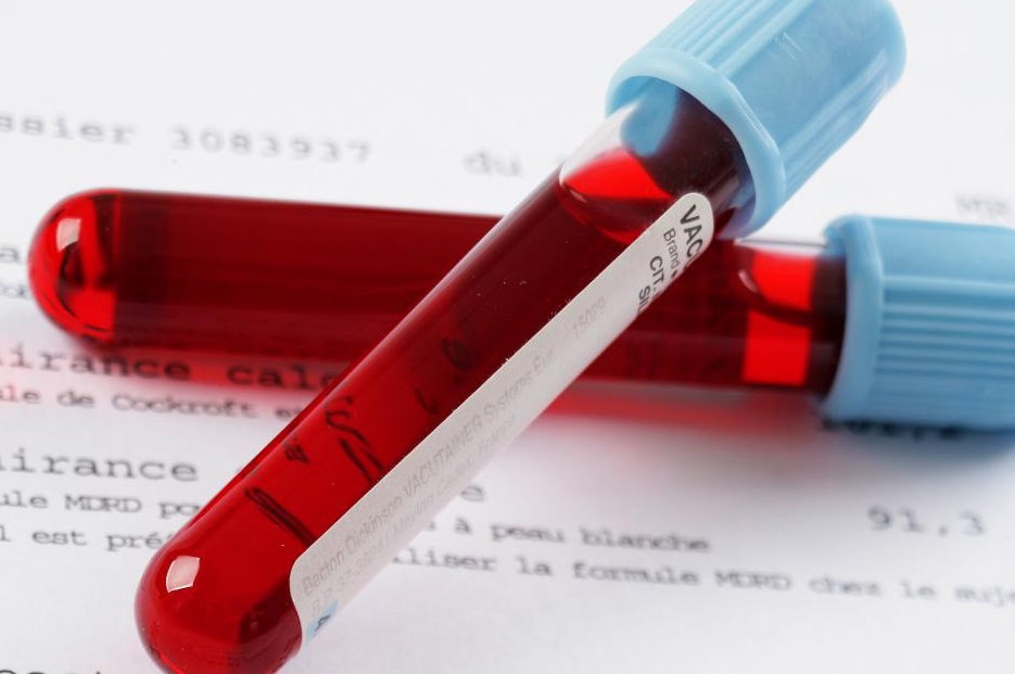
As part of a screening survey, they not only study a child for an ultrasound scan, but also conduct a laboratory biochemical blood test to determine the concentration of an important hormone for pregnancy. HCG and plasma protein PAPP-A. All information is evaluated in a complex and allows you to judge whether a woman has increased risks to give birth to a child with severe genetic abnormalities.
If the screening has already been passed, then at the 14th week there may be other indications for ultrasound:
the threat of termination of pregnancy, pain, the appearance of bleeding;
unsatisfactory screening results requiring follow-up examination;
clarification of the period of pregnancy, if the obstetrician-gynecologist has doubts, if the size of the uterus does not correspond to the specified obstetric period;
elimination of delayed fetal development, non-developing pregnancy.
Some pregnant women this week can go on an ultrasound scan and without a referral from a doctor in order to find out the sex of the child, since it is not always possible to see the sex of the fetus during the first screening at 11-12 weeks.


Diagnostic features and preparation
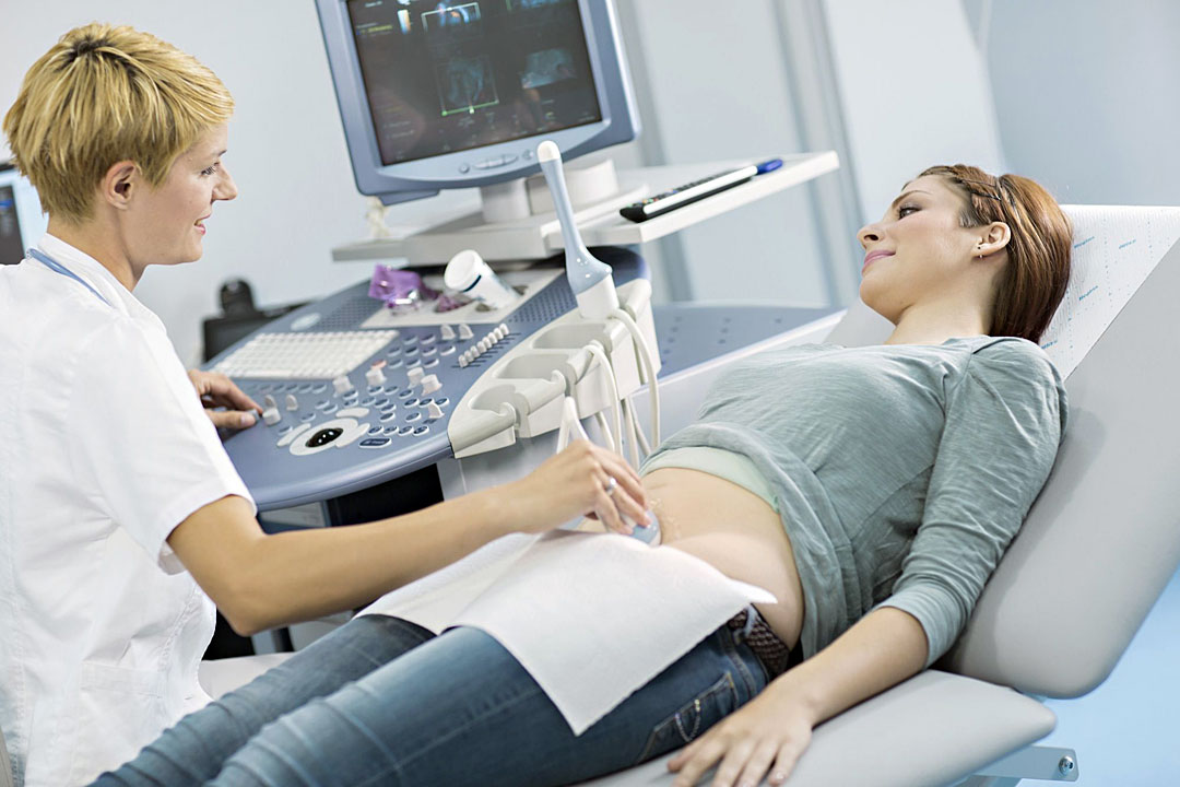
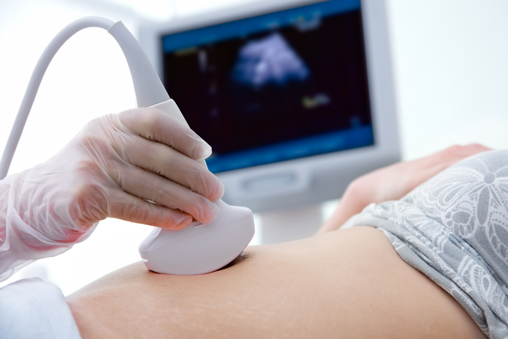
In this period, future mothers in the vast majority of cases, diagnosis is carried out abdominally - the scanner sensor is located on the abdomen. The size of the uterus and the amount of amniotic fluid allow a good view of the baby. However, in exceptional cases, they can use the intravaginal method of examination. With it, the sensor is inserted into the vagina and the uterine cavity is examined through the thinnest wall of the uterus.
The doctor will most likely use the vaginal sensor if the woman has luxuriant forms, if she has fat deposits that make it difficult to examine, and if there is a probability of spontaneous miscarriage, because this condition requires careful examination of the cervix, cervical canal, and through the vagina the most reliable.
Some specific preparation for ultrasound on this period is not necessary. Before a vaginal (internal) ultrasound scan, a doctor may ask a woman to empty her bladder.You should make sure in advance that the intestine is not filled with fecal masses and intestinal gases - before visiting the diagnostic room should go to the toilet and take a dose of drugs that prevent increased flatulence - Smectu or Simethicone.
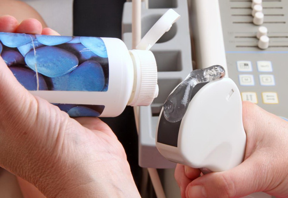
You should take a diaper with you to the ultrasound room to put it on a couch, as well as disposable medical wipes to remove the remnants of the gel from the skin, which is used during ultrasound scanning through the abdominal wall to facilitate the passage of ultrasonic waves.


What can be seen?
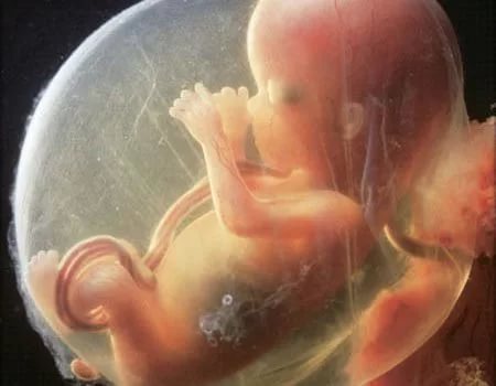
The baby has grown up - the weight of the fetus at week 14 is already from 30 to 50 grams, the size of the fetus is 12-14 centimeters. The formation and development of the spine is in full swing during this period, the muscular and bone systems are being improved. The child is actively moving, waving his arms and legs, tumbling, making swimming movements, but the woman cannot yet feel them, because there is enough space in the uterus and the baby does not push the walls of the uterus. But the somersaults and flares are clearly visible during the passage of the ultrasound examination.
This week, the baby clearly distinguishes between periods of wakefulness and sleep. However, he continues to move even in a dream, because neural connections are formed between the muscles and the brain, and therefore movements in a dream are reflexive. During this period, the baby comprehends another important "science" - he learns to smile and use facial muscles.
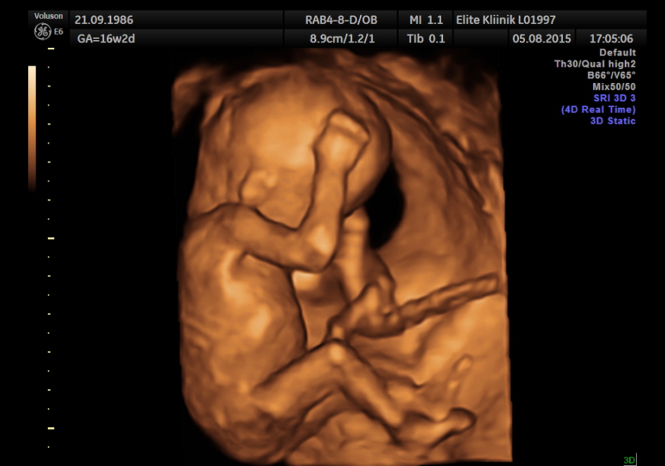
Therefore, on an ultrasound scan performed with a high-resolution scanner - 3D, it is possible, if lucky, to see the first grimaces that the crumb depicts on its face.

The very face this week, too, is beginning to undergo changes - the eyes, widely spaced on the sides, begin to converge, the auricles "slip" into their proper place. Baby is well versed in sounds. He hears the soundtrack of his mother's body - a heartbeat, breathing, bowel work, and also quite emotionally responds to sounds from the outside. On ultrasound baby can respond to the unfamiliar voices of the medical staff and light pressure on mom's belly sensor. The reaction can be of two types: either the crumb revives and begins to move actively, or it “gets scared” and hides. Sometimes a shy little kid can start running from the sensor, trying to hide intuitively.
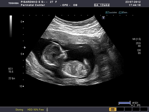
Many mothers are alarmed when they see on the monitor not a plump crumb how they painted it to themselves in the imagination, but a thin “goner” with short legs, thin and thin hands and a still big head. This is the absolute norm, just such a child should be on this date. He will begin to accumulate subcutaneous fat closer to childbirth.
Formed external genitals. With a good overview you can already see the sex of the child. The child actively swallows the amniotic fluid, in which it floats, and pisses into it. The composition of the waters is updated every three hours. In general, the baby looks already quite “humanly”, it no longer looks like an embryo.
Decoding results
First of all, the number of fruits and their position in the uterus are determined. If at this time the doctor indicates that the child is in pelvic or transverse presentation, there is nothing terrible in this, since the baby is actively turning over, and more than a dozen times change position in space until he is old enough to accept a single presentation.
On the development of the baby at this time speak the main dimensions, which are described in the conclusion under the heading "Fetometry". The standards for this period (13-14 weeks) are as follows:
BPR (bipartial size), mm | Head circumference, mm | Abdominal circumference, mm | DBK (thigh length), mm | WPC (humerus length), mm | Estimated weight, gr |
20-31 | 73-110 | 58-90 | 9-15,8 | 7-15 | 31-52 |
(The table shows the minimum and maximum value of the indicator for this period).

Among the anatomical indicators, the doctor examines and evaluates the structure of the brain, a large cistern, indicates the size of the cerebellum. For 14 weeks, the normal size of the cerebellum is from 1 to 1.5 cm.Examine the spine, heart, stomach, intestines of the baby, his lungs.
The sizes considered important at this time the length of the bones of the nose and the thickness of the collar space. Both of these indicators may indirectly indicate existing pathologies of genetic origin. TVP at week 14 is normally 1.7 mm (deviations in the range of 0.8-2.7 are permissible). The bones of the nose should be determined, their size at this time - 2.0-2.9 mm.
Such size as TVP after 14 weeks loses its diagnostic value, and if you go on an ultrasound scan a week later, at 14-15 weeks, you will not be able to measure TVP.
In addition, the doctor assesses condition of amniotic fluid, their number, place of attachment of a young, newly formed placenta, counts the number of vessels in the umbilical cord. Normally they should be 3.

Possible problems
Among the most common problems that can be identified on an ultrasound at this time is the discrepancy between the size of the fetus and the gestational period. If the lag or advance of the existing norms is insignificant, nothing terrible happens - the child may already have individual features of appearance (someone has a bigger head, someone has a longer nose, etc.).
However, if the backlog from the norms exceeds 2 obstetric weeks, a detailed analysis and additional examination are required to find out if the baby has developmental delays, congenital anomalies, or if the gestational age is correctly set.

Excess of norms for 2 weeks and more can speak about an error in calculating the term, possible genetic problems, edema and intrauterine infection, a tendency towards a large or giant fetus. In both cases, the woman can get referral for a consultation to genetics. Do not refuse.
Timely problems can be corrected in some cases. If we are talking about chromosomal pathology, then a woman may be asked to terminate a pregnancy for medical reasons. How to proceed, it's up to her and her family to decide.

Snapshots
Ultrasound images on this period look like. You can well consider the profile of the baby, his arms, ribs, spine. More detail the child can be seen in the three-dimensional image. Such a “photo” can already decorate a family album. Enough clearly presented in the pictures of this term and twins.

If you want to get a baby scan on ultrasound, request an electronic copy on any carrier. For this, of course, will have to pay, but it's worth it. Paper photographs are very short-lived, and within a year they will lose their color and contrast.
Electronic files will allow you to return at any time to the beautiful time of waiting for the baby, and then show him how small he was at week 14.

About what happens to the mother and baby in the 14th week of pregnancy, see the next video.


