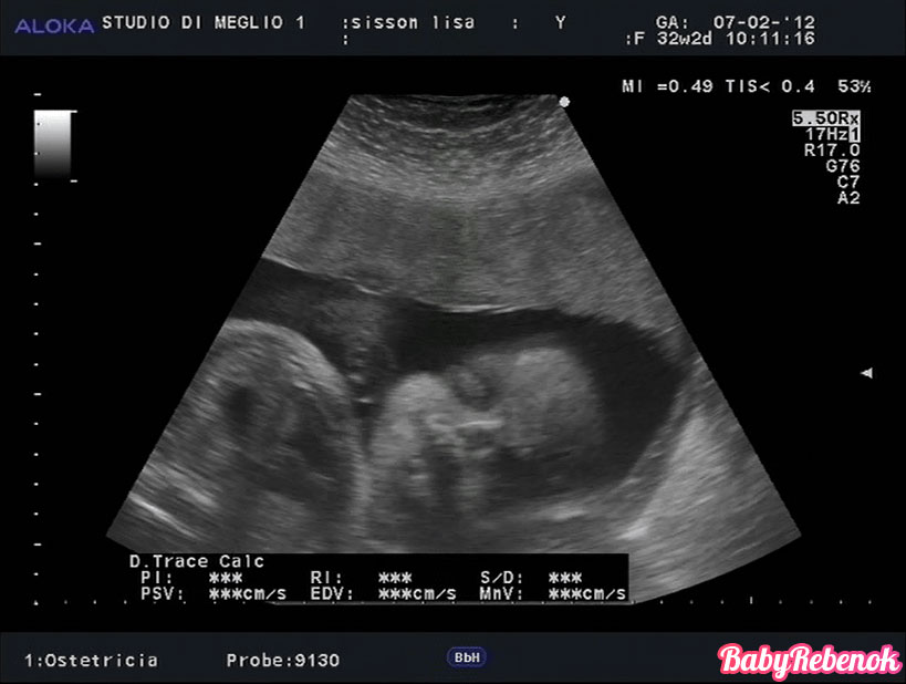
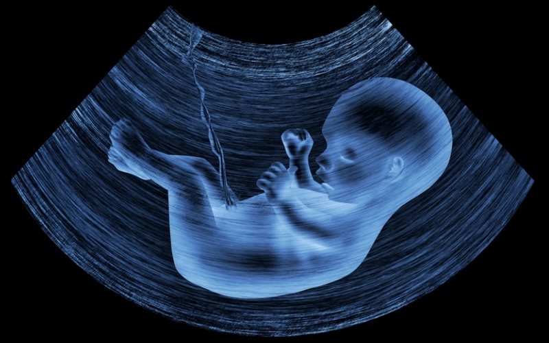
Ultrasound at 32 weeks gestation: fetal size and other features
An ultrasound examination in the third trimester of pregnancy is carried out for certain medical reasons. Typically, such a study is assigned to a pregnant woman whose fetus has certain developmental pathologies.
Purpose of
The research in this period of pregnancy often coincides with the time of the third screening. Usually this examination is performed at 30-32 weeks of fetal development.
It should be noted that carrying out an ultrasound at this time of pregnancy may not be shown to all women.
Using ultrasound examinations, doctors can determine main indicators of fetal development. This method allows for a complete integrated fetometry. This study includes quite a few different parameters.
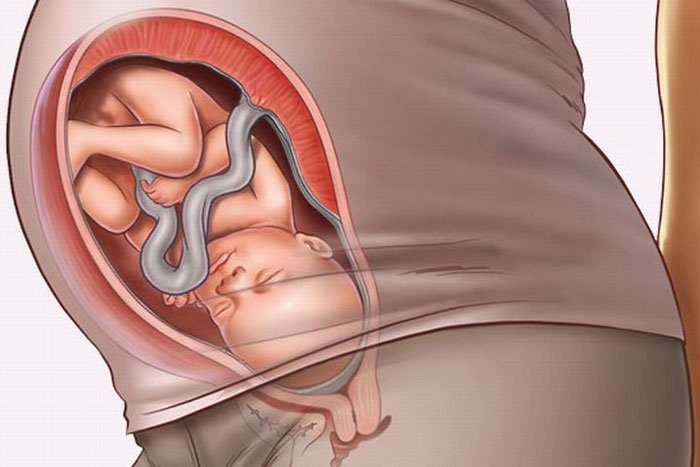
32 obstetric week is the period when the baby’s body is almost fully formed. This physiological feature allows you to determine quite a few different indicators.
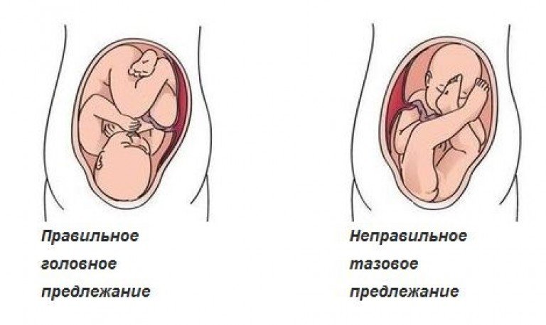
Quite often, doctors prescribe the passage of ultrasound to determine the final tactics of obstetrics. Mothers who have a pelvic presentation of the fetus at this time may also need to re-conduct an ultrasound examination a couple of weeks before delivery.
Ultrasound at 31-32 weeks of pregnancy can also be prescribed with multiple pregnancies. Especially often this situation occurs if one of the fetuses revealed intrauterine developmental defects. In difficult clinical cases, doctors may resort to the appointment of an expert-level ultrasound examination.
Of course, during the ongoing ultrasound examination, you can also determine the sex of the future baby. This is usually done when the pregnant woman missed the previous screening. In most cases, the sex of the unborn child is determined in the middle of the second trimester.
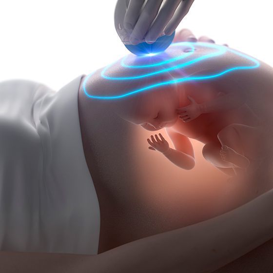
Ultrasound at this stage of pregnancy can be done in different ways. Doppler sonography is used to determine blood flow pathologies. Using this method, diagnostics specialists can determine various pathologies of the placental and uterine blood vessels feeding the fetus.

Doppler sonography is also used successfully. to identify emerging heart defects in the fetus.
It should be noted that expectant mothers who, during an ultrasound scan at this gestational age, doctors found any deviations from the norm, should not immediately start to panic.
One conclusion of the ultrasound test is not a diagnosis at all. Interpretation of the result must be carried out by an obstetrician-gynecologist who observes a pregnant woman during the entire period of pregnancy.

The main parameters studied
The third trimester is the final stage of intrauterine development of the baby. To assess the pathologies during this period of pregnancy, doctors have developed a number of different clinical indicators.
For a comprehensive conclusion, a comparison of all the results obtained is required.
Fetometry - basic research, which is actively conducted during this period. This method allows to evaluate the main indicators of fetal anatomy.To do this, during the study, the doctor ultrasound evaluates several parameters. These indicators include biparietal size and circumference of the fetal head, as well as the abdominal circumference.
Required sizes of the main tubular bones. For this, their length is estimated. Too short tubular bones may be a sign of some genetic diseases or emerging pathologies of the musculoskeletal system.
During the study, which is carried out at this stage of pregnancy, it is also estimated that abdominal circumference. Doctors found that this indicator is often associated with head circumference. If the baby’s tummy is too large in diameter, then this can be a manifestation of a very dangerous pathology - ascites. It is characterized by the accumulation of an excess amount of fluid in the abdominal cavity of a child.
At this stage of pregnancy can be determined and estimated weight of the baby. An experienced ultrasound specialist will determine how much the fetus weighs up to tens of grams. Too large fruit is, as a rule, an indication for a cesarean section, especially if the mother’s pelvis is too narrow. Underweight usually occurs in one or both babies with multiple pregnancies.
Evaluation of the performance of all vital systems of the fetus - A very important step in the conduct of ultrasound. At this time, the doctor is already well defined fetal heartbeat. Excess or decrease of this indicator below normal values indicates the formation of pathologies in the work of the cardiovascular system. Such manifestations are especially dangerous if the mother has a history of heart disease.
Detection of heart defects is necessary. Modern techniques allow to detect such defects quite effectively. Pathologies of development of heart valves and the appearance of pathological blood flows (regurgitation) on them can become an indication for surgical correction immediately after the birth of a child.
With the help of ultrasound at this stage of pregnancy can also be identified various abnormalities in the development of the kidneys and urinary tract in the fetus. Qualified professionals can also identify and emerging pathology of the pelvis and bladder. Along with the study of the urinary system, the doctor necessarily evaluates the work of the gastrointestinal tract and respiratory system in the fetus.
Examination of the spine in a baby is carried out only if there is a suspicion of the presence of genetic or chromosomal diseases. For this, an ultrasound doctor examines a linear section of the spine. Spinous processes are also studied. If, during the study, an ultrasound specialist revealed any significant defects and he had suspicions about the presence of signs of chromosomal diseases, he would send the future mom for a consultation to genetics.
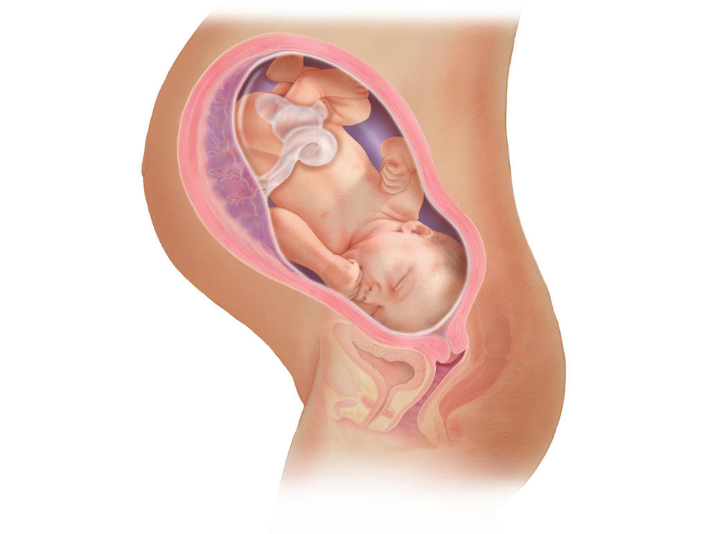
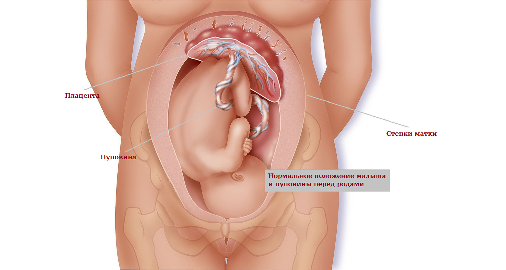
The placenta, or "baby seat" - is a very important organ during pregnancy. Through it, the baby receives all the necessary nutrients, as well as oxygen. This is due to a single uteroplacental blood flow system with the mother.
Assessment of the thickness of the placenta, as well as the tone and structure of its blood vessels is necessarily carried out in the study during this period of pregnancy.

During any ultrasound examination, the doctor will necessarily evaluate and the main parameters of the internal genital organs of the future mother. This is necessary for the timely detection of pathologies that can lead to disruption of pregnancy and difficulties during childbirth.
For this, the inner surface of the uterus is examined, and the size of its appendages and ovaries is determined. During the ultrasound, you can identify tumors and cysts. If these pathologies in a pregnant woman were identified before the onset of pregnancy, then in such a situation, the dynamics of their growth are necessarily evaluated. In case of unfavorable course of fibroids or cysts, careful selection of the necessary obstetrician tactics may be required.

The norms of the studied parameters
Normal doctors consider headache presentation. Too active babies can change their position in the womb several times. Usually by the end of the third trimester, the presentation is already becoming permanent. In some situations, doctors may prescribe and re-conduct ultrasound in 2-3 weeks.
The location of the placenta on the back wall is the most optimal. Doctors also determine how high this organ is from the internal os of the uterus. If the placenta is attached to it too low, then this may be due to its increment or lead to various pathologies.
During the study also required estimated cervix. Normally, this indicator should be more than 30 mm. The shortening of the cervix is an extremely unfavorable symptom of isthmic-cervical insufficiency. In this case, additional suturing is required in order to eliminate this pathology.
It is very important during the time of such a study to evaluate and tonus of the uterus. Hypertension can lead to premature labor. This condition also contributes to the formation of placental insufficiency.
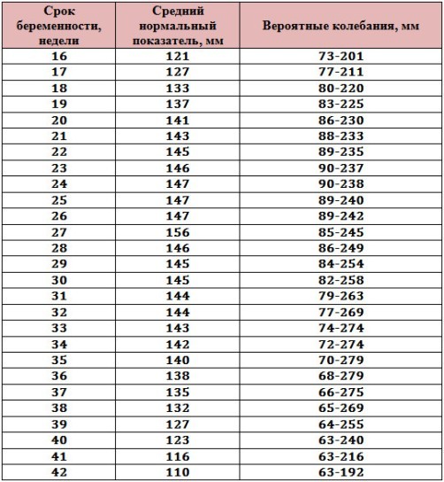
The amniotic fluid index is a very important indicator, which is also estimated during this period of pregnancy. Too much of its accumulation is a manifestation of polyhydramnios. Normally, this figure is 140-269 mm. A significant decrease in this indicator indicates the manifestation of low water.
Fetal size
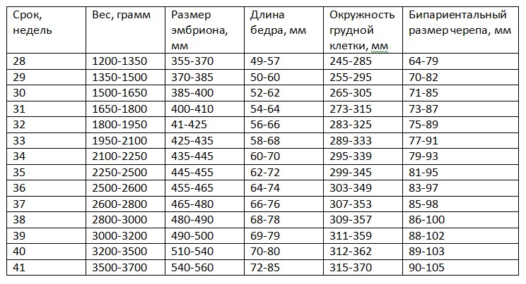
For the convenience of assessing intrauterine development of the fetus at this stage of pregnancy, doctors use a special table, which presents all normal values of the main studied parameters:
Evaluated criterion | Norm |
Biparietal head size | 77-91 |
Frontal-nuchal size | 97-116 |
Abdominal circumference | 26,6-32,5 |
Head circumference | 29-33,3 |
Humerus length | 53-62 |
Femur Length | 56-66 |
Forearm length | 45-53 |
Weight | 1790-1890 |
Growth | 42,5-43,6 |

Decoding results
Expectant mothers should remember that the obstetrician-gynecologist conducts the interpretation of the obtained values, and not the ultrasound specialist. One conclusion of the ultrasound test is not yet a diagnosis. To confirm or exclude the pathology of the fetus, a comparison of ultrasound findings with biochemical analyzes, which were carried out during different periods of pregnancy, is also required.
Tachycardia - an unfavorable symptom that may indicate fetal hypoxia. The development of this condition can lead to the entanglement of the baby’s neck by the umbilical cord. Heart palpitations are also found in some heart defects.
Bradycardia detected in this period in the fetus may be due to a lag in the intrauterine development. This symptom also appears in the presence of certain cardiovascular abnormalities. Lowering the heartbeat of a fetus below 130 beats per minute requires mandatory careful diagnosis. to identify various valvular heart disease.
At this stage of pregnancy, placental maturity is usually of the first degree. At earlier stages, it is zero. With the passage of pregnancy, maturity of the placenta increases. If the placenta does not "ripen" to childbirth, then this is already a pathological condition.
The study of the thickness of the placenta is also necessarily conducted. The smaller thickness of this organ is found with the possible development of placental insufficiency. Too fat placenta may indicate the development of edema or inflammation. Various viral and bacterial infections lead to the development of such pathological changes.