Ultrasound in the 22nd week of pregnancy: fetal size and other features
Contact between mother and baby at 22 weeks of pregnancy can be considered established. A woman already feels a stir, can monitor the activity of the crumbs and even understand what he likes and what does not. There is a second trimester, behind the first two screenings. At week 22, ultrasound will be assigned to those who did not have time to make it a week or two earlier. That the survey will show, we will tell in this article.
Purpose of the survey
Ultrasound at week 22 completes the second prenatal screening, which is conducted from 18 to 21 weeks of pregnancy. If for a number of reasons a woman could not be examined at that time (she was sick, she left), now is the time to visit the ultrasound diagnosis room.
Blood for biochemical analysis is usually given earlier - from 16 to 18 weeks. An ultrasound scan is not so tied to a blood donation as it was during the first screening, which the woman underwent in the first trimester. That is why there is time to choose the most suitable day for meeting your baby. For now - on the monitor of the ultrasound machine.
The purpose of the survey is to identify possible pathologies and abnormalities in the development of the fetus, both genetic and for other reasons.
If the mandatory prenatal examination has already been completed, then there may be other reasons this week to go to the diagnostician. The control ultrasound is sent to women who have second screening revealed increased risks of pathologies, as well as those who are carrying twins or triplets.
Ultrasound scanning will be shown to those who have the threat of abortion, there were complaints of pain and atypical for the "interesting position" allocation. If the pregnancy is due to the IVF procedure, the ultrasound is also prescribed more often than usual, and the 22nd week may not be an exception.

On an ultrasound this week, women who have previously had frozen pregnancies or miscarriages at this time can go. Ultrasound scanning may be needed if you have doubts about the exact duration of pregnancy.
Some women go to ultrasound for 22 weeks by own decision, for example, to find out the sex of the child, if this question is fundamentally important for the family or simply out of curiosity.
The sex of the baby leaves no doubt, it can be easily seen, because the crumb is not yet so large as to curl up and close the view, and is no longer so small that the genitals have microscopic dimensions.

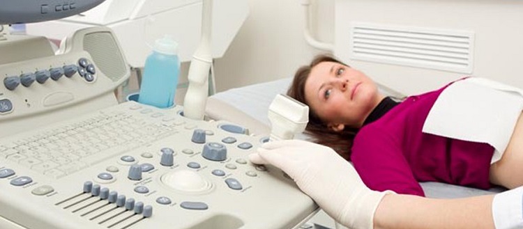
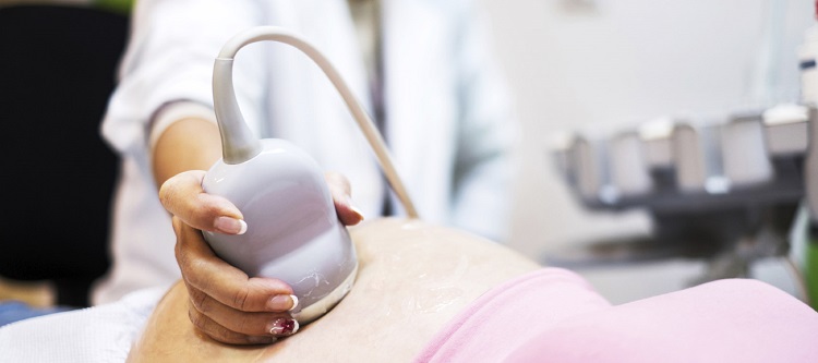
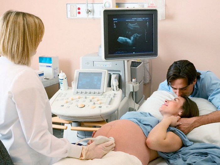
How is the examination?
You can make an ultrasound this week in two ways - external (transabdominal) or internal (intravaginal). For most expectant mothers, the examination is carried out through the front abdominal wall, the child is quite clearly visible at this time. But if the visualization is difficult (with low water or overweight in the mother), the doctor can scan through the vaginal wall, it is thinner and better permeable for ultrasonic waves.
If a woman comes to the diagnosis for a reason the threat of spontaneous abortion, then the examination will be carried out by a vaginal sensor, since such a method makes it possible to carefully study the signs of a threat, the condition of the uterine walls and cervix.
If a woman responsibly prepared for ultrasound in the first trimester, refused food that promotes gas formation, and also filled her bladder, if she was to be examined through the anterior abdominal wall for a short period, then Now prepare for ultrasound scanning is not required.
The amount of amniotic fluid is already sufficient so that the review with external ultrasound is quite clear, and the presence of possible gases in the intestines of the future mother no longer plays any role, because the uterus has increased in size, has gone beyond the pelvis, and cannot squeeze its bowel loops .
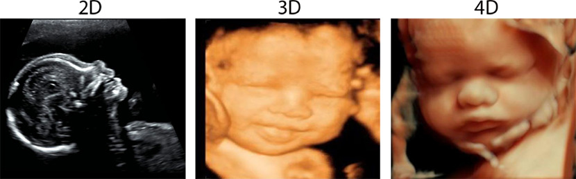
If you want to undergo an ultrasound scan in three-dimensional format, the so-called 3D ultrasound scan, then you should be prepared for the procedure to be several times longer in time than a conventional ultrasound scan, and therefore you should not drink a lot of liquid so that you don’t want to go to the toilet. Two-dimensional conventional ultrasound at week 22 will last about 7-10 minutes, and three-dimensional - from 40 minutes to an hour.
A woman should take a passport, a policy, an exchange card, as well as a diaper to put it on a couch, and removable shoes.
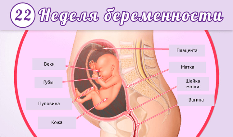
What will the research show?
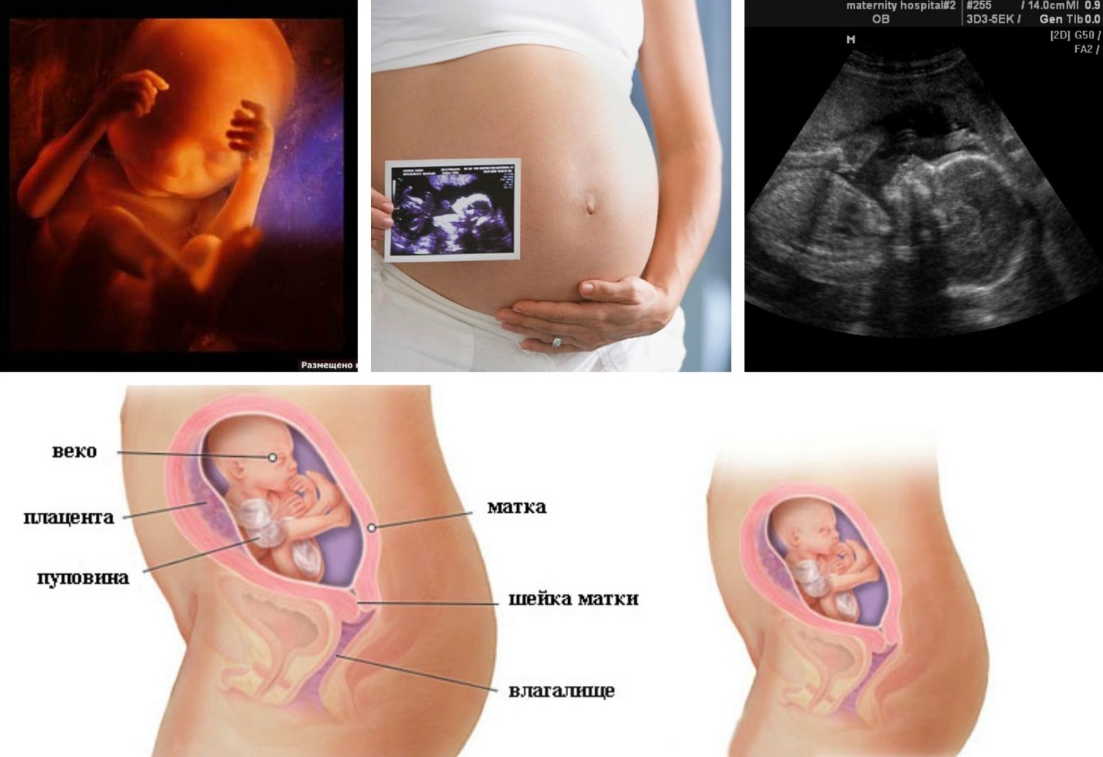
The baby has changed a lot since the last date with his mom on the ultrasound monitor in the first trimester, he grew up. Now the size of the fetus is already sufficient, so that you can more closely examine the crumbs. Its height is about 25-27 centimeters, and its weight approaches 400-450 grams. All internal organs and systems are formed, now they can only ripen and grow.
The woman “stepped over” the equator of her pregnancy, the first half is over. Now there is less chance to lose a child, mother becomes calmer. But the baby is more and more mobile every day, as the nervous system develops, the baby learns to control its body - limbs, mimic muscles. On ultrasound, the future mother will be shown how the baby has learned to move. At the same time, he already touches the walls of the uterus, and the vast majority of women already feel the movements of their crumbs.

The brain of the baby at week 22 "acquires" the first gyrus. The formation of the spine is completed - the vertebrae and the discs between them are almost functional. The heart of the baby becomes larger in size, it rhythmically and loudly knocks on the ultrasound a woman can hear it.
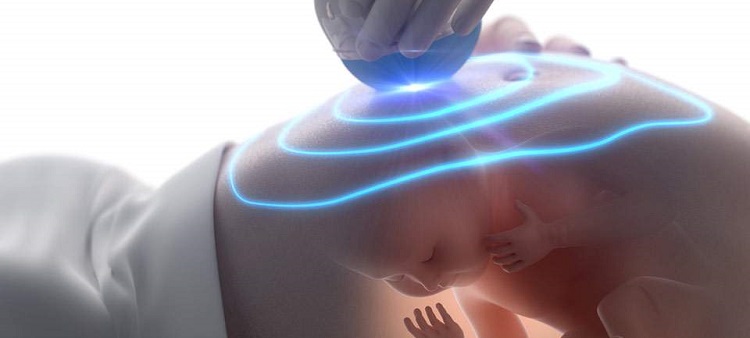
Cilia and eyebrows appear, but they are so thin that it is impossible to examine them even on a high-resolution device. The child, although still very small, distinguishes well between "his" and "strangers". If the mother puts her hand on the belly during the ultrasound, the baby will approach the palm of her hand, and the sensor of the scanner and the doctor’s hand will react to someone else’s patient to the contrary - it will begin to move away.

Decryption and norms
In the result of an ultrasound scan received in her hands, a woman will see a large number of numerical values. To understand how the fetus develops correctly and according to the duration of pregnancy, doctors use special tables. Fetometry kid will understand whether everything is fine with him. During this period, all children grow at about the same pace, and therefore the values given in the tables are relevant for most expectant mothers.
The doctor measures the transverse and longitudinal dimensions of the head. They are called bipariate and fronto-occipital. These are the most important indicators of the development of the baby at this time. The proportions of the body are indicated by the dimensions of the paired bones - the femur, tibia, and the humerus and forearm bones.
About how well the baby eats, whether he has internal edema or hypotrophy, say the circumference of the abdomen, the circumference of the head, chest.
The average fetometric standards at 21-22 week of pregnancy:
BPR, mm | LZR, mm | DBK (thigh), mm | DKG (calf), mm | WPC (shoulder), mm | DKP (forearm), mm | Abdominal circumference, mm |
51-54 | 66-70 | 36-39 | 33-35 | 33-35 | 28-30 | 157-169 |
Amniotic fluid normally have a transparent consistency, their normal amount at this time - 88-97 mm. The thickness of the placenta is 22.8-23.6 mm, the degree of maturity of the “children's place” is still zero. The position of the child in the space of the uterus does not yet have a great diagnostic value. The pelvic or transverse position of the fetus, which is determined by ultrasound, should not disturb either the expectant mother or her attending physician, because the crumb will turn over many times before it becomes cramped and its movements are restricted.

Possible problems
The most common problem that the expectant mother may encounter on the results of an ultrasound scan at week 22 is the mismatch between the size of the fetus and the obstetric period. A slight deviation does not cause concern, but a significant excess or lag may be signs of possible pathologies of the child’s development. Considered significant difference in 2 weeks.
Obstetric period is considered from the first day of the last menstrual period. From the embryonic, actual, it differs by about 2 weeks. The difference in parameters, therefore, may be due to an error in the establishment of a term. This is not uncommon in women with irregular menstrual cycles, as well as in women who do not remember the exact date of the last menstruation.
If all sizes of the fetus at the same time differ from the norm in a greater or lesser direction, doctors may also consider the option of a symmetric intrauterine growth retardation. And then additional examinations will be needed to find out whether the baby receives enough nutrients, vitamins and minerals, or if he has an intrauterine infection.

The growth of children in the second trimester may be spasmodic, and therefore It is possible that on the control ultrasound in a week or two the baby’s parameters will return to normal. If not, then treatment will be prescribed to improve the uteroplacental blood flow, which will enrich the future mother's blood with vitamins and other important substances useful for the child.
If, according to the doctors, the increased or reduced sizes of the baby’s body parts are related to possible developmental pathologies, then biochemical blood analysis, which was given earlier, and studies of anatomical features will indirectly confirm this. Genetic abnormalities in 99% of cases are accompanied by defects of the internal organs, and they are already clearly visible at this time.
Among other problems of ultrasound diagnostics at week 22 is the inability to consider the sex of the child. This is only possible if the baby is located with his back to the sensor, his bottom is sitting down, and then the doctor does not have the physical ability to examine the external genitals of the baby. In this case, if the definition of sex is of great importance, the woman will be advised to come to the ultrasound later, after a couple of weeks. Perhaps the baby will change its position in the space of the uterus, then the diagnosis of the child’s gender is not difficult.
Another common problem is the threat of abortion. A woman will be told about it if an ultrasound examines a thickening of the uterine walls, hypertonicity of the uterus muscles, as well as changes in the cervix and cervical canal.
Forecasts in most cases, positive, time taken measures to preserve pregnancy guarantee the birth of a completely healthy and strong baby on time.


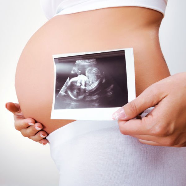
Snapshots
In the ultrasound pictures, the baby is already clearly visible. He is still thin, and it is completely normal. Three-dimensional ultrasound allows you to see the baby in more detail. Twins this week looks like this.
If you plan to get a snapshot for the family archive, It is worth asking to give it on electronic media. The paper on which ultrasound images are printed is short-lived, the “photo” of the kids quickly loses clarity and fades. Pictures are available for a fee.