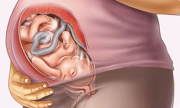
How often and at what time do ultrasound during pregnancy?
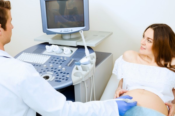
Ultrasound diagnosis is an important tool for doctors in the management of any pregnancy. This method allows you to get a large amount of information about the baby’s condition, development and growth, to make sure that he has no pathologies, and also to find out if his mother’s reproductive health is in order.
For future moms, according to experienced obstetricians, ultrasound is the best medicine, because each such “meeting” with a baby improves mood and even improves well-being.
About how often you can do ultrasound in the period of carrying a baby, we will tell in this material.
The essence of the method
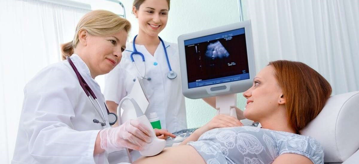
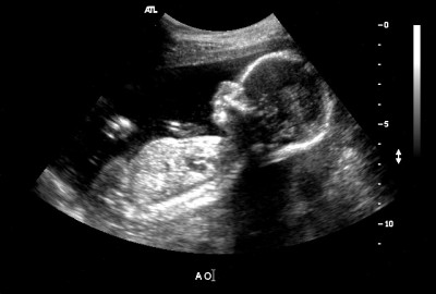
Ultrasound, which is also called sonography, is a gentle non-invasive method of examination, in which ultrasound waves help to examine the internal organs of the human body.
The devices used for research are equipped with ultrasonic wave generators. These waves pass through fabrics and liquids of different speed and consistency at different speeds. Fabrics tend to inhibit the penetration of ultrasound.
Waves colliding with liquids easily pass through them, and bumping into tissues and organs are reflected at different speeds, which depend on the density of the tissues. So the signal sent by the sensor goes back to the sensor and transforms into a picture. This is how the image on the scanner monitor is obtained.
The devices themselves are different - both entry-level and expert class. Diagnostic doctors are also different.whose task is to decipher and measure what is seen on the monitor image. Ultrasound diagnosis today is no longer considered an exotic method, it is carried out everywhere. During pregnancy, ultrasound helps in solving a variety of medical problems.
Types of ultrasound during pregnancy
Types of ultrasound diagnosis in the management of pregnancy are used different. It all depends on the specific purpose of the study and the circumstances.
Here are the main types of research:
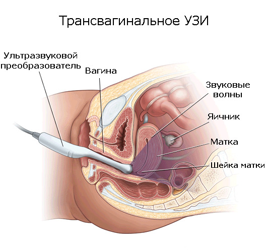
Intravaginal (transvaginal)
This method of obtaining information in the people is simply called internal ultrasound. For examination, the vaginal oblong sensor is used. Inspection is carried out through a relatively thin vaginal wall. This study is used when you need to examine the pelvic organs of a woman., identify any gynecological problems.
During pregnancy, this method is used to conduct an examination in the diagnosis of an “interesting position” in the early stages and with the threat of miscarriage, in order to better assess the condition of the internal genital organs of the expectant mother.
In a transvaginal way, ultrasounds will be done to women who have excess weight and different body fat deposits on the abdomen, which make it difficult to visualize, in later pregnancy.

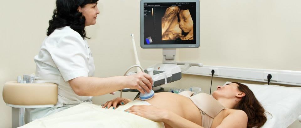
Transabdominal
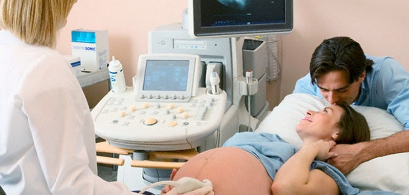
Doctors use this method in the second and third trimester of pregnancy, as well as before and after childbirth. An ultrasonic wave sensor is placed along the outer wall of the peritoneum, after having previously lubricated the abdomen with a special gel, providing tighter contact between the sensor and the skin at the molecular level.
In the early stages of such an examination is impractical because the uterus is not filled with a well-conducting wave of fluid, it is much easier to assess the condition through the vaginal wall.
As a child grows, the amount of amniotic fluid increases, which are an excellent environment for ultrasonic waves.

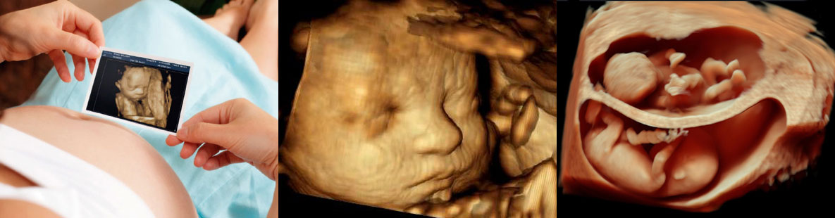
3D
This is a modern method that has gained wide popularity among doctors, and especially among future parents, who Such a study provides an opportunity to see the unborn baby in all its glory. and even understand who he looks more like - mom or dad.
In fact, such an ultrasound does not differ from the standard two-dimensional, the other is only the sensor, which forms on the screen not a flat two-dimensional, but a three-dimensional three-dimensional image.
Such an ultrasound is best done on a period of 20 weeks, because it is after this period that the child is clearly visible, he has formed facial features, no difficulty in determining his sex.
4D
This is an even more modern method, which allows not only to obtain an image, as with a two-dimensional equivalent, and not just to get it three-dimensional, as is done in 3D-diagnostics, but also see not just a static picture, but a baby in real time. That is, one more important parameter is added to all characteristics of ultrasound diagnostics - time.
As a result parents can get not only a beautiful picture of the baby in the family album, but also video - A small film about the intrauterine life of a son or daughter.
Such a “movie” would be a good gift for the child himself, for example, at his majority.

5D
This is an innovative development that is already beginning to be actively introduced into medical practice. Special devices-scanners with 5D support can do research in any format - two-dimensional, three-dimensional, four-dimensional. The difference is that the image will not just be volumetric with movement in real time, but the program itself will measure the main indicators - the length of the bones, TVP, etc.
This is convenient for specifying data, for diagnosing, if the baby is located in the uterus so that an ordinary two-dimensional sensor cannot show some parts of its body. 5D in this case, it will show and measure with great accuracy.

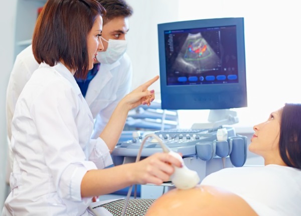
USDG
This is a conventional two-dimensional or three-dimensional ultrasound, which determines the rate of blood flow through the uterine vessels, the measurement of the placenta and the study of its structure. In the people, this diagnosis was called "Doppler ultrasound," and such a study is prescribed from about 20-22 weeks of pregnancy, when the placenta is already well developed. A study is conducted, as usual, two-dimensional, in the technique of execution there are no differences.
USDG is used when there is a suspicion of impaired uteroplacental blood flow, malformations of the baby, hypoxia, fetoplacental insufficiency or the lag of the baby in development.
All women at least once during pregnancy such examination is appointed. Some do it more often if the pregnancy is problematic, and there are concerns about the condition of the child.
Dates of mandatory diagnosis
Mandatory ultrasound during pregnancy can only be considered conditionally. The Ministry of Health of Russia recommended future mothers to conduct such studies, and therefore they are considered to be planned.
A woman has the right to refuse to undergo an ultrasound scan at any time without giving reasons.. But before making such a decision, it is necessary to carefully weigh all possible risks, because the refusal of ultrasound is the inability to establish whether the child is developing correctly, or if he has gross anomalies.
During the period of carrying a child to a healthy woman with an unrequited history, three planned studies were shown.

First trimester
Ultrasound scans are performed as part of the first prenatal screening aimed at identifying the risks of the birth of a child with gross genetic and chromosomal abnormalities. In addition to scanning, a woman has to donate blood from a vein for biochemical analysis.
Ultrasound scan held from 10 to 13 week. It allows you to see some signs (doctors call them "markers") of possible defects, such as Down syndrome, Patau, Turner disease, Cornelia de Lange syndrome, and various pathologies of the brain and spinal cord (nervous tube).

Second trimester
Ultrasonography in the middle of pregnancy is part of the second prenatal screening, which also includes a blood test and an ultrasound scan. The tasks of screening are the same - searching for markers of probable pathologies.
In addition, the second planned study assesses the pace of development of the baby and features of pregnancy. Such a survey is conducted. for the period from 18 to 21 week.

In the third trimester
The third planned ultrasound is the last antenatal screening that is performed. from 30 to 34 weeks of pregnancy. It also assesses the development and growth of the child, its intended weight, position in the uterus relative to the exit from it, after all, childbirth is not far off, and doctors need to decide on the delivery tactics.
Indications for unscheduled ultrasound
If necessary, ultrasound diagnosis can be used at any time. For example, at the very beginning - to establish the fact of pregnancy itself, to establish the fact of multiple pregnancy, when a woman has two or three babies under her heart.
Women often go to an ultrasound at their own request - to find out the sex of the child, to see how he grew up. Doctors have certain indications for unscheduled ultrasound studies.
Among them:
- Refinement gestational age. If the woman does not remember the date of the beginning of the last menstruation, if she has an irregular cycle, then the obstetrician will need the conclusion of an ultrasound specialist to establish the duration of the expected birth.

- Threat of interruption. If a woman complains of pain, discharge from the genitals, which should not be normal, abdominal tension, tone, if a manual examination of the gynecological chair determines the smoothness and shortening of the cervix, the opening of her external os, the ultrasound scan is done urgently.
- Suspected baby developmental delay, intrauterine death. The referral in this case is issued by the obstetrician on the basis of the difference between the size of the uterus, the height of its bottom, which are determined at the reception, and the actual period of gestation. Also, out-of-schedule ultrasound sends pregnant women who have suddenly changed the character of the child's movements - it became more aggressive and frequent, rare and apathetic, or the movements of the crumbs in the mother's womb stopped altogether.

- ECO. Women who could become pregnant through in vitro fertilization need to be monitored continuously for their pregnancy. They can prescribe ultrasound more often, just to make sure everything goes well.
- Evaluation of the effects of disease and treatment. If a woman has had an infectious disease in the period of carrying the child, if she has received the treatment prescribed by the doctor, a control ultrasound scan is done to assess whether the baby is not injured and that everything is fine with him. If a woman underwent any operations, for example, cervical closure, periodic ultrasound control is shown almost to the birth.

How to prepare for the survey?
As such, preparation for ultrasound is needed only in early pregnancy, up to 10-12 weeks. As already mentioned, ultrasound penetrates the liquid better, and therefore, when preparing for an external study performed by the transabdominal method, this should be taken into account.
A woman needs to drink about half a liter of water about an hour before the diagnosis in order to fill her bladder. In later periods this will not be necessary, since the amniotic fluid will be more than enough to conduct an ultrasonic signal.

In the early stages, regardless of which type of examination is to be performed - external or vaginal - care should be taken that the intestine is not overfilled with fecal masses and intestinal gases.
Swollen intestinal loops can squeeze the pelvic organs, distorting the true size of the latter. therefore before ultrasound for a couple of days it is not recommended to eat foods that promote increased gas formation. These include legumes and peas, beans, sweet pastries, black bread, any sweets, especially factory production, sparkling water, dairy products in large quantities.
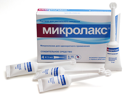
On the morning of the scheduled examination, you need to empty your intestines, if there is no urge to defecate, you should use a microclyster, for example, "Microlax"As well as a drink Activated carbon or "Smektu"(Also suitable"Espumizan"And" Simethicone ") to release the intestines from gas bubbles.

Such preliminary preparation will not be necessary in the second trimester and in late pregnancy, when the uterus grows so much that it will move the intestines itself. Then no gases can distort the true picture of what is happening with the baby.
You should take an exchange card, passport, medical policy, paper napkins to remove excess diagnostic gel from the abdomen, as well as a condom, if you have a transvaginal method.
On any kind of ultrasound scan should bring a clean diaper with youwhich can be laid on the gynecological chair (with transvaginal examination at an early date), on the couch (with other types of ultrasound on other terms). It is advisable to have changeable shoes with you and some money in case the baby’s photos are printed in this office on a paid basis.
Planned ultrasound and unscheduled examinations prescribed by a doctor are done free of charge. But some services are not included in the insurance claims provided by the medical policy. So, screening at the time of screening - paid service, as well as printing a photo or copying a video file from the device to electronic media.


You can find out the rates for these ultrasound possibilities that are important for future mothers in a particular medical facility where she will be examined.
When can pregnancy be determined?
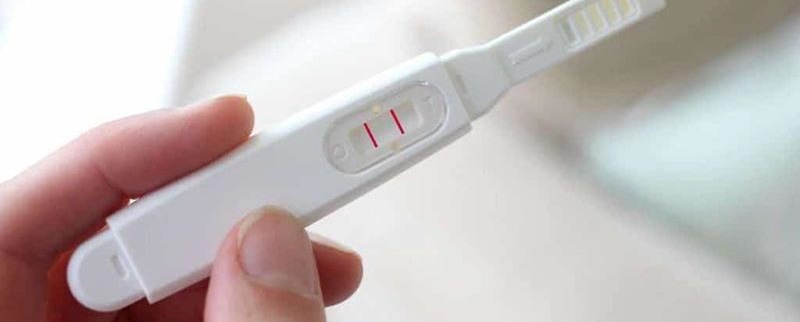
This question interests all women who dream of having a child and are planning it. Two weeks after ovulation In the blood of a woman, the level of the specific hormone, hCG, is already rising, and pharmacy tests and laboratory tests of the blood for the chorionic gonadotropic hormone are beginning to give positive results. but Ultrasound on this period does not show anything, even if the test pleased with a double strip.
The size of the baby at this time is only about 1 millimeter, and no modern apparatus or doctor of the highest category can see it. But after a week this possibility theoretically appearsHowever, much of this depends on the quality of the scanner and the level of readiness of the diagnostician.
Baby grows up to 4 millimeters, he has the heart starts beating. On the ultrasound, there is still no heartbeat in the full sense of the word, but the pulsation should not escape the sight of an experienced doctor.
Thus, the earliest time at which a pregnancy can be seen is 7-10 days after a delay or three weeks after ovulation, of course, if it occurred on time and the baby’s implantation into the uterine cavity was not delayed.
Usually, ultrasound shows the presence of the ovum in the uterus from 4 weeks after the delay, from 5-6 obstetric weeks.
In addition to the usual female curiosity, which, as we know, is stronger than common sense, in the first weeks of pregnancy, an ultrasound scan may also be necessary for well-founded medical reasons.
Such the need arises if the delay is accompanied by bleedingwhich are not related to regular menstruation, pain. After conception, performed by IVF, ultrasound control is also necessary in the first weeks.
To make sure that the pregnancy has come, and the fertilized egg has sunk into the uterus, as it is prescribed by the laws of nature, it is also necessary if the woman has previously had miscarriages in the early stages, ectopic pregnancies, missed pregnancies at the very beginning of gestation, and also surgery, tumors and other gynecological problems that may affect the normal development of pregnancy.
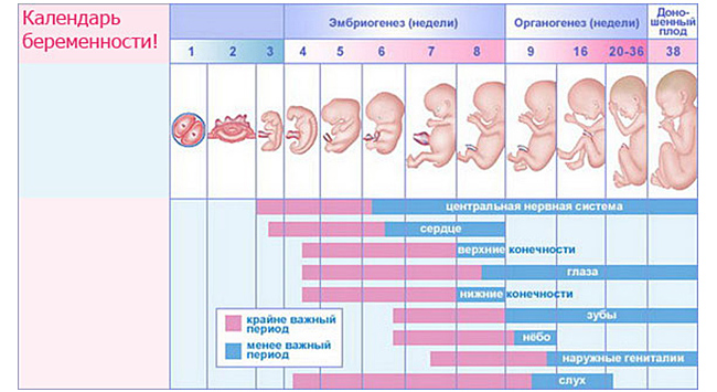
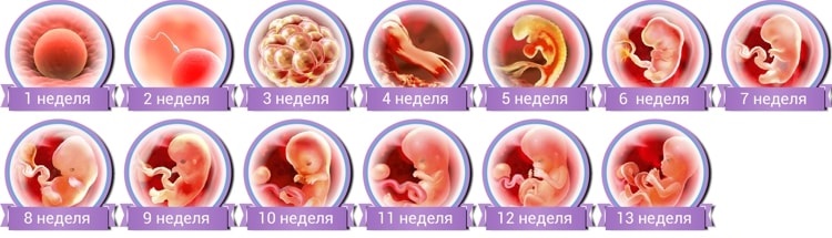
The development of the baby by week
If the future mother did an ultrasound every week, starting from the first week of pregnancy, when the baby was not yet conceived, she would be able to see the whole evolution of humanity on the example of one baby.
Before ovulation, ultrasound shows how the follicle matures, after it - how the corpus luteum is formed in the ovary - a temporary gland, whose task is to help the embryo in the very first weeks of its development with a shock hormonal background.
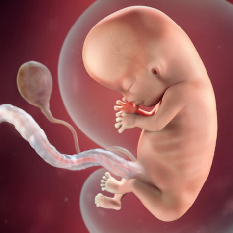
You can determine the fertilized egg from 5 weeks of pregnancy. The baby's heartbeat can be heard already 3 weeks after conception. At this time, the laying of the internal organs begins, but they will not be seen yet. At 5-6-7 weeks of gestation, an ultrasound scan shows only a fertilized egg — one or two, if there are several babies. The doctor can measure it, assess the viability.

8 week The baby's pregnancy already looks like a little man, though it is only the size of a grape. He has a big head and soon the embryonic tail will fall off. The face is actively formed, the ears, the formation of the genital organs begins, but it’s impossible to see them on the ultrasound scan, as this is all about the formation of the internal sex glands.
On week 9 the baby has all the organs, although they are in their infancy. An ultrasound examination at this time makes it difficult to examine them in detail, but in general terms it becomes clear that the baby has two arms, two legs, a head, a big belly, there is a fairly well-formed heart, liver, kidneys, and lungs.
10 week The ultrasound scanner captures the baby’s movement well. They are still chaotic, sometimes involuntary, but a good sensor already allows you to "peep" what a crumb in the womb does.
At week 11 the baby still has pronounced imbalances of the body and head - the head is large, and the body looks small and very thin compared to it. A good device with high image detailing will allow to see the face of the baby during this period. with eyes still wide apart, ears drooping below our usual level of perception. Every day the baby's facial features change, soon the ears and eyes will fall into place.
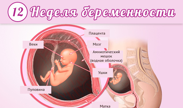
At week 12 baby is the size of an average lemon. He is no longer an embryo, but a fetus. Now the crumb can please the doctor and the future mother, who is looking at him through the monitor of the ultrasound machine, with more active facial expressions, more “conscious” and precise movements. If the scanner is good, then you can count the fingers on the baby’s pens.
On week 13 the child begins to acquire sexual characteristics. The genital tubercle, absolutely identical up to this point in both boys and girls, becomes either a penis or labia. In theory during this period it is quite possible to determine the sex of the baby using ultrasound diagnostics, but sex differences still look too small, and it is not always possible to accurately recognize the boy or girl.
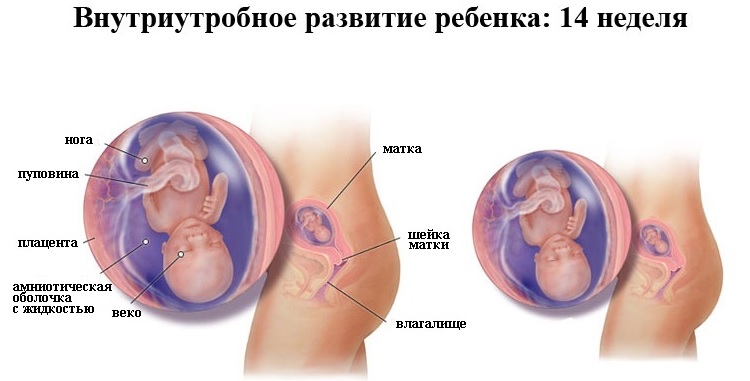
On week 14if pregnant comes on ultrasound, her baby can show everything that he has already learned. And he can do a lot - he sucks his finger, plays with the umbilical cord, reacts to loud sounds, unfamiliar voices. Moreover, it is already possible to determine the character of the baby. One of the crumbs at the sound of a doctor's voice, alien to him, begins to move briskly, while the other one stops, trying to hide.
On week 15 formation of the cerebral cortex begins, grooves and convolutions appear. Now every day the little ones will "grow wiser". In the meantime, the baby is busy actively training his lungs - he inhales and spits out the amniotic fluid. At the same time, the digestive system “trains”, the baby drinks, pees, the formation of original feces in the intestines begins.
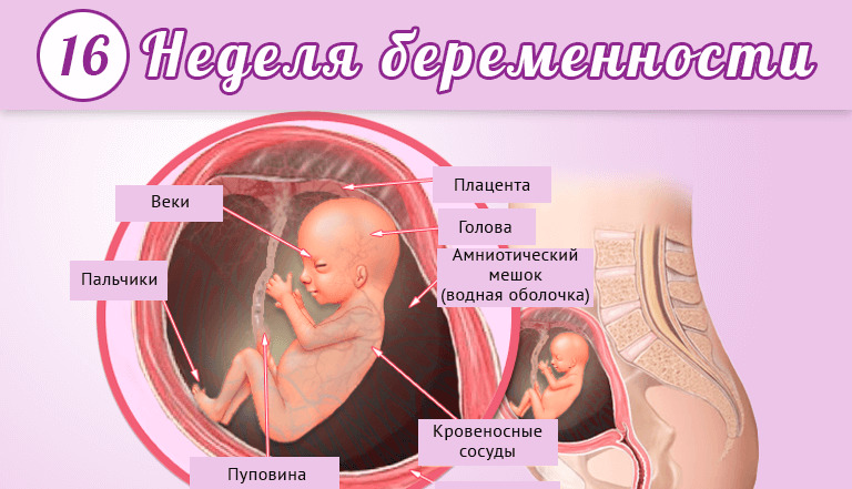
On week 16 the baby reaches impressive size - its height is now almost 11 centimeters. With an ultrasound scan, the doctor may clearly consider all the internal organs of the child, evaluate them and detect some malformations, if any.
Mom will be able to see how the baby turns his head and makes swimming movements. His bone and muscular systems are almost formed, and now neural connections are established between muscles and the brain. Soon the baby will get the opportunity to coordinate their movements.
At 17-18 week pregnancy is already possible determine the sex of the child on ultrasound. Moreover, the accuracy of this definition will be higher than ever.. At an earlier period, the genitals were almost invisible, in later ones, the baby will become cramped, and he will take a pose with his legs tucked to his stomach or sit down, and it will be even more difficult to consider sex differences.
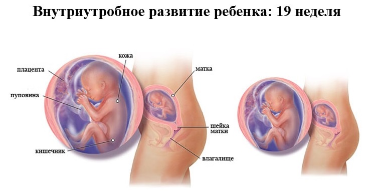
On week 19 pregnancy, the baby begins to be covered with a protective lubricant that will protect its skin right up to the birth and during the first hours after birth. His hair is growing, his fingernails and legs have grown. He hears, dreams, smiles, yawns, hiccups, plays with his legs or umbilical cord when he is not sleeping. Parents will be able to see any of these actions themselves if an ultrasound scan is performed on this period.
20-21 weeks the crumb is fully formed and is already starting to resemble a newborn. Now all his organs and parts of his body will only grow, there will be no new ones. Growing up baby fast now it reaches 25 centimeters from crown to heels.
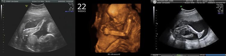
On the ultrasound diagnostics during this period, the sex of the baby is almost unmistakably determined. If you make a volumetric color ultrasound, then you can see the child’s amazing mimicry, which is amazing in its manifestations - he knows so many grimaces that you can admire them for a very long time.
22-23 week a child can demonstrate a brain that is ten times larger. Its formation was completed, now the “debugging” of the central nervous system will be in full swing.
The baby begins to gradually accumulate subcutaneous fat, but still looks quite thin. This should not scare the future mother, very soon there will be both “cheeks” and “dimples”.
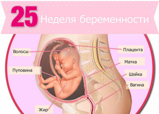
24-25 week pregnancy on ultrasound you can see a fully developed, large child, whose weight is approaching a kilogram. At this time, it is determined whether the child is left-handed or right-handed., because he already gives preference to one handle, making his movements.
The ultrasound shows how the baby clenches the fists, puts them under the cheek in a dream, at these times the doctors are already more closely studying the structure of the placenta in order to recognize its early aging, if it occurs.

26-27 week the baby is actively growing and gaining weight. His endocrine system works almost autonomously. the formation of immunity begins. During the passage of the ultrasound scan, the child will no longer show somersaults and somersaults, as he becomes crowded in the uterus.
At 28-30 week baby for the first time can please mother with chubby cheeks, because he has already accumulated a sufficient amount of subcutaneous fat.Enjoy this spectacle can be on a conventional, two-dimensional ultrasound, but the impressions received from the three-dimensional, do not go to any comparison. The crumb has grown up, its weight is more than a kilogram, and the height is already about 40 centimeters.

30-33 week It is worth visiting a color ultrasound to see how the baby’s skin has changed. It ceased to be red, wrinkles smoothed out due to subcutaneous fatty tissue. Research on this term is of great importance, because most babies by the 33rd week already occupy that position in the uterus, from which they will soon be gathered to the world.
At 34-37 week the baby sleeps more, gathers up with strength, soon an important event is coming up for him - his own birth. Externally, the child at this time changes little, its growth slows down, only the weight gain remains intense. An ultrasound examines the placenta, the umbilical cord, and much depends on their condition in the last weeks of pregnancy.
38-40 week baby looks exactly like at birth. Many mummies save photos with 3D ultrasound on this period and compare them with photos in the first days after the birth of the baby.
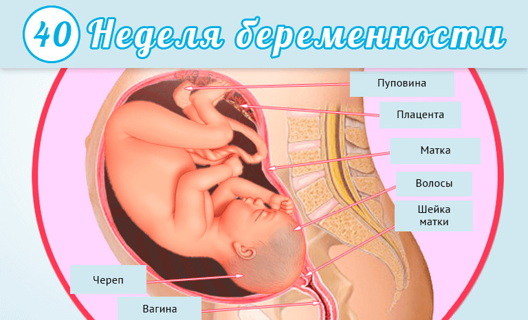
Motor activity on the examination from the baby does not have to wait, he is very closely. But here mimicry and grimaces literally save the situation - if you manage to capture them in the picture, it will be one of the best shots in the family album.
Explanation of ultrasound - the basic rules and terms
Questions interpreting the results of ultrasound excite all pregnant women without exception. You always want to be sure that the child develops according to the term, that everything is in order with him.
To understand the protocol ultrasound, you do not need to have great knowledge in the field of medicine. It is enough to understand what terms the diagnostician uses, and what they mean. We publish all the submitted data solely for information; it is not recommended for the future mother to make diagnoses for herself. Ultrasound should only be decoded by a doctor.

First trimester
In the first third of the gestation period in the ultrasound protocol a special role is assigned to the location and size of the ovum. It is his development in the initial stages that gives the most accurate picture of the course of pregnancy.
To characterize this parameter, a certain value is used - SVD (mean internal diameter of the ovum). It is measured until about 10 weeks of pregnancy, after this period, the main parameter is a different size - KTR.
SVD is very important in the diagnosis of non-developing pregnancy, anembryonywhen the baby died for some reason in the very early stages of pregnancy, if there is no embryo in the fetal egg, as well as with the threat of a miscarriage or a miscarriage that has begun.
The coccyx-parietal size of the fetus is a segment from the tailbone to the crown. It is the main parameter indicating the growth rate of the crumbs up to 15-16 weeks. In the early stages, the diameter of the yolk sac is also measured, which serves as a food storehouse for the baby.
In the first third of the term of carrying a baby The main task of ultrasound scanning is to identify possible problems. in the formation and development of the baby, so that the woman has a choice - to leave the child or have an abortion for medical reasons, if pathology is detected in the baby.
To do this, measure the length of the bones of the nose (DNA) and the thickness of the collar space (TVP). These are the very markers that can talk about the risk of developing pathologies.
In the second and third trimesters
From the second trimester in the protocol ultrasound includes new concepts and obscure at first glance, the abbreviations - BPR, DBK, etc. These abbreviations indicate the main dimensions, because the baby has grown, and the doctor no longer measures his height from the tailbone to the crown of the whole. Get an idea of the proportions of the body of the baby can be on its individual parts.
The main parameters are the longitudinal and transverse dimensions of the head.They check the terms of pregnancy, specify the date of the expected birth, their proportions can talk about possible problems with the health of the child.
The longitudinal dimension from the frontal to the occipital bone is called the frontal-occipital bone (LZR), and the transverse dimension, from the temple to the temple, is called bipariented (BPR). They cannot be considered separately, when decoding the ultrasound protocol, both sizes are taken into account collectively.

Also, paired bones are to be measured - these are the bones of the thigh (DBK), the bones of the tibia (DKG), and also the humerus (KDP) and the bone of the forearm (DKP). On the development of the baby indirectly can talk about the circumference of the tummy of the baby (OJ), and the diameter of his chest.
All these dimensions together allow the program embedded in the scanner to calculate the estimated weight of the fetus, which can be specified in the protocol under the abbreviation PMP.
Also investigated the thickness of the placenta, the amniotic fluid index - the amount of water (IAG). On seventh month with the help of USDG, blood flow in the uterine vessels and placenta is examined.
Closer to childbirth, these parameters become crucial, as well as the anatomical features of a woman - the features of the pubic symphysis, the size of the pelvis.

How is the rate determined?
Compliance or non-conformity of the baby’s size with the norms is determined by special diagnostic tables used by all diagnostic physicians. In most modern scanners, this information is embedded in the program, so she herself indicates which values correspond to the term, and which values do not.
Small deviations from the indicated values usually do not have a diagnostic weight.. In order to say that the growth rate of the baby lags behind the average parameters, the backlog should be significant - two or more weeks. Therefore, if the baby is behind the ultrasound scan for a week, no one will beat the alarm, and expectant mothers are encouraged to do so.
Identified deviations and defects necessarily need to be confirmed, based on ultrasound alone, no diagnoses are made.. If the doctor suspected hypoplasia of the nasal bones in a baby, a consultation of genetics and more accurate, for example, invasive diagnostic procedures will be required.
If the study showed different length of the shin bones or the double contour of the head, another one is necessary - an expert ultrasound to exclude the human factor and the banal errors of outdated technology.
When determining whether a child is normal, his possible hereditary features of appearance are taken into account, because there are large and miniature parents, with long or snub noses, respectively, and children are all different, and this difference becomes especially noticeable in the 2nd and 3rd trimester of pregnancy.
Therefore, a long or short nose, long or short legs is an individual trait, the main thing is that the body of the baby should be proportionate, and the internal organs should work without interruption. The tables themselves, according to which data are compared, are presented below.

Rates of parameters by week in the table
Term week | BPR, mm | LZR, mm | DBK, mm | DKG, mm | Humerus, mm | Forearm bone, mm | Coolant, mm | Head circumference, mm |
14 | 18-26 | 17-33 | 8-14 | 4-11 | 7-15 | Not defined | 66-90 | 83-110 |
15 | 21-32 | 25-38 | 10-17 | 7-17 | 10-18 | Not defined | 78-90 | 90-111 |
16 | 26-38 | 32-49 | 13-24 | 11-21 | 13-23 | 12-17 | 88-116 | 112-135 |
17 | 29-42 | 37-58 | 16-27 | 14-25 | 16-26 | 15-20 | 93-130 | 121-148 |
18 | 32-46 | 43-65 | 18-32 | 16-27 | 19-31 | 17-22 | 104-144 | 131-160 |
19 | 36-51 | 48-70 | 21-34 | 19-31 | 21-33 | 20-25 | 114-153 | 142-173 |
20 | 39-55 | 53-75 | 23-37 | 21-34 | 24-36 | 22-29 | 124-165 | 154-186 |
21 | 42-59 | 57-78 | 26-41 | 24-35 | 26-38 | 24-32 | 137-176 | 165-200 |
22 | 45-63 | 61-82 | 28-44 | 26-38 | 28-41 | 26-34 | 148-190 | 177-212 |
23 | 48-65 | 65-86 | 31-47 | 28-40 | 30-33 | 29-36 | 160-202 | 189-224 |
24 | 51-68 | 69-90 | 33-50 | 39-42 | 33-45 | 31-38 | 172-224 | 201-236 |
25 | 53-70 | 72-93 | 36-52 | 32-44 | 35-48 | 33-41 | 183-229 | 214-249 |
26 | 56-74 | 75-95 | 38-55 | 34-46 | 37-49 | 35-42 | 194-240 | 224-261 |
27 | 59-75 | 79-98 | 40-57 | 36-48 | 38-51 | 37-44 | 205-253 | 235-272 |
28 | 62-78 | 82-102 | 43-59 | 38-51 | 40-52 | 39-47 | 217-265 | 245-285 |
29 | 64-81 | 85-104 | 45-60 | 40-53 | 42-56 | 40-48 | 228-279 | 255-295 |
30 | 67-83 | 88-108 | 47-62 | 42-55 | 44-55 | 42-50 | 238-290 | 365-305 |
31 | 69-85 | 90-110 | 50-66 | 43-56 | 46-58 | 44-52 | 247-301 | 273-315 |
32 | 72-87 | 93-112 | 52-67 | 45-57 | 47-60 | 45-52 | 258-314 | 283-324 |
33 | 74-89 | 96-115 | 54-68 | 47-59 | 49-61 | 46-53 | 267-325 | 289-333 |
34 | 76-90 | 99-116 | 57-70 | 48-61 | 51-63 | 48-56 | 276-336 | 295-339 |
35 | 79-92 | 101-120 | 56-73 | 50-63 | 52-63 | 49-56 | 285-343 | 299-345 |
36 | 81-94 | 104-122 | 61-74 | 50-64 | 53-65 | 50-57 | 292-354 | 303-349 |
37 | 83-96 | 106-124 | 63-76 | 53-65 | 55-67 | 51-59 | 299-361 | 307-352 |
38 | 86-97 | 109-126 | 95-78 | 55-67 | 55-68 | 52-60 | 304-367 | 309-356 |
39 | 88-100 | 111-128 | 68-79 | 56-68 | 59-70 | 53-60 | 310-373 | 311-358 |
40 | 90-101 | 113-129 | 70-82 | 57-70 | 60-71 | 54-62 | 313-380 | 312-362 |

Table of norms in the first trimester
Term week | SVD, mm | KTR, mm |
5 | 18 | Not determined |
6 | 22 | Not determined |
7 | 24 | 6-10 |
8 | 30 | 8-18 |
9 | 33 | 18-27 |
10 | 39 | 24-38 |
11 | 47 | 34-50 |
12 | 56 | 42-59 |
13 | 65 | 51-75 |
The length of the bones of the nose and TVP in the first trimester
Term week | Nose bones, mm | TVP, mm |
10 | LOCATED | 1,5-2,2 |
11 | LOCATED | 1,6-2,4 |
12 | More than 3 mm | 1,6 -2,5 |
13 | More than 3 mm | 1,7-2,6 |
About the dangers and complications
About the dangers of ultrasound for pregnant women, disputes have been going on for more than a dozen years. It must be said that official evidence of the harm or harmlessness of this diagnostic method does not exist in nature.
For many years, doctors have been watching pregnant women who undergo such procedures, and so far they have not been able to establish the facts of the negative impact on the development of the child. Therefore, it is believed that ultrasound can be done as many times as the situation requires.
However, to this day, the effect of ultrasonic waves on the further development of the child, on his condition 20, 30, 50 years after birth, remains unexplored. It is not possible to check this with scientific methods, it is precisely this fact that opponents of ultrasound diagnosis during pregnancy refer to.
Traditional medicine has its own view on everything that happens. Until harm is proven, the method is considered conditionally safe.

Numerous reviews and questions of future mothers in the women's and parent forums on the Internet relate to the problems of complications after passing this type of diagnosis. Transabdominal ultrasound usually has no negative effects on the condition of the woman and her baby.
Rarely, after an external examination, a small and briefly elevated uterine tone appears.which passes quickly. Its specialists are not inclined to associate it with ultrasound, most often the cause of increased tone - in the excitement and nervous experiences with which the expectant mother was sent for research.
Transvaginal ultrasound may cause some "side" phenomena. Usually future mothers complain that the stomach hurts after the examination, there are brown discharge, as well as scanty pink and clear discharge.
If their number is small, and they last only a few hours after the diagnosis, there is nothing to worry about. The reason for unscheduled treatment to the doctor is bleeding after ultrasound, the appearance of pain of a pulling, cutting or cramping nature.

What can be connected with such “consequences” is unambiguously difficult to answer. Experts tend to believe that the majority of the fault is pregnancy itself. Under the influence of the hormone progesterone the mucous membranes of the vagina become more loose, vulnerable. Even an ultrasonic sensor can cause microtrauma..
It would be wrong to blame the ultrasound scan for a miscarriage that has taken place after the study or for a threat if the bleeding does not stop for two or more days after the passage of diagnostic measures. If this happened, then, according to most doctors, ultrasound diagnostics simply coincided with the onset of pathological changes in the pregnant woman's body.
At women's forums, frightened pregnant, of course, they will immediately start telling horrors about real and fictional characters, as well as citing as an example articles on the Internet that ultrasound has a detrimental effect on DNA. Official science, such data are not known. Therefore to draw a parallel between pain and secretions and the ultrasound examination completed the day before is not worth it.

Diagnostic accuracy
Ultrasound, although it is considered one of the most informative methods for examining pregnant women, is not considered a high-precision diagnostic method. Its accuracy, depending on the quality of the device and the qualifications of the diagnostician, varies between 70 and 90%.. Do not assume that such a diagnosis will answer all questions that may arise.
Whether fetal hypoxia is visible, whether cerebral palsy or diabetic fetopathy is visible on ultrasound - all these are questions that have nothing to do with the real goals of the study. Ultrasound does not make a diagnosis. The technique allows only to detect some warning signs that may indicate the presence of disorders, pathologies, anomalies. And it is not at all the fact that suspicions are confirmed.
Any ambiguous or alarming conclusion should be checked and rechecked by specialists using other diagnostic measures, laboratory diagnostics, invasive procedures.
In the next video you will find more information about ultrasound.


