Is ultrasound harmful to the fetus during pregnancy?
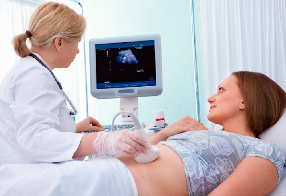
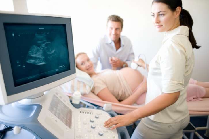
Ultrasound examination during pregnancy refers to the baseline examinations that are carried out to all pregnant women. This method has been used in medicine for many years, but still causes various myths and personal opinions about the need to use it. This article will help expectant mothers to understand whether they can do ultrasound during pregnancy, and whether it has any negative impact on their unborn child.

The benefits of carrying
Of course, one should not underestimate the possibilities offered by the use of this method in modern gynecology. Timely diagnosis of many pathologies that occur during pregnancy helps save thousands of new lives around the world. Without ultrasound, in many cases it is simply impossible to do.
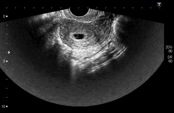
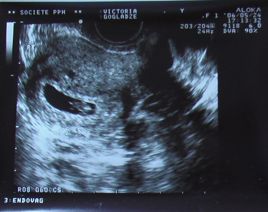
This study helps determine the presence of the ovum. This criterion is the earliest manifestation of pregnancy. A gestational (fetal) egg is revealed already in the first weeks after conception. To determine it, only the most modern devices are used, which have a fairly high resolution.
Ultrasound helps to establish signs of the viability of the embryo at the earliest stages of its intrauterine development. Using this method perfectly detected "frozen" or "frozen" pregnancy. In this case, as a rule, further development of the fetus is impossible and urgent gynecological surgery is required.
With the help of this study, you can establish the expected duration of pregnancy. If the future mom bears several babies at the same time, then the use of ultrasound in this case is practically indispensable. This study is especially necessary if the pregnancy occurred after in vitro fertilization. In this case, ultrasound helps to assess the viability of each of the implanted embryos, as well as monitor their development and growth.

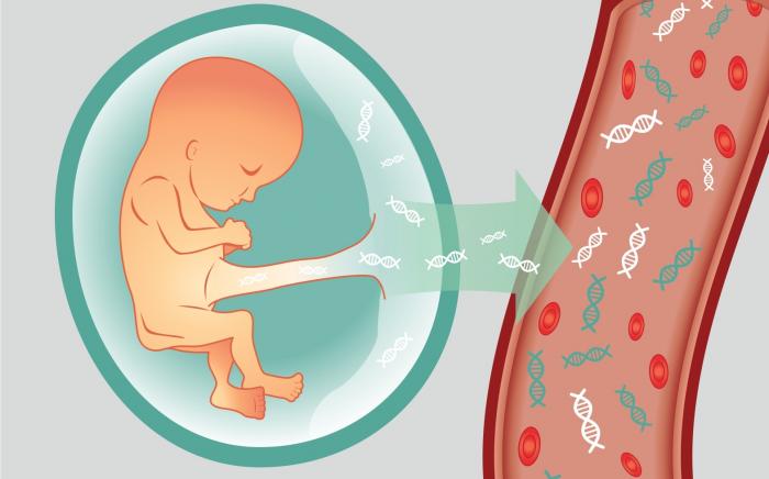
With the help of ultrasound, it is possible to determine various genetic and chromosomal abnormalities. These diseases can occur in the fetus at different periods of its prenatal development. At later stages, ultrasound diagnosis helps to identify anatomical defects in the placenta, as well as to identify signs of low water.
For so many years, obstetricians have determined the position of the fetus in the uterus only with their own hands. Quite often they were wrong. This led to the fact that during childbirth doctors resorted to the wrong gynecological techniques. In the end, all this contributed to the severe birth injuries of the babies.

Currently, it is possible to determine the position of the future baby in the uterus using ultrasound. Such a study, conducted before birth, helps doctors decide on the best tactics for future obstetrics.
Is it harmful to the fetus?
There is no denying that frequent ultrasound may harm the health of the unborn child. In the scientific world there is a huge amount of various studies proving this statement. Scientists from Ireland argue that the abuse of ultrasound at the stage of gestation may lead in the future to the development of various tumors.True, they made this conclusion on the example of laboratory mice.
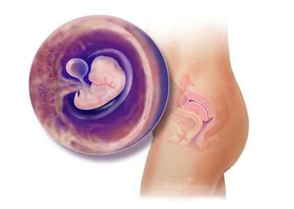
The harm of ultrasound in the early stages of pregnancy may be more likely. At this time there is a very important process in the body of the future baby - organogenesis. All the internal organs and systems begin to form in the fetus. Any physical effects during this period have undesirable effects on this process. In this case, frequent ultrasound has a negative effect on fetal development.
A negative effect may also be associated with a certain temperature effect. This effect is possible during the contact of the ultrasonic sensor with various organs. The longer the study is conducted, the more pronounced the effects after it.
Scientists note that while pointing the ultrasound sensor to a specific anatomical zone, its temperature rises by several degrees. Under the influence of ultrasound in the cells of internal organs also increases the permeability of cytoplasmic membranes. The composition of the ions that participate in their structure also varies. This leads to the fact that cell membranes become more permeable for penetration into the cells of various substances.

Ultrasound diagnostics specialists also note that during such studies in different cells of the body, even the rate of various enzymatic processes may change. Prolonged exposure to ultrasound in this case may trigger the formation of various deviations.
Especially unfavorable in this case is the performance of a study on the term of intrauterine development of 5-6 weeks, when there is an active laying of all the internal organs of the unborn child.
European scientists note that the use of ultrasound may even lead to conditions associated with impaired cellular respiration and metabolism. Some experts believe that it is these disorders that further lead to the possible formation of various chromosomal abnormalities in the fetus. However, it should be noted that all assumptions are only scientific theories and are not recognized by the entire medical world.
According to most experts, the most It is dangerous to conduct ultrasound using focused ultrasound. In this case, the impact on a specific area of the skin becomes the most pronounced. If the treatment is carried out for a long time, it can even lead to significant violations.
For many years, scientists have tried to identify the internal organs that are most vulnerable to the effects of ultrasonic waves. They concluded that the anatomical structures with a fairly good blood supply and innervation are most susceptible to this effect. The brain is also among the most vulnerable organs. The impact of ultrasound waves on this organ can damage it.

Some scientists believe that the frequent use of ultrasound leads to a significant increase in the number of left-handed children in recent years. They also believe that this is a consequence of the direct effect of ultrasonic waves on actively growing brain cells - neurons. Experts believe that such children in the future may experience various difficulties with schooling or, conversely, develop some ingenious abilities.

American experts note that the incidence of autism is increasing in their country every year. They suggest that there is a pattern between frequent vaginal ultrasound and the appearance of various neurological and mental disorders in the future children.
First autism signsAs a rule, they manifest themselves in preschool children. In the appearance of adverse symptoms, a disturbance of the coordinated work of the cerebral hemispheres cortex has an enormous effect.Such babies have various behavioral disorders and speech changes. Some American scientists believe that the appearance of such deviations in babies is influenced by frequent ultrasound during pregnancy, but they have not yet conducted any serious studies.

Some researchers believe that conducting an ultrasound at an early stage can even lead to a miscarriage. This theory has no scientific evidence. All the results were also carried out on laboratory animals. Some studies have found that performing An ultrasound scan at gestational age from 9 to 11 weeks can cause the mother to reject the fetus. The probability of such a situation, as a rule, is 20-25%.
Another theory implies that the abuse of ultrasound during pregnancy can lead to an increase in the birth of babies who have any further tumors of the blood and blood-forming organs. To prove this, supporters of this scientific hypothesis give statistics on the incidence of leukemia in children. The incidence of blood neoplasms in children in recent decades has indeed increased significantly.
Debunk Myths
It is important to note that not only ultrasound conducted during pregnancy can lead to heat exposure. Ultrasound of the kidneys or heart also has a pronounced effect on the woman's body. However, the effect of ultrasound during pregnancy causes the greatest excitement in future moms.
Many of the scientific assumptions are myths, since they have no real evidence.
Most of them are made only on laboratory animals. In this case, it is impossible to talk about a clear correlation with the child population. Many theories exist in medicine for quite a long time, but have not been confirmed.
Opinions of parents on the conduct of ultrasound are also significantly different.

There is currently no clear correlation between the effects of frequent ultrasound and various developmental defects. Such judgments are mostly subjective.
One of the most common myths is the assumption that during an ultrasound scan, the baby in the womb experiences some discomfort. In the very early period of its development, the embryo practically does not feel such an impact at all, or it has a slight effect on it. In the later stages of pregnancy, the amniotic fluid protects the baby from the direct effects of ultrasound, which supposedly brings strong discomfort to the baby.
Many mothers believe that in order to make their future baby move more actively during the study, they should definitely drink coffee before the ultrasound. This is a real myth. Coffee has no pronounced effect on the motor activity of the fetus. The baby in the womb begins to move more actively not from caffeine, but during the change in the position of the mother's body. Mommy's inconvenient posture causes the fetus to move more actively, which is manifested on ultrasound.

Some parents believe that during an ultrasound, the future baby sees different light effects and even recognizes sounds. This opinion does not currently have any scientific evidence. The nervous system of the child in the early stages of its development, as a rule, is still not able to perceive the irritations that are provoked by the ultrasonic sensor.
How often can you do?
Currently, obstetrician-gynecologists adhere to existing regulations in their work. Given such regulatory medical documents, the future mommy, in whom the pregnancy proceeds without any pathological disorders, should undergo ultrasound at least three times during the entire childbirth. Such a number of procedures carried out, according to representatives of official departments, can not cause pathology in either the mother or her unborn child.
It should be noted that if a pregnant woman has any chronic diseases, and there are defects in the development and growth of the unborn child, then she will need to undergo an ultrasound scan more often. In this case, the need for additional research is established by the attending physician.

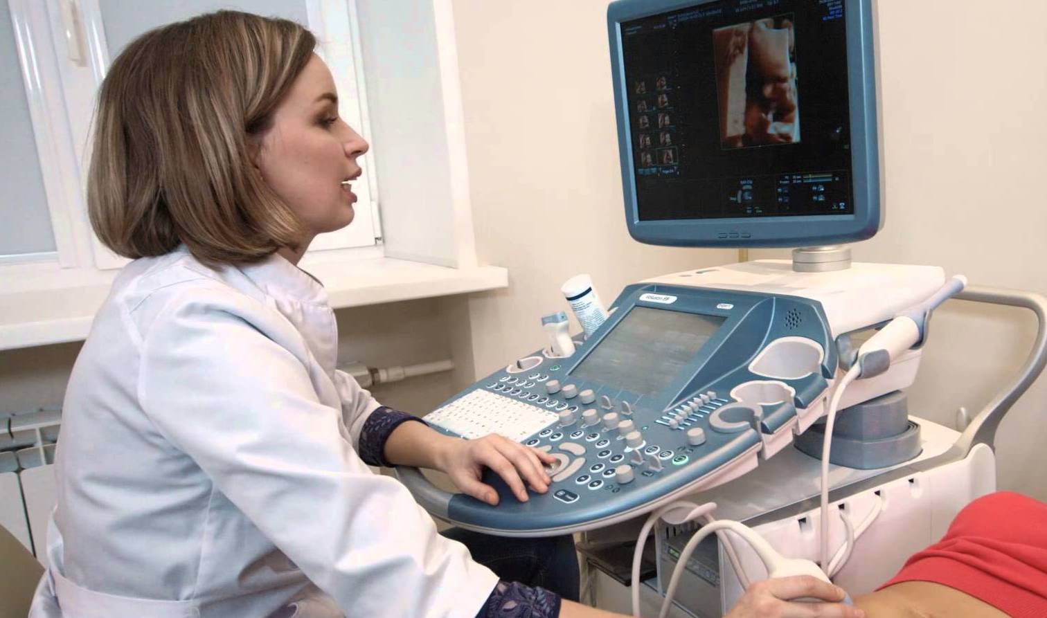
Currently, ultrasound techniques are constantly being improved. Become popular 3D and 4D research. With the help of these studies, you can get not only a three-dimensional and spatial image, but even watch the active movements of the fetus while it is still in the womb.
Such an examination is usually very like future dads and moms. The first movements that their baby makes, brings parents into a real delight and gives them pleasant exciting memories in the future. However, their joys are not shared by the little man who is in the womb. For him, such an examination is a real test of "strength."
Ultrasound examination performed in this mode has a very strong effect on a growing small organism. If ultrasound is performed only in special M and B modes, then in this case it is carried by the fetus much more easily.
Parents should remember that ultrasound is not a fun procedure, but is carried out only for the diagnosis of various pathological conditions and control over the course of pregnancy.

Doctors recommend that you always conduct an ultrasound in the first trimester. Usually The first examination is performed for up to 12 weeks. Such a primary screening is conducted at 13 and 14 weeks of pregnancy.
Carrying out ultrasound in earlier periods should be carried out only under strict medical indications. To do frequent research absolutely all pregnant women can not.
Unjustified conduct of ultrasound in the early stages of childbearing can contribute to the development of adverse events that will manifest in a child in later life.
The next regulated deadline for this study is the 2nd trimester. Usually examination held on 20-22 week intrauterine development of the baby. This type of ultrasound can also be called anatomical. At this time, experienced ultrasound diagnostics doctors may notice various pathologies and abnormalities in the development of a baby.
If the course of pregnancy in a future mom is normal, then additional ultrasound may not be necessary in the third trimester. This decision is made by an observant gynecologist. It should be noted that the normal healthy course of pregnancy is now extremely rare. This situation explains why so often ultrasounds are performed before delivery in pregnant women.

On whether it is safe to do an ultrasound during pregnancy, see the following video.


