3D ultrasound during pregnancy

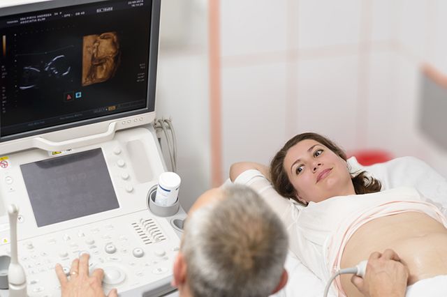
About 3D ultrasound you can hear a lot of positive feedback from expectant mothers. In three-dimensional format, you can view the child in detail and get amazing pictures for your home collection of family photos. Is it possible for everyone to do such an ultrasound, what this examination represents and what are its advantages and disadvantages, we will tell in this article.

What it is?
3D ultrasound (sonography) is an echographic study that has found wide application in various fields of medicine and especially in gynecology and obstetrics. During pregnancy, ultrasound is done to get a three-dimensional image of the fetus.
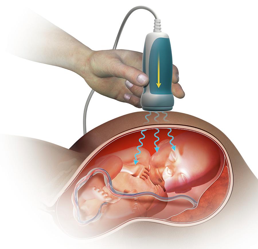
The principle of operation of the apparatus for this study is identical to the already familiar two-dimensional ultrasound - ultrasound waves, which are produced by the ultrasound generator embedded in the equipment, pass through tissues and fluids in the human body in different ways. They are repelled from some tissues, absorbed by others. The sensor "catches" the reflected waves and makes a detailed picture of the internal space. That is how the image is formed on the monitor of the ultrasound scanner.
With a three-dimensional scan, the frequency of the ultrasonic waves is unchanged, it does not differ from the frequency used in two-dimensional ultrasound. Others are only the sensors themselves, which capture the signal reflected from the internal organs. Due to this procedure in 3D format during pregnancy is essentially no different from a similar procedure in two dimensions. Significant differences in the result.
Three-dimensional ultrasound is considered to be the newest method of research. Few people know that Japanese researchers were the first to announce the possibility of a three-dimensional review as early as 1984. Then they received the first volumetric images. To manufacture devices for such a scan in Japan began in the mid-90s of the last century. The method has already reached Russia in the middle of the “zero”. Recommendations for the study of the fetus, the calculation of its mass, the detection of malformations in 3D format were written by British and Austrian doctors. Today, the opportunity to do not just ultrasound, namely, three-dimensional have any pregnant in any Russian city.

Indications for
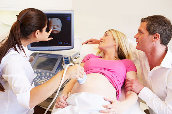
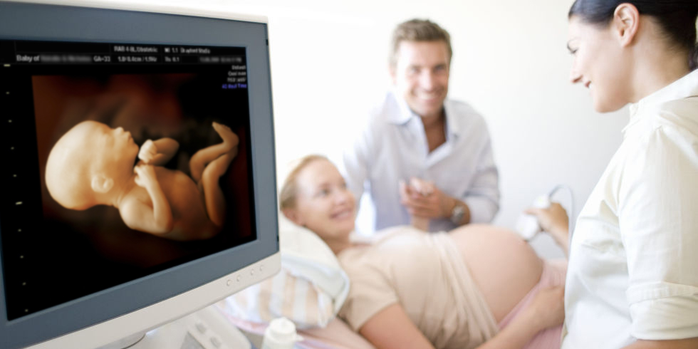
Most often, future moms go to 3D scanning voluntarily and voluntarily. Literally from the first days of pregnancy, a woman wants to see how the baby grows and develops inside her. In the middle and second half of pregnancy, they can not see who the child looks like. Indeed, the technique allows you to get very detailed and emotional pictures in which the baby shows itself in all its glory.
However, there is a three-dimensional scan and clear readings. Certain categories of pregnant women who are at risk receive directions for such an ultrasound, which is also called expert. These include:
women whose pregnancy is complicated by the threat of termination, Rh-conflict, diseases and defects of the reproductive system, with a burdened obstetric history;
women who are at high risk of having children with chromosomal abnormalities, developmental abnormalities;
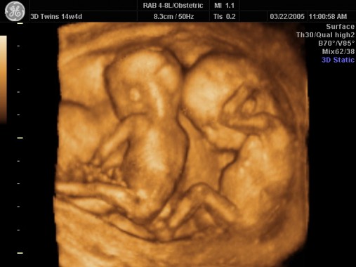
pregnant women bearing several fruits at the same time;
women who become pregnant through in vitro fertilization (IVF);
surrogacy cases;
women who have identified and confirmed fetal developmental pathologies — to choose tactics for assisting the child if the mother refused to terminate the pregnancy and decided to give birth to the baby;
future mothers who are diagnosed with a high risk of preterm birth, as well as women with rapid early aging of the placenta.
You should know that a three-dimensional ultrasound study, if it is given a referral by the attending physician for medical reasons, is free of charge and fully covered by the insurance policy.
If a woman decided to look at her baby from all sides out of sheer curiosity or wants to receive the first intrauterine pictures of a future son or daughter in a photo album, then she will do the examination in a paid clinic entirely at her own expense.
The cost of the procedure in different regions may vary. To this we can safely add the cost of a snapshot, video, copies to electronic media, which are offered for a fee. It turns out in general a very significant amount, it is not the most economical examination.

How long do you do?
In principle, a woman can make a three-dimensional ultrasound scan at any time, the main thing is that the fetus is already visualized, that is, has dimensions of at least 4 mm. However, according to established practice, such a study is usually prescribed after 20 weeks of pregnancy. There are three important periods for ultrasound diagnosis in the development of a baby. During these periods, by order of the Ministry of Health, mandatory examinations of pregnant women - prenatal screenings are carried out.
From 11 to 13 weeks (more often at 12 weeks), the best examination is considered the usual two-dimensional method.. At this time, a simple two-dimensional image is more than enough to find out how the fetus develops and grows, whether it has signs of possible chromosomal abnormalities, as well as other defects.
The second examination is carried out for the period from 16 to 21 weeks. Most pregnant women are also given a conventional two-dimensional ultrasound. During this period, women from the risk group listed above, as well as in some cases women who, for a number of reasons, have missed the first screening in general, can receive directions for 3D diagnostics.
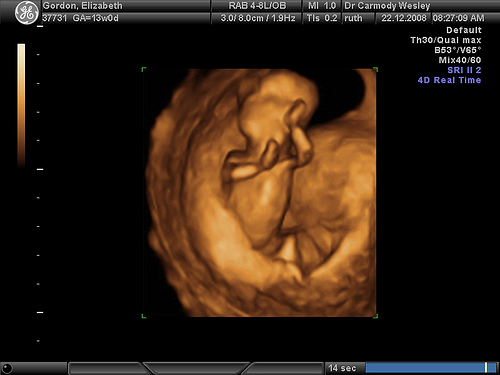
The most interesting results are obtained by a 3D survey during the third screening, which is conducted from 30 to 36 weeks of pregnancy. During this period, three-dimensional ultrasound shows a clearer picture of possible deviations in the development of the child.
If three-dimensional ultrasound is not due to medical necessity and there are no problems with the child and carrying, then 3D ultrasound is better to do in the period from 20 weeks, for example, from 24 to 28 weeks.
During this period, the sex of the child is easily determined, the three-dimensional images of the baby’s gender do not leave room for doubt. In addition, the fetus at this time is not yet so large as to be in a “crooked” pose, which is inconvenient for inspection by the sensor, as it happens in the last weeks of pregnancy.
This does not mean that a woman who applied to the three-dimensional ultrasound room for a period of 9 or 10 weeks will be denied diagnosis. It’s just that in paid medical centers, where a pregnant woman will go for it at an early term, they don’t like to tell potential clients that the method will show little interesting things in the first trimester, because each client is a contribution to the income of a private medical institution.
To save your money and not be disappointed, you should not go to this procedure before 20 weeks.
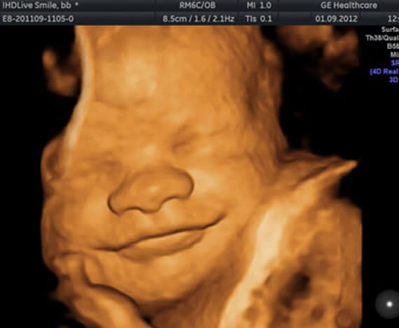
If you want "milots", then it is better to go to such an examination after 30 weeks, for example, at 32 or 34 weeks of pregnancy. At this time you can get great shots with smiles, yawns, funny grimaces, besides, it becomes clear what the kid looks like.
Advantages and disadvantages
3D ultrasound has important advantages over the procedure, which is carried out in a two-dimensional format:
The image obtained can be easily understood not only by professional doctors, but also by people unprepared for this, parents of a child, grandparents, family friends who are far from medical subtleties;
three-dimensional image allows you to consider those areas of the body that are not visible during two-dimensional diagnostics, respectively, the effectiveness of the assessment of development increases;
The pictures taken during the three-dimensional study have a pronounced psychotherapeutic effect - the expectant mother, having personally counted all the fingers on the hands and feet of her child, having seen his serene facial expression, calms down, takes it for granted that the kid feels great and stops “terrorizing »Doctors and relatives with their anxieties and doubts;
Two-dimensional ultrasound for the successful early diagnosis of heart defects and pathologies of the central nervous system requires the presence of a highly qualified specialist diagnostician. A young doctor with insufficient experience may not see some cardiac pathologies on a conventional ultrasound. A three-dimensional study gives a more detailed picture, the diagnosis of heart defects and the central nervous system becomes much more accurate;
3D research allows to diagnose with great accuracy external defects, for example, clefts of the facial bones, “cleft lip”, “cleft palate”;
The three-dimensional scanning procedure does not require an increase in the length and power of the ultrasonic rays, and therefore it cannot be argued that a volume examination is more harmful than a normal one.
Like any other diagnostic method, 3D ultrasound also has its drawbacks. Consider them in more detail:
Three-dimensional ultrasound gives less accurate results when examining such internal organs of the baby as the stomach, liver, gallbladder, intestines and lungs. Ordinary two-dimensional ultrasound, although it cannot boast such a clear and beautiful picture of the image of the child himself, gives a more detailed idea of how his internal organs are arranged;
- the cost of a three-dimensional survey is significantly higher than the cost of a conventional scan;
- A normal ultrasound scan does not take more than 5-7 minutes at the doctor’s and pregnant To obtain a three-dimensional image and carry out all diagnostic actions in 3D format, it will take at least 45-50 minutes.


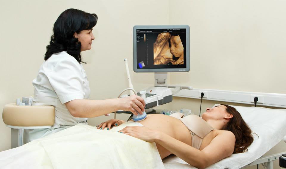
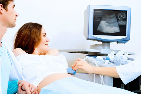
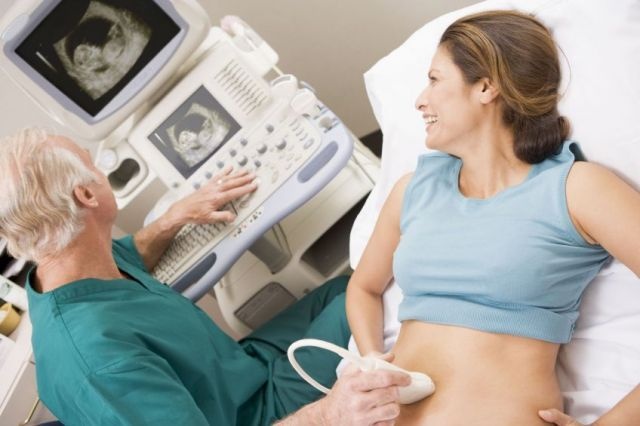
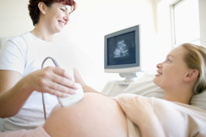
How is the examination?
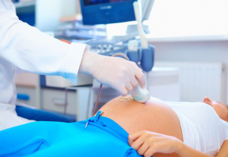
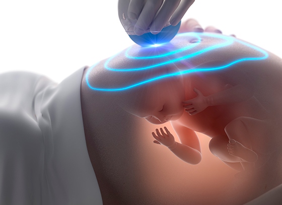
The procedure for passing a three-dimensional ultrasound examination is no different from a similar two-dimensional procedure. In the early stages, vaginal sensors are used, by means of which the vision is carried out through the vaginal wall. In later periods, doctors use a transabdominal method of research, in which the sensor is located on the belly of a pregnant woman, the review is carried out through the wall of the peritoneum.
Separate preparation for 3D ultrasound is not required. It should take into account all the same factors that affect the picture of the study and two-dimensional diagnosis. For a short time in a transabdominal examination it's worth taking care that the bladder is full. To do this, a couple of hours before the survey is to drink a few glasses of water or juice. When transvaginal examination filling the bladder is not required, on the contrary, it is better to release it before the procedure.
Ultrasonic waves are not very well reflected from the cavities filled with gases, in addition, intestinal loops overflowing with gases in the early period can compress the pelvic organs. Therefore, it is better to go on an ultrasound examination before the 12th week of pregnancy, after emptying the intestines and taking a dose of drugs that prevent flatulence, such as "Espumizana"Or" Simethicone. "

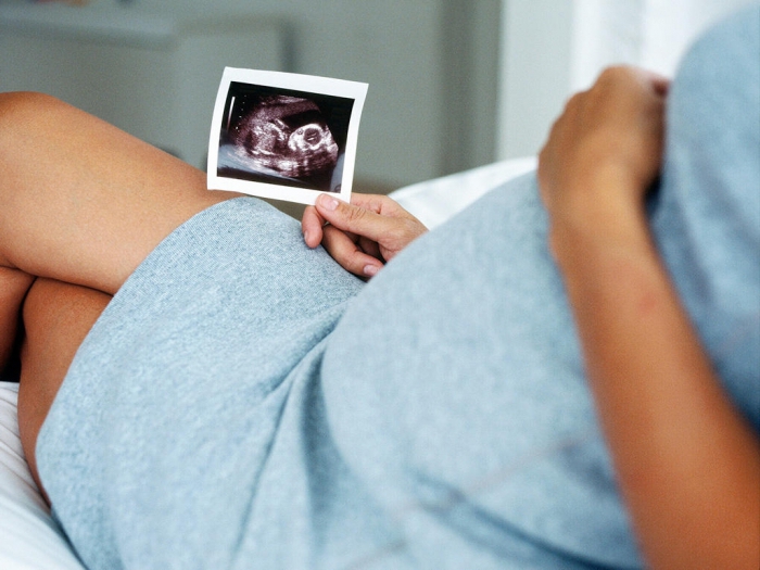
After the end of the examination in a volumetric format, the doctor always invites the woman to take pictures and video files home (for a fee, of course).
Method safety
Disputes about the safety of ultrasound in general and three-dimensional ultrasound in particular, do not subside. The fact is that science has left enough “gaps” for conjectures and hypotheses.The exact effect of ultrasonic waves on a fetus growing in the mother’s womb is not fully understood, and therefore it would be unfair to call it 100% safe.
However, many decades of using ultrasound diagnostics in obstetrics suggest that such use is quite successful, and the procedure is relatively safe. The allegations of any particular harm that a three-dimensional ultrasound can cause are not substantiated or substantiated.
Scientists from all over the world carried out entire campaigns for examining pregnant women - they studied the patterns between the frequency of ultrasound during pregnancy and the evils of their development. No direct connection was found. However, the study of the issue is considered insufficient due to the fact that now there is no way to evaluate how the effect of ultrasound on the growing tissues of a child’s body can affect the long term - in 10, 20, 30 years.
Those who oppose ultrasound in a three-dimensional format (and the usual two-dimensional study, by the way, too), cite such arguments as the uncertainty of the distant effect of ultrasound waves on humans, as well as the duration of the procedure. With three-dimensional diagnosis, it is almost 9 times larger, respectively, the fetus is exposed to radiation longer, which, according to some, is harmful and even dangerous.
It should be noted that it is impossible to consider a three-dimensional ultrasound as an independent method during screening. it is usually prescribed in case of problems or doubts in the quality of specifying diagnosticsbut because the opponents of such a survey emphasize that the baby, for screening, experiences the effects of ultrasound twice - on a planned routine ultrasound, and then on a long three-dimensional expert study.
It is not yet possible to confirm or deny the truth of these statements and hypotheses, science does not have enough convincing facts and proven results. Therefore, to do or not to do a three-dimensional ultrasound - it is up to the most pregnant.
3D or 4D?
The main difference between 3D and 4D ultrasound is that the four-dimensional image allows the doctor to see the crumbs from different angles in motion in real time. The picture on the monitor of the scanner turns out just amazing, clear and detailed, like a small film, which can not fail to impress future moms and dads. The principle of operation completely coincides with the principle of other types of ultrasound examinations, the procedure is carried out similarly, it has no features.
Most clinics that offer this service position it as 3D / 4D, this is due to the fact that the spectacular image in motion is of low diagnostic value, because to make measurements of individual parts of the baby’s body, you need a static picture, freeze-frame.

The cost of a four-dimensional study is much more expensive. The recommended timing of the survey coincides with the recommendations for three-dimensional ultrasound. 4D-diagnostics - a new method, but already beloved by many. After all, he most fully allows you to explore the appearance of an unborn child. This is what is sometimes necessary for pregnant women who are worried for any reason, worry about and are very worried about their baby.

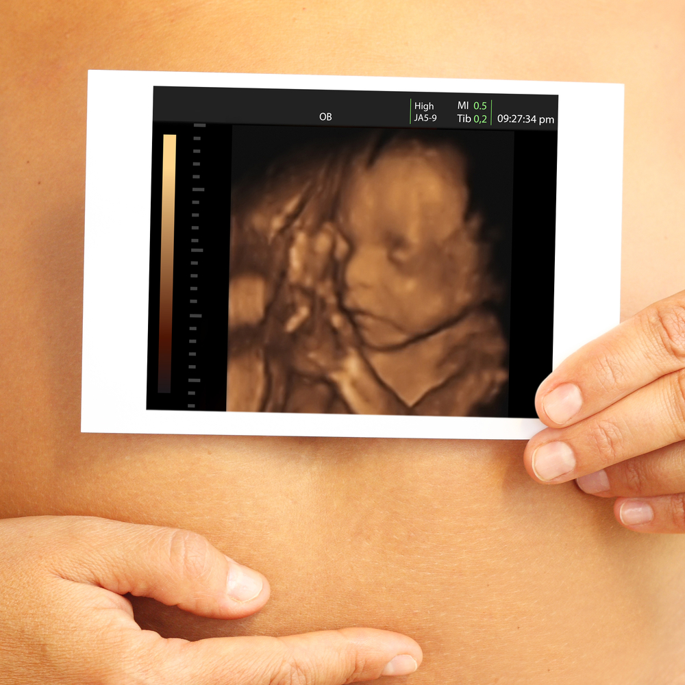
Snapshots
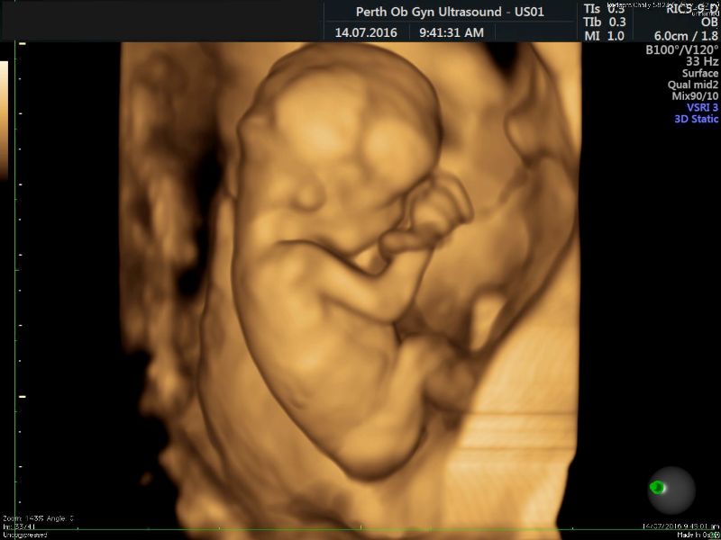
The sex of the child, captured in images obtained as a result of 3D diagnostics, is obvious to all. The boy is without a doubt a boy, and the girl is a girl, and to think about the error associated with hitting the umbilical cord between the legs or the genitals sandwiched between the legs, which most often lead to an incorrect interpretation of the floor on a two-dimensional examination, in this case is not necessary.
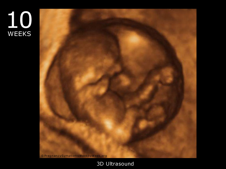
Pictures at different stages of pregnancy are different in terms of technical performance and emotional color. For example, in 10-11 weeks, it will be difficult enough to see the forming face, but a big head and hands will not leave future parents indifferent.
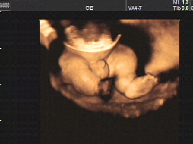
A three-dimensional image at 14-15 weeks is also not very clear, but it allows you to see the developed limbs, facial profile, ears.Of course, the child still looks thin now, but this is an absolute norm.

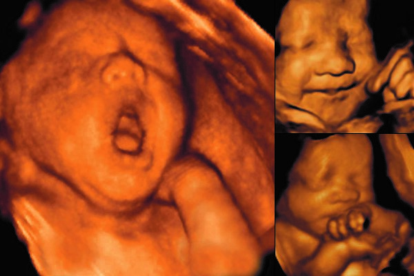
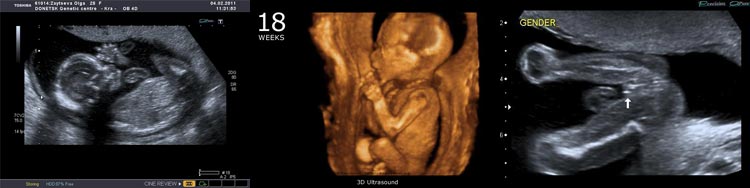
The pictures taken at the second screening at 18 weeks, on the usual ultrasound and in three-dimensional form are already quite different - a more three-dimensional image allows you to better imagine the baby growing inside, and also helps to determine the external signs of genetic pathologies.
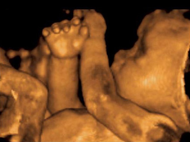
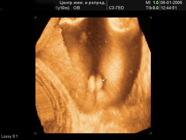
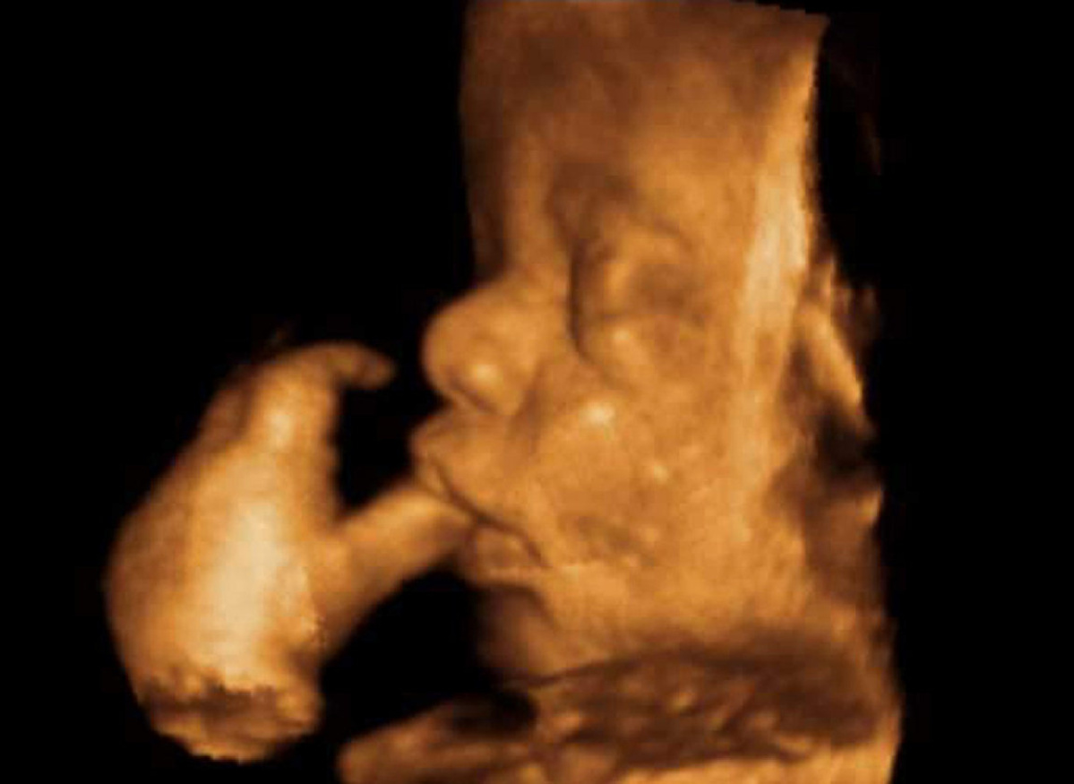
22 week on a three-dimensional ultrasound looks like. You can already not only see the floor and the structure of the baby’s body, but also his “activities” - sucking fingers, playing with the umbilical cord.

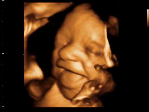
Emotions that will never be forgotten can be seen in 3D pictures already for a period of 27-28 weeks. The little ones are yawning and smiling, angry and frowning. Even more entertaining is the three-dimensional “photo session” for a period of 34 weeks. If you are lucky, parents will receive as a souvenir an extensive gallery of various faces and grimaces, funny and touching at the same time.
Reviews
Numerous reviews of future mothers who have undergone the procedure of 3D ultrasound, mostly positive. Women note that this is a real event and the mass of pleasant discoveries, because the baby ceases to be a flat dark image, as in a conventional device, it becomes realistic, almost as it appears before mom immediately after birth.
Among the shortcomings, some parents noted the concern of the child during the study. However, the reason for the child’s dissatisfaction most likely lies not in the reaction to ultrasonic waves, but in the fact that the mother has to stay in one position for quite a long time (about 45 minutes), lie on her side or back.

Other disadvantages noted by women are the high cost of the examination, as well as the insufficiently high quality of the images, if the procedure was carried out on equipment of the not very high class.
You can find out more information about 3 D and 4 D ultrasound scan from the following video.


