Ultrasound in the 5th week of pregnancy: fetal size and other features
The first trimester of fetal development is a very important period of intrauterine development. At this time, the future baby, who is already actively living in the womb, is beginning to quickly develop internal organs and systems. This article tells about the purpose for and how the fetal ultrasound is performed at 5 weeks of gestation.
What is it for?
Conducting ultrasound in the early stages of fetal development is used mainly to establish pregnancy. To calculate the period of prenatal development of the child, gynecologists use the definition of the obstetric period. It is usually less than 14 days less than the gestational period of pregnancy.
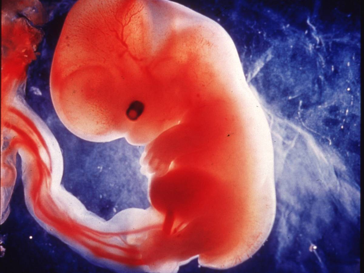
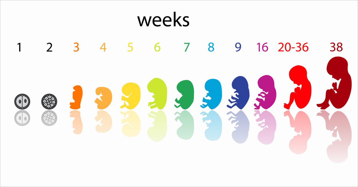
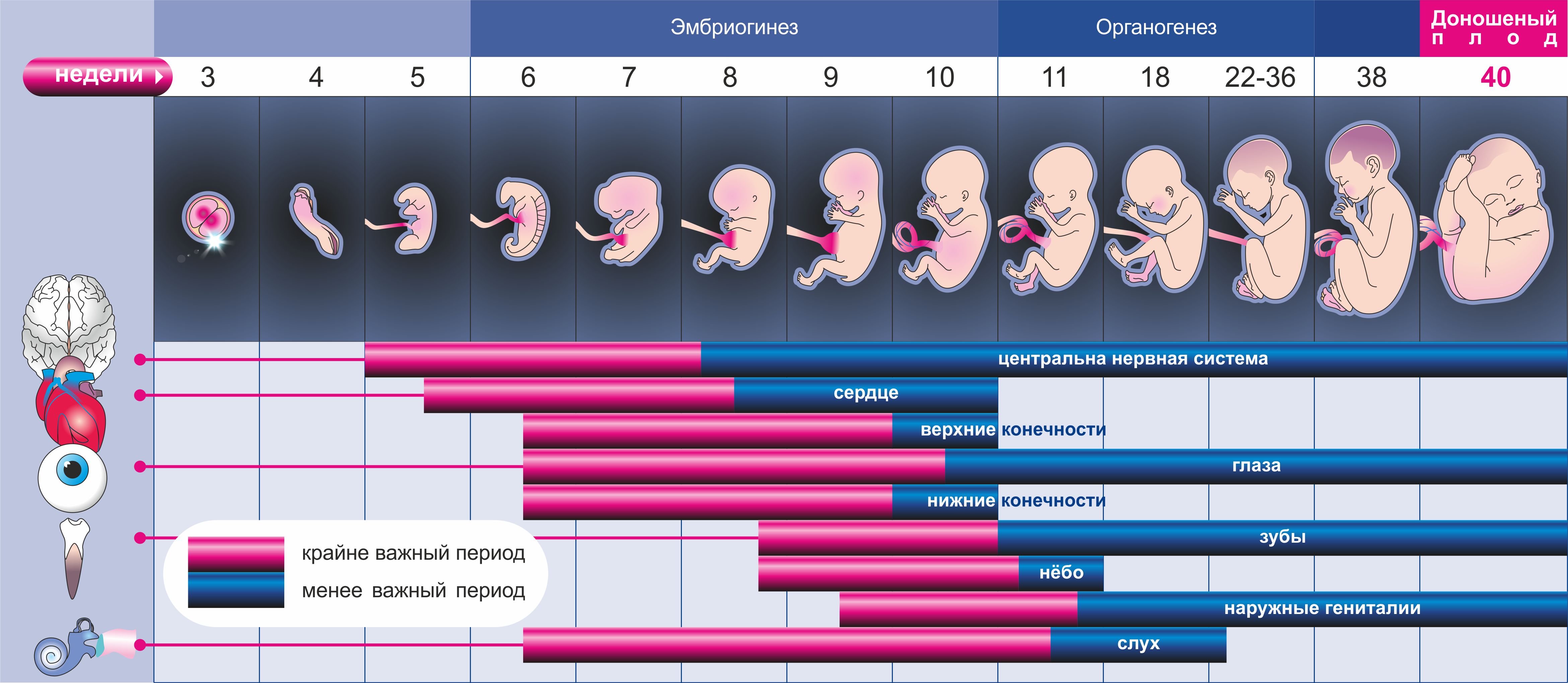
4-5 weeks of intrauterine development is a very important period for the embryo. At this time, the fetus begins the most important process - organogenesis. This period is characterized by the differentiation and development of many internal organs and systems of his body.
In most situations, ultrasound is not performed at this early gestational age. The exceptions are cases when a pregnant woman independently insists on carrying out this method.
Such a limitation of the performance of ultrasound in the early stages of gestation is very important. The fact is that ultrasound, especially used in the focal mode, in the early stages of pregnancy can harm the unborn baby.
Doctors-gynecologists allocate certain medical indications. If they are available, they can send the expectant mother to the ultrasound room to perform such a study.

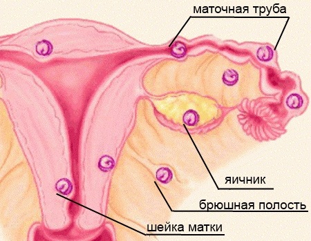
An ultrasound scan at such an early date is required, as a rule, in the event that when doctors want to establish the exact location of the ovum. This situation occurs, as a rule, with the exception of ectopic pregnancy. This pathological condition is usually very clearly visible on ultrasound.
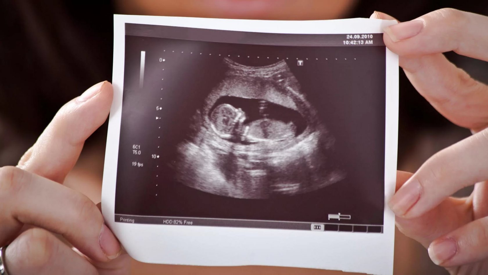
If, despite the clinical examination of the woman and the laboratory tests performed by her, the doctors find it difficult to establish a pregnancy, then ultrasound also comes to their aid. During such a study, you can also take the first shot of the embryo. As a rule, it is already possible to determine the size of the embryo well.
Some pathological conditions that may occur in the very first period of pregnancy can also be detected using ultrasound. Usually, future mothers are sent for such a study if they suspect a spontaneous abortion or spontaneous miscarriage.
If before the pregnancy, the woman suffered from any pathology of the genital organs, then she could also be referred to an ultrasound of the pelvic organs. Such studies help to establish the degree of existing reproductive disorders, as well as eliminate exacerbations of chronic diseases of the genitourinary system in the future mother.
Many future moms are interested in whether to undergo ultrasound in such early terms. Most doctors in this case will say that without a strong need or in the absence of medical indications for ultrasound in the early period of fetal development to perform such an examination not worth it.
Currently, there are other methods to establish pregnancy.

What will the research show?
Ultrasound, performed in this period of development of the future baby, not yet highly accurate. This is not at all due to the conduct of research on poor-quality equipment or the lack of clinical experience of the specialist conducting this test. As a rule, in the early period of embryo development, most clinical indicators of doctors simply can not interpret.
During the procedure, a specialist ultrasound examines only the basic parameters of fetal intrauterine development. It should be noted that there are far fewer such values in the early life of the embryo than during the third trimester of pregnancy.
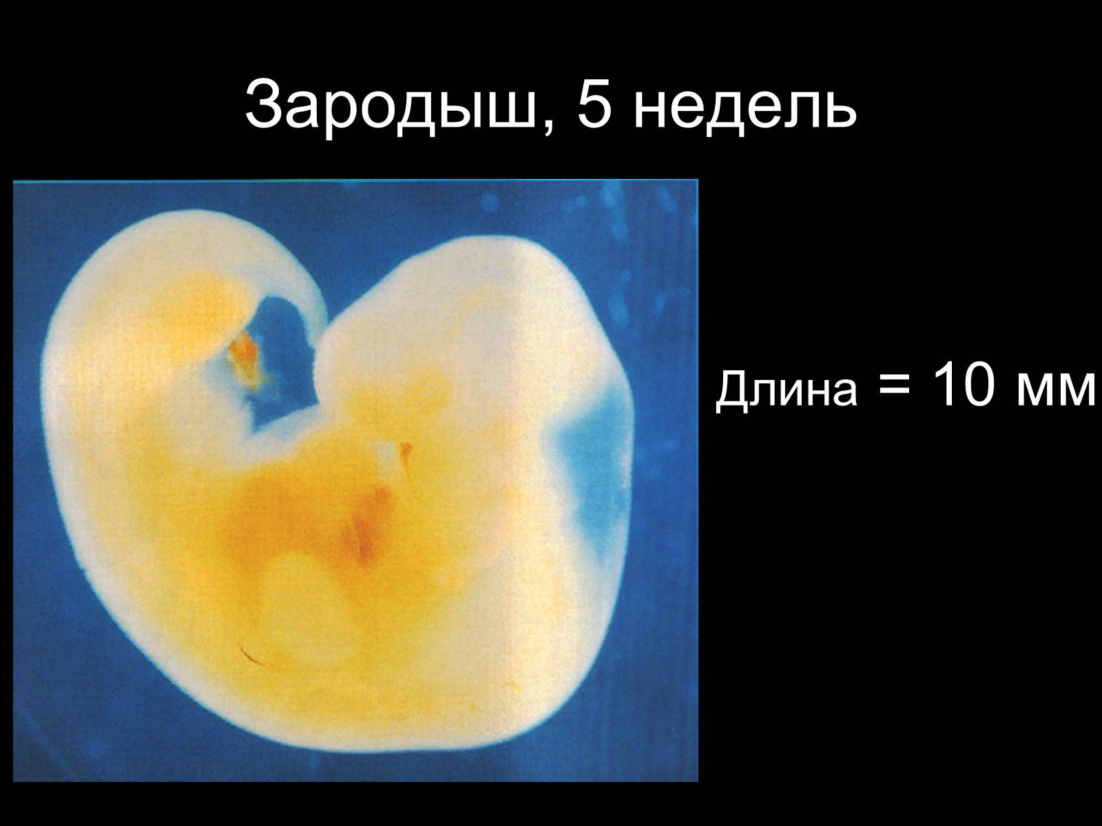
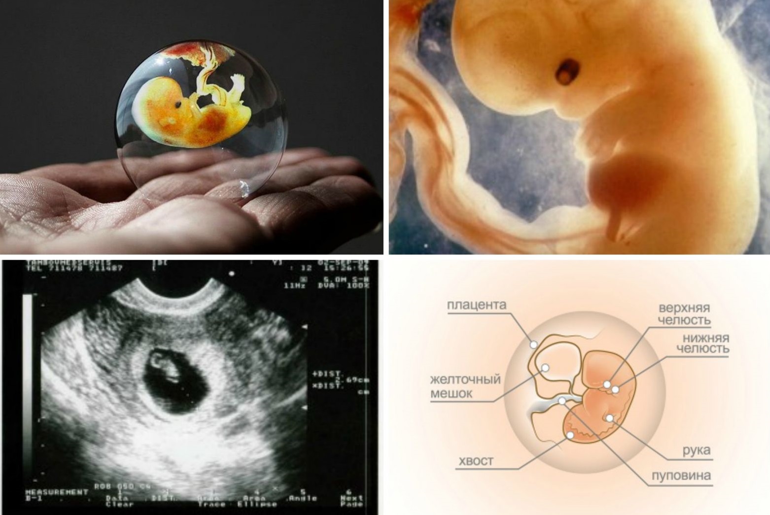
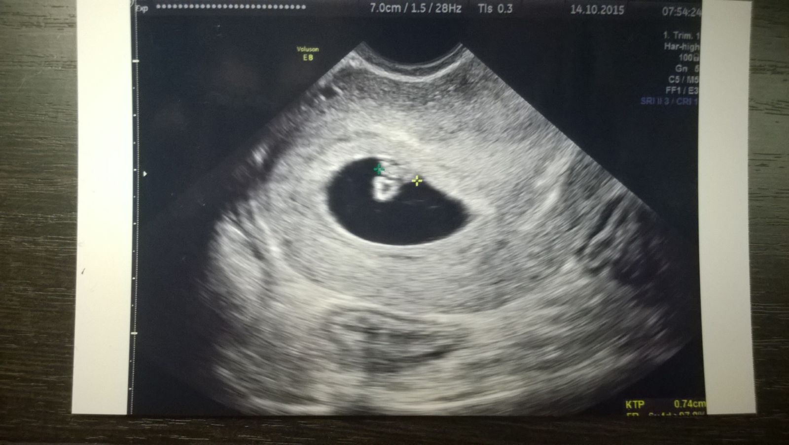
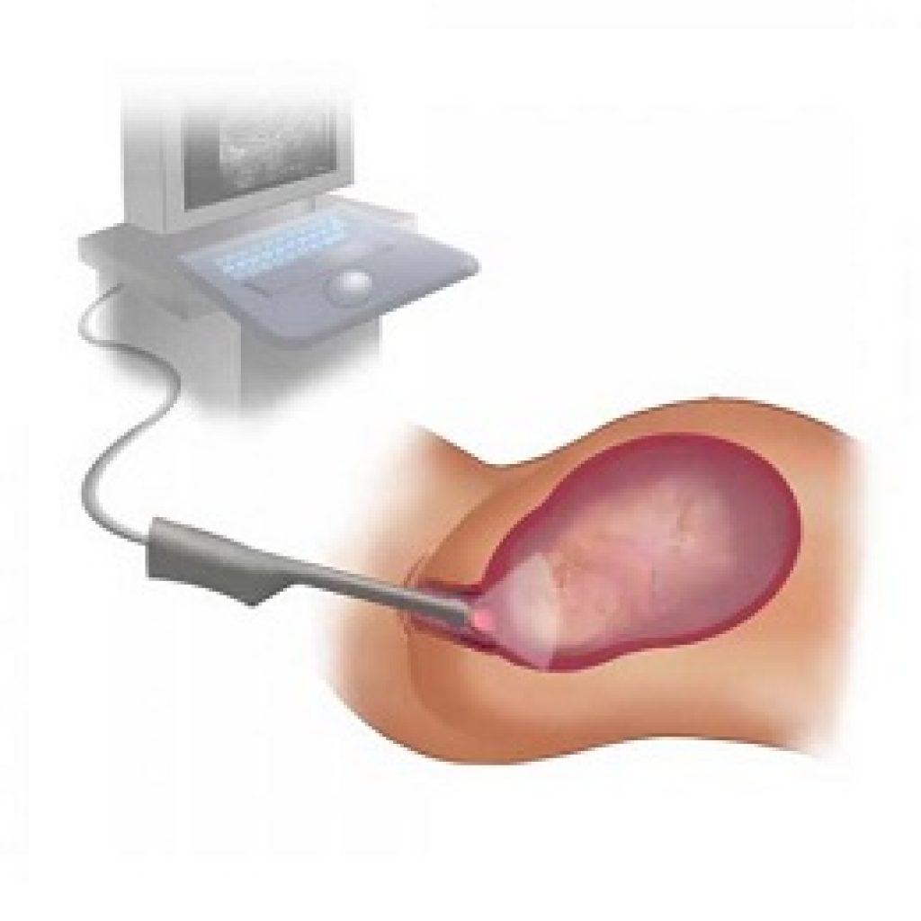
In each period of prenatal development of the future child, doctors call him various medical terms. At week 5, it is called an embryo. The size of such an embryo, as a rule, does not exceed 3 mm. Such a crumb can not "consider" on ultrasound.
Doctors do not detect the embryo, but the presence of the ovum. This formation is like a "protective shell" in which the future baby develops. In the future, various fetal membranes will form from the shells of the gestational egg, which will also protect the fetus from various adverse factors.

By the end of week 5, two very important anatomic formations of pregnancy begin to form. They are called amnion and yolk sac. The embryo is located in the amniotic vesicle. This formation is also well visualized using an ultrasound transducer.
At its core, the yolk sac is the fundamental organ responsible further for the development and formation of all elements of the cardiovascular system in a future baby. From it, as the embryo develops, a heart will form. Baby's heartbeat will appear a little later.
Little by little, small blood vessels begin to form in the microscopic villi of the chorion. During pregnancy, they will develop and grow in size. This type of blood supply is very important for the embryo developing in the womb.
The villi of the chorion of the fetus are in direct contact with the maternal blood vessels. Through them, the baby receives nutrients for its growth and development, as well as oxygen dissolved in the blood.
Some experienced specialists already at week 5 can discern many anatomical structures of the embryo. Usually, this also requires not only sufficient experience of the ultrasound diagnostic doctor, but also the use of modern high-precision devices.
Focusing the ultrasound sensor allows the examiner to quite clearly distinguish the egg of the egg itself. It looks like an oblong formation that attaches to the endometrium - the inner epithelial lining of the uterus.
The internal element of this formation is the amnion. The doctors also call this education the camera, which is located inside the amniotic vesicle. It contains a liquid component.
The outer shell is also quite well visualized using an ultrasonic sensor. It surrounds the amnion from the outside and is, in fact, the fetal membranes of the embryo.
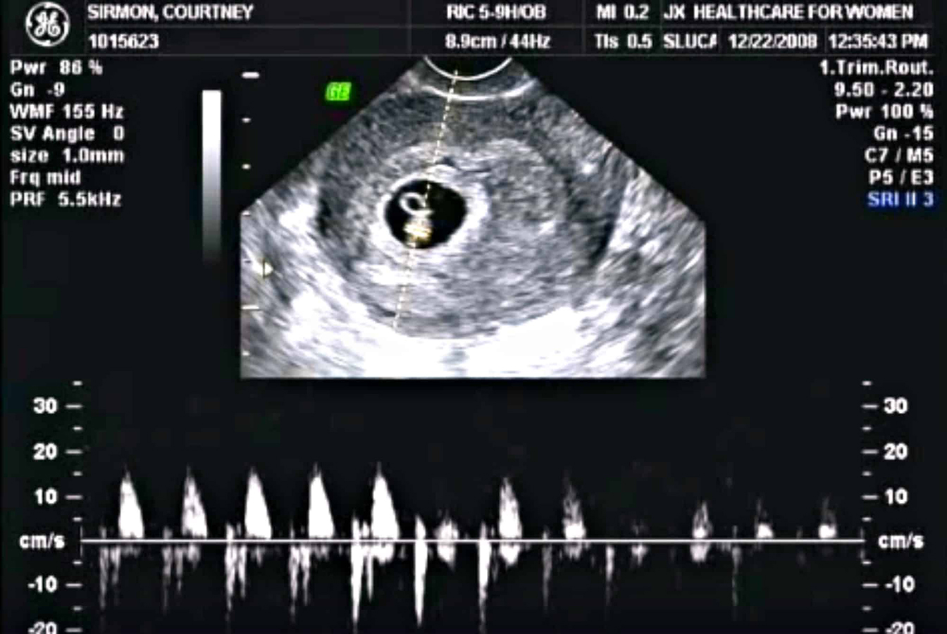
A fruit or gestational egg in this case looks like an echo negative element filled with liquid from the inside. On the posterior surface of such anatomical formation there is dorsal amplification of ultrasound. The shape of a gestational egg can be either oval or round.
On closer inspection, an echopositive bezel is determined around this formation.
The norms of the main parameters studied
Doctors conducting early examination of the fetus use various obstetric concepts and terms.Usually they are used throughout the period of carrying a baby.
The size of the gestational egg at 5 weeks of pregnancy in most cases is ½ cm To measure this indicator, doctors use special medical criteria. The normal diameter of a gestational egg is determined only with one measurement.
There is another, more accurate criterion. It is called the average diameter. To measure it, an ultrasound specialist should conduct at least three measurements carried out in different planes. This parameter in combination with others may become a criterion for assessing the first weeks of fetal development of the embryo.
There are situations when the size of the ovum obtained during the study is substantially less than ½ cm. In this case, doctors usually recommend repeating the examination after 6-8 days. Sometimes this indicates "Frozen" or "suspended" pregnancy. To exclude this pathological condition and repeated research is carried out.
During an ultrasound examination in early pregnancy, it is not only fetal development that is evaluated. The condition of the internal genital organs of the future mother is also very important for making a prediction of the childbearing.
Ultrasound helps to detect various neoplasms of the ovaries, cysts, as well as fibroids. Quite often, these pathologies lead to miscarriage or spontaneous abortion at the earliest time.
Methodology
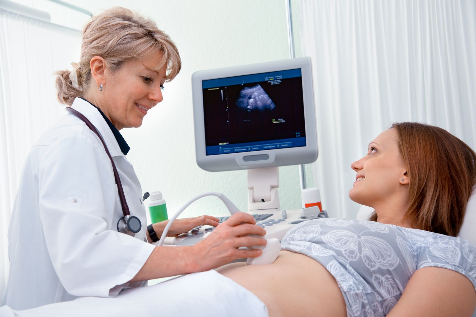
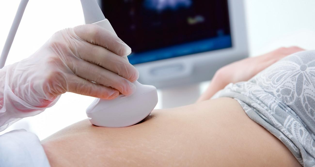
Ultrasound in early pregnancy can be performed at once in several ways. The choice of methodology depends on many original reasons. Typically, the method of conducting such a survey is determined by the gynecologist who observes the woman.
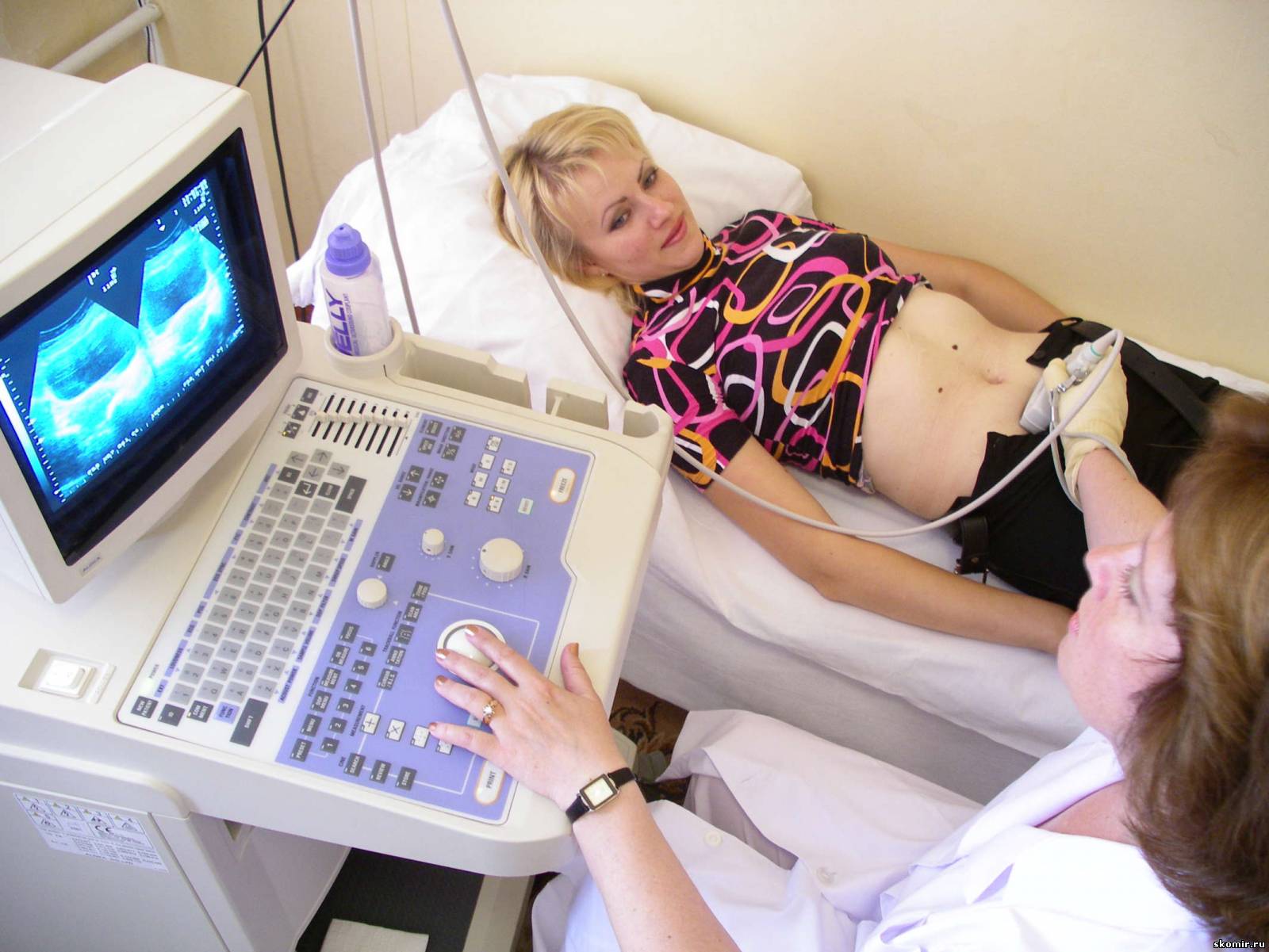
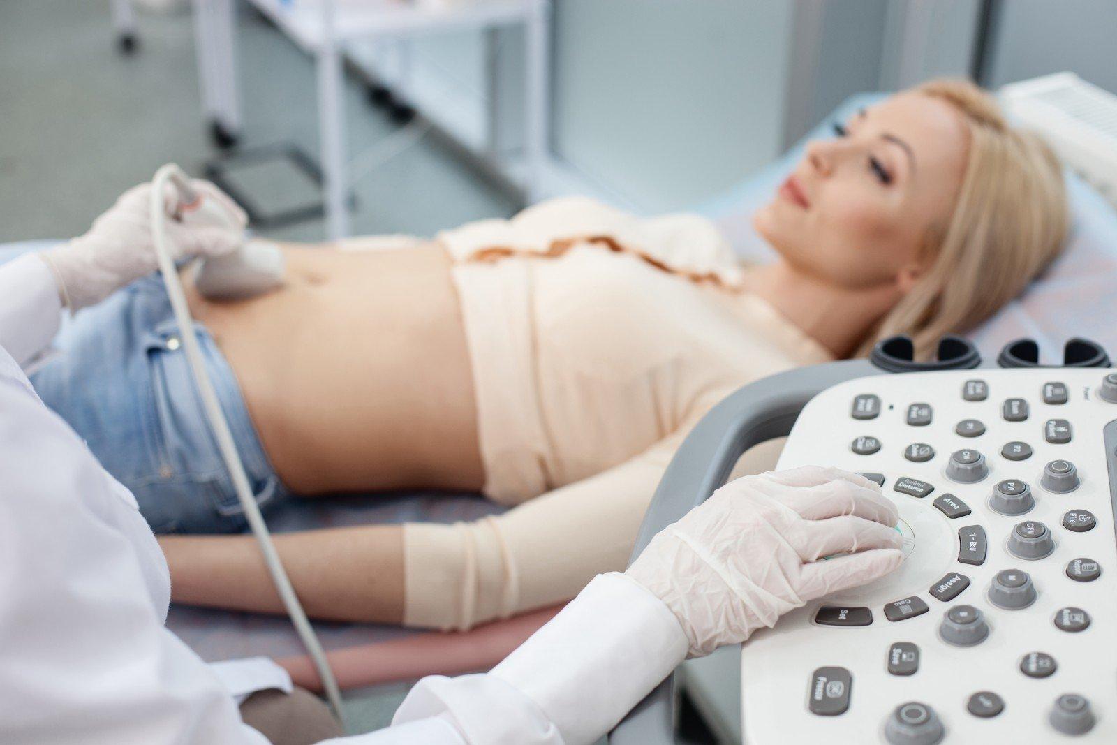
You can examine the pelvic organs transabdominal method (through the anterior abdominal wall), as well as transvaginally (through the vagina). In most cases, obstetrician-gynecologists prefer to use transvaginal method of research, as it allows to obtain more accurate results at this stage of pregnancy.
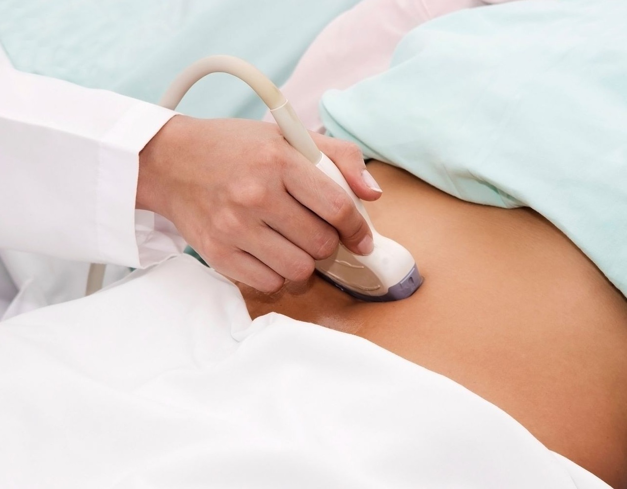
If the patient has signs of exacerbation of chronic obesity or vaginitis, accompanied by various vaginal secretions, then doctors may recommend that she conduct a study in a transabdominal manner. In this case, no pain or discomfort during the examination will not occur.
In order to get a bright picture on the monitor of the device, the doctor conducting the study uses special gel. It is applied directly to the skin. This gel is very hypoallergenic in composition. It can be safely used even in younger children, as well as in pregnant women without the risk of developing allergies or other unpleasant consequences.

Many women who are sent for an ultrasound scan at the 5th week of pregnancy are interested in whether certain training is required before the study. I must say that this is quite an early date. In this case, no special training is required. However, there are some exceptions.
In some situations, the doctor may ask a pregnant woman to come for an ultrasound scan. with a full bladder. This is required in certain cases in order to better view the inner wall of the uterus and the ovum. The filled bladder presses against the uterus, making it more accessible for visualization.
Absolutely all expectant mothers do not need such preparation.
A couple of days before the study, doctors recommend that women do not eat foods that can cause severe gas formation. Legumes, various types of cabbage, carbonated drinks, fruits and vegetables 2 days before the ultrasound scan should still be limited. This is especially necessary for women who have an increased tendency to gas formation on the background of their associated diseases of the gastrointestinal tract.
During the research procedure itself, the woman is on the couch. Starting position - on the back. During the study, the doctor may ask the pregnant woman to turn to the left or right side. This is usually necessary if the future mom has any pathological changes or abnormalities in the development of internal organs.
If the test is performed transvaginally, then the ultrasound probe is inserted into the vagina. During the study, the doctor constantly monitors the general condition of the patient. If during the procedure she developed some kind of severe pain, the examination can even be stopped. In this case, transabdominal ultrasound is performed.
If a pregnant woman has twins or identical twins, the ultrasound scan will have a number of features. In this case, one or more fetal eggs are determined. Of particular importance is how the conception took place - by a physiological route or by IVF.
If the pregnancy is due to in vitro fertilization, then the day of embryo transfer to the uterus is also taken into account. This is necessary to establish the obstetric period of pregnancy.

After the examination procedure has been completed, a special medical form with a conclusion is given to the patient. In it, the doctor who conducted the ultrasound, indicates all identified deviations, as well as fixes the basic dimensions and diameters of the ovum and its anatomical components.
Moms-to-be should definitely remember that The conclusion of an ultrasound examination is not yet a diagnosis. With this form, a pregnant woman goes to a gynecologist. The doctor summarizes all the results and only after that can establish any diagnoses. In some cases, it may be necessary to conduct a re-examination if the doctor doubts the accuracy of the examination.


