Ultrasound on the 7th week of pregnancy: fetal size and other features

Mom's mood at 7 weeks gestation changes, like the weather by the sea. Attacks of fun on the background of hormonal adjustment can be replaced by periods of increased anxiety and even panic over the health of the future baby. Dispel doubts and calm the woman will help safe for both ultrasound procedure. In what cases such a diagnosis is required at this time, how is it carried out and what can be seen on the ultrasound during this period, we will tell in this material.

Objectives of the survey
At 7th obstetric week, an ultrasound scan is not considered mandatory. If a woman feels good, then there is no need for it. However, during this period of time, many women think about registration in the antenatal clinic. From the moment of conception is 5 weeks, from the first day of the delay has passed 3 weeks. There are practically no doubts in the “interesting situation”, and the onset of toxicosis leaves no room for speculation. It's time to see a doctor, get registered and start to undergo the first tests.

Among them there may well be a recommendation to visit the ultrasound room. A doctor may send a pregnant woman to this examination at such an early date for a number of very good reasons:
confirmation of the fact of pregnancy is required if the size of the uterus during a manual examination does not indicate this;
there is reason to believe that multiple pregnancy;
conception occurred through IVF;
increased risk of adverse outcome - previously, women had miscarriages, frozen, ectopic pregnancies, prenatal fetal death in the early stages;
A woman complains of severe toxemia, pain in the lumbar region and lower abdomen, as well as secretions uncharacteristic for pregnancy.

The seventh-week ultrasound is sometimes used to find out the exact dates of gestation.
Such measures may be necessary if the woman’s cycle is irregular; she does not remember the date of the last menstruation. Finding this out now is important in order to know exactly when to direct the future mother to the first screening. It runs from 11th to 13th week, and even one extra or missing week can affect its accuracy.
Do I need training?
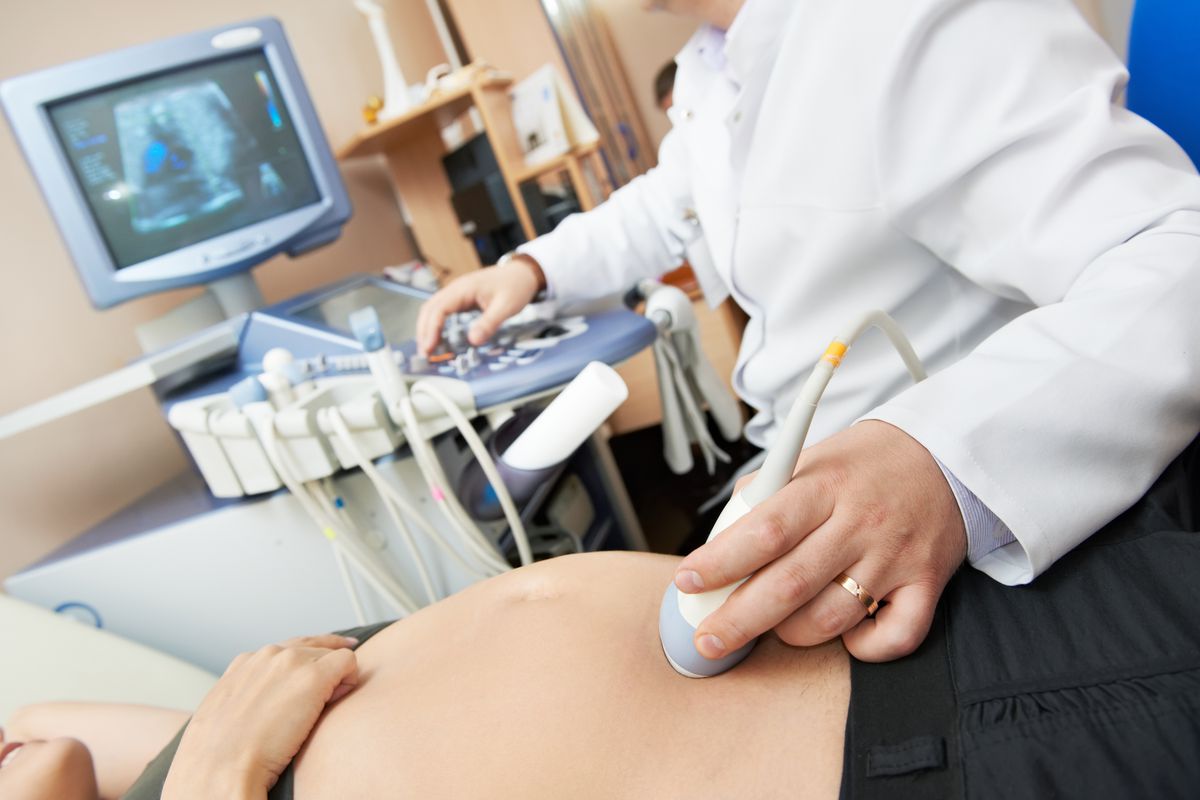
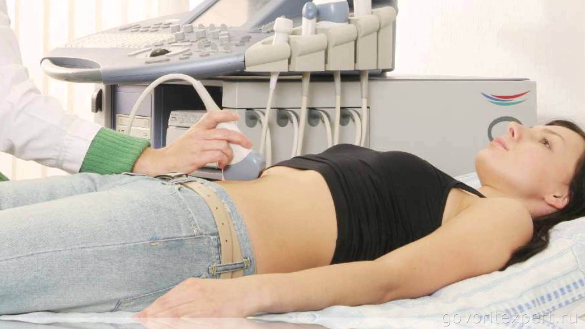
Examination at 6-7 weeks of pregnancy is most often carried out by the intravaginal method. The term is still too small for the child to be clearly seen through the anterior abdominal wall. But through the vaginal wall the embryo is perfectly visible and you can find out all the questions of interest to the doctor concerning this pregnancy.
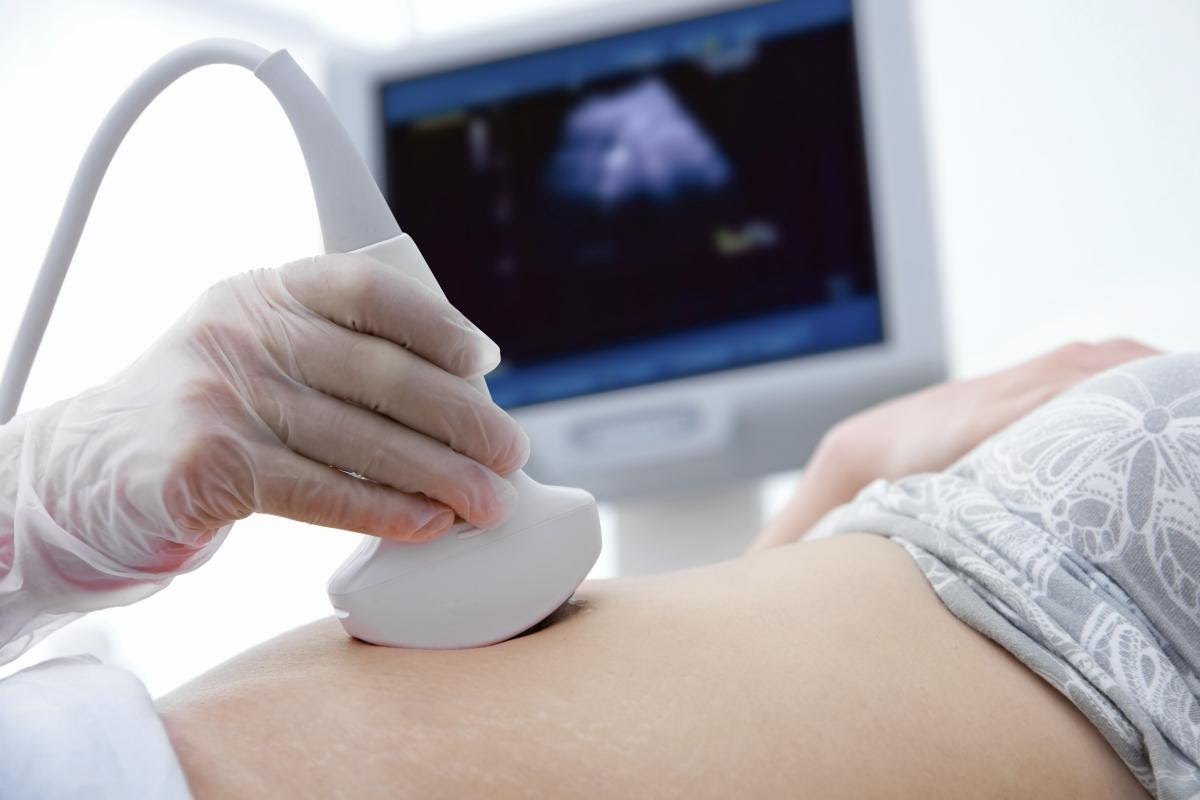
Transabdominal method (through the abdomen) ultrasound at this time is also carried out, but only in exceptional cases. With this method, you can consider the baby in a woman of thin build, but for the owner of extra pounds and curvaceous forms, visualization will be insufficient.
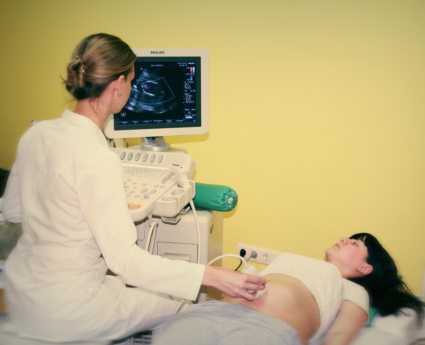
Preparation for ultrasound on such a short term of pregnancy includes bladder fillingif the examination is performed externally through the anterior abdominal wall.A few hours before visiting the doctor’s office, the expectant mother should drink tea or juice, mineral water without gas. It will be enough 2-3 cups, so that by the time the diagnosis starts, the bladder is sufficiently filled.
You do not need to drink fluids before performing a transvaginal ultrasound scan. On the contrary, if you want to go to the toilet, it is better to go so that both the bladder and the intestine are freed from the contents and do not interfere with the inspection. Intestinal gases can also distort the results if they accumulate in large quantities.
Increased flatulence - not uncommon during the carrying of the child at any time, and therefore it is desirable for several days there are no products that can contribute to flatulence. Such products include cabbage and yeast pastries, legumes, carbonated drinks, sweets, as well as high-fat dairy products.


Swollen intestinal loops from the gas can squeeze the pelvic organs. To avoid this, the survey results were more reliable, a few hours before the ultrasound is recommended to take a dose of "Simethicone" or "Espumizana».
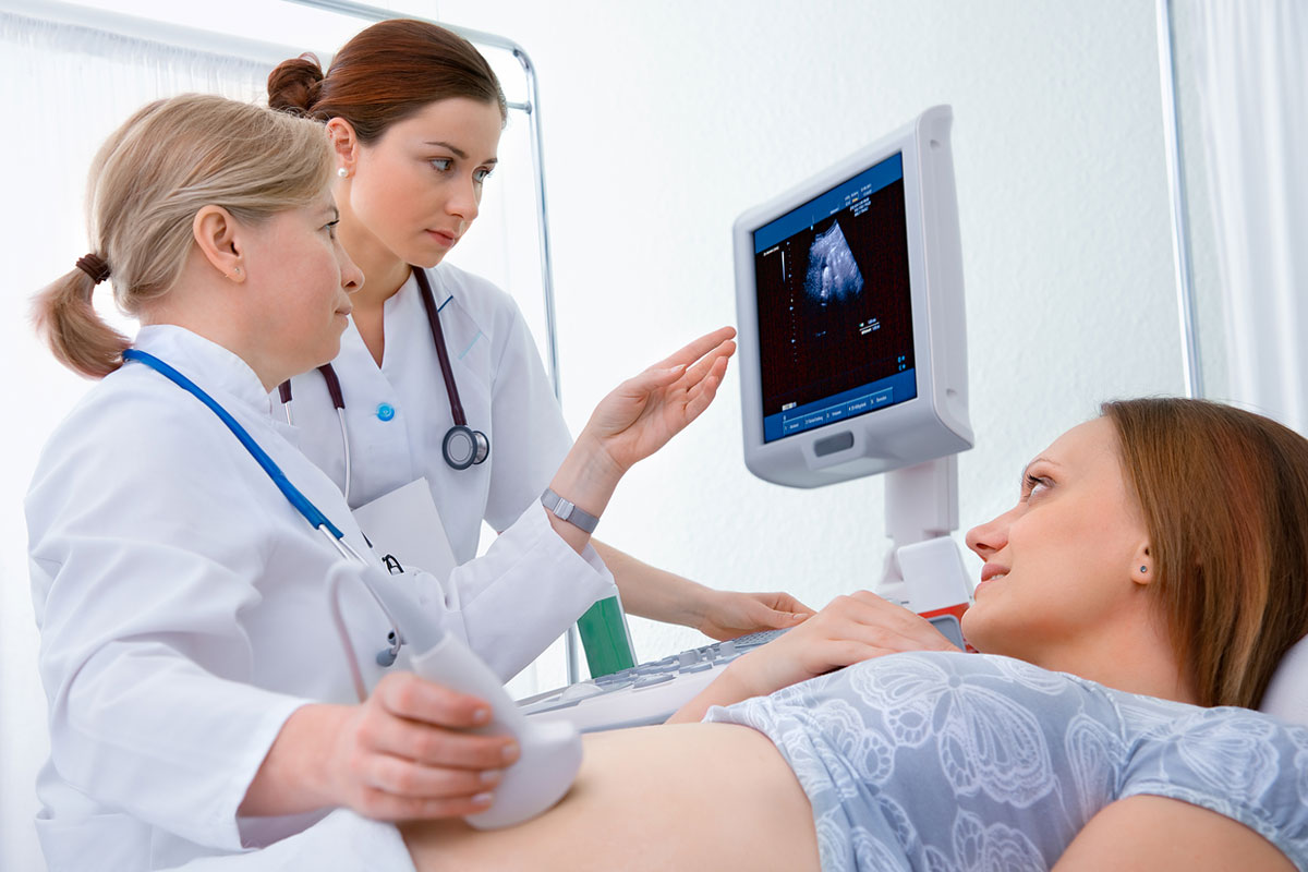
The survey takes about 5-7 minutes. Everything goes smoothly. The results are handed out immediately.
What shows?
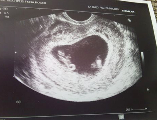
In the seventh week, the baby is officially still considered an embryo, although only a few days remain until it becomes a fetus. A week earlier, it was difficult to examine anything except for dark spots and spots on the ultrasound. Now the embryo has grown approximately twice and is already well visualized on the screen of the scanning equipment. The size of the fruit is now from 2.5 to 5 mm. Its weight is close to 1 gram. To imagine the true parameters of the baby, it is enough to imagine a white bean. This will be the size of your child with shells.
On ultrasound, the ovum with the embryo inside looks like an oval spot. However, the rich life boils inside. The kid began to unbend this week from the posture of the embryo, and also learned to move. Brushes appear on the handles at week 7, and feet form on the legs. Instead of fingers, the rudiments are still flaunting.
The embryonic tail, which was a logical continuation of the line that will become the spinal column, gradually decreases. In large compared with the rest of the body, the head begins to form the brain, both hemispheres. The formation of a spout begins on the face.
The heart of the baby is already four-chamber, just like Mom and Dad. The formation of the bronchi began.
It is clear that to see all these interesting and numerous processes on ultrasound will not work. Also It makes no sense to ask the doctor a question about the sex of the child. Gender, although predetermined from the very moment of conception, cannot yet be considered - there are no external genital organs. The formation of the genital tubercle began between the legs, from which the main sex differences subsequently formed, but for now the female embryo is absolutely no different from the male embryo.
On ultrasound in the seventh week you can see the place of attachment of the ovum, and if the device is good, you can listen heartbeat baby If the equipment in the ultrasound study leaves much to be desired, then the doctor will still be able to determine the pulsation in the heart area, and he will note that the fetus is alive.
Rules and interpretation of results
The number of measurements performed by the diagnostic ultrasound scanner is as small as the baby itself. Therefore, a huge number of numbers and abbreviations in custody after passing the diagnosis, as in later terms, will not. Only the most basic parameters, which so far are the only and decisive.
First of all, the doctor describes how many fetal eggs are located in the uterus and exactly where they are entrenched. If a woman bears twins, then this week it is already clearly seen whether they are identical twins or not.
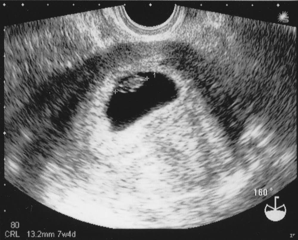
With respect to the embryo, the viability is determined - the heartbeat is recorded, and if lucky, the motor activity.The development of the baby indicates the size of the ovum. It is measured between the inner walls, and therefore it is called the SVD - the average inner diameter. Normal values of the ovum increase rapidly.
Svd
The results are decoded in accordance with the table:
The diameter of the ovum (SVD, mm) | Matching gestational age |
12 | 6 weeks + 1 day |
13 | 6 weeks + 2 days |
14 | 6 weeks + 3 days |
15 | 6 weeks + 4 days |
16-17 | 6 weeks + 5 days |
17-18 | 6 weeks + 6 days |
18-19 | 7 weeks exactly |
The doctor is not limited to measuring SVD, he also evaluates the contours of the ovum. In normal pregnancy, it has the correct oval or rounded shape, clear and smooth contours, does not look squeezed and deformed.
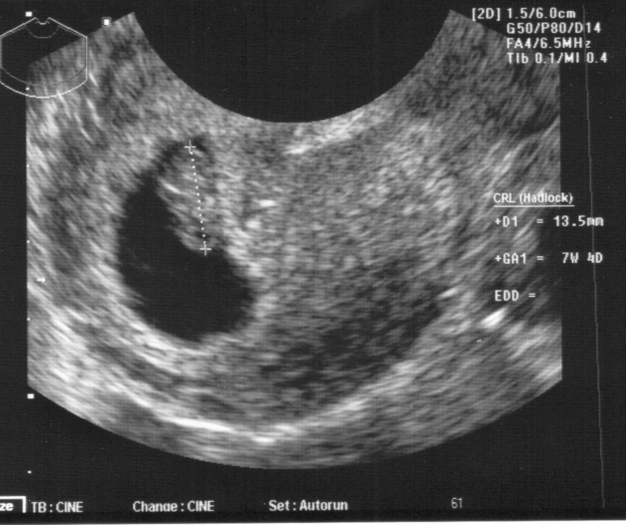
From this week, another parameter appears, which from now on will be more informative in terms of adjusting the exact timing of pregnancy. it coccyx parietal size - distance from coccyx to crown, CTE. Now that the baby has learned to unbend, this parameter can be measured on an ultrasound machine of any level.
KTR
The results are decoded in accordance with the table:
KTR, mm | Matching Date |
6,5-7 | 6 weeks + 2 days |
7 | 6 weeks + 3 days |
8 | 6 weeks + 4 days |
8,5 | 6 weeks + 5 days |
9 | 6 weeks + 6 days |
10 | 7 weeks exactly |
The yolk sac, which provides the embryo with food, in the seventh week has dimensions not exceeding 4 mm. The heart of the baby is gaining momentum and the heart rate (heart rate) this week is in the range from 126 to 149 beats per minute.
In addition, the doctor examines the uterus, fallopian tubes, ovaries for pathologies and signs of threatened abortion.

Possible problems
As a result of an ultrasound scan at the 7th week of pregnancy, some problems can be found that cannot be said about. The most common ones.
Anembrionia
This problem is the second name - the empty egg syndrome. When anembryonic fertile egg is present in the uterus, but there is no embryo in it. This happens not so rarely - about every fifth pregnant woman, according to statistics, may have an embryo. The reasons for science are not known for certain, but doctors suspect that the death of the embryo at the very initial stage is caused by severe genetic “mistakes”, past infections, sudden jumps in the hormonal balance, unhealthy lifestyle of a woman. Incidentally, it is unbelievable, but the fact is that professional athletes have an embryonia quite often.
Doctors say that blame serious physical exertion. It is at week 7 that diagnosticians usually give the final verdict - is there a baby in the womb or is it not. Unfortunately, medicine in this situation is powerless. Fetal egg to be scraped.

Fading Pregnancy
About such an equally sad result of ultrasound a woman can not even guess. On her health, the death of the baby can not be reflected in any way. At the study, the doctor notes that the size of the ovum is far behind the norm, the fetal egg is deformed, no heartbeat and signs of movement of the baby.
If after a few days on the second ultrasound the result is confirmed, and the drop in the level of the hormone HCG in the blood additionally “signals” about the death of the baby, then curettage of the uterus cavity is performed. Embryonic tissues and particles of fetal membranes are sent to the genetic laboratory to find the true cause of the incident.

Ectopic pregnancy
The fact that the embryo entrenched in the fallopian tube or in the cavity behind the uterus can be said if at the 7th week the ultrasound doctor does not find in the uterus of the ovum, although the level of hCG in a woman’s blood is high enough to judge that pregnancy not only available, but also evolving. This is a very dangerous condition for a woman. requiring emergency surgery to save a woman’s life.

Risk of miscarriage
The thickening of the uterine walls in the seventh week, according to the results of ultrasound, may indicate the existing threat of termination of pregnancy, the presence of hypertonia.In this case, the woman is assigned appropriate supportive treatment, which she can receive at home or in the hospital - this will be decided by the doctor.
Retrochorial hematoma
On the ultrasound, it is visible as a darkened spot. Essentially, this detachment of the ovum. The size of the hematoma determines the prognosis. In most cases, the pregnancy can be maintained, if the woman strictly follows all the recommendations of the doctor.
Among the main reasons are considered increased physical exertion, stress, lack of the hormone progesterone, infectious diseases that a mother could suffer in the earliest periods after conception.

Snapshots
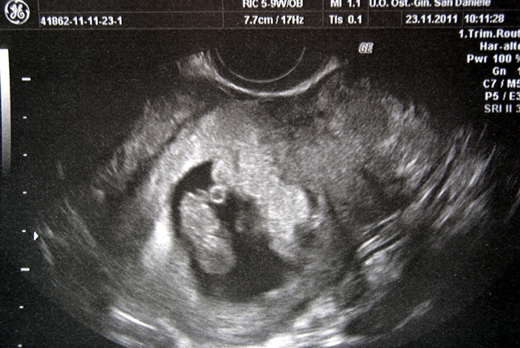
In the seventh week of pregnancy there is no need for a three-dimensional ultrasound. The image is still quite difficult to understand and decipher to an unprepared “viewer” like the future father. If you want to have in the family album the first “photo” of the crumble- “bean”, then it will look something like this.

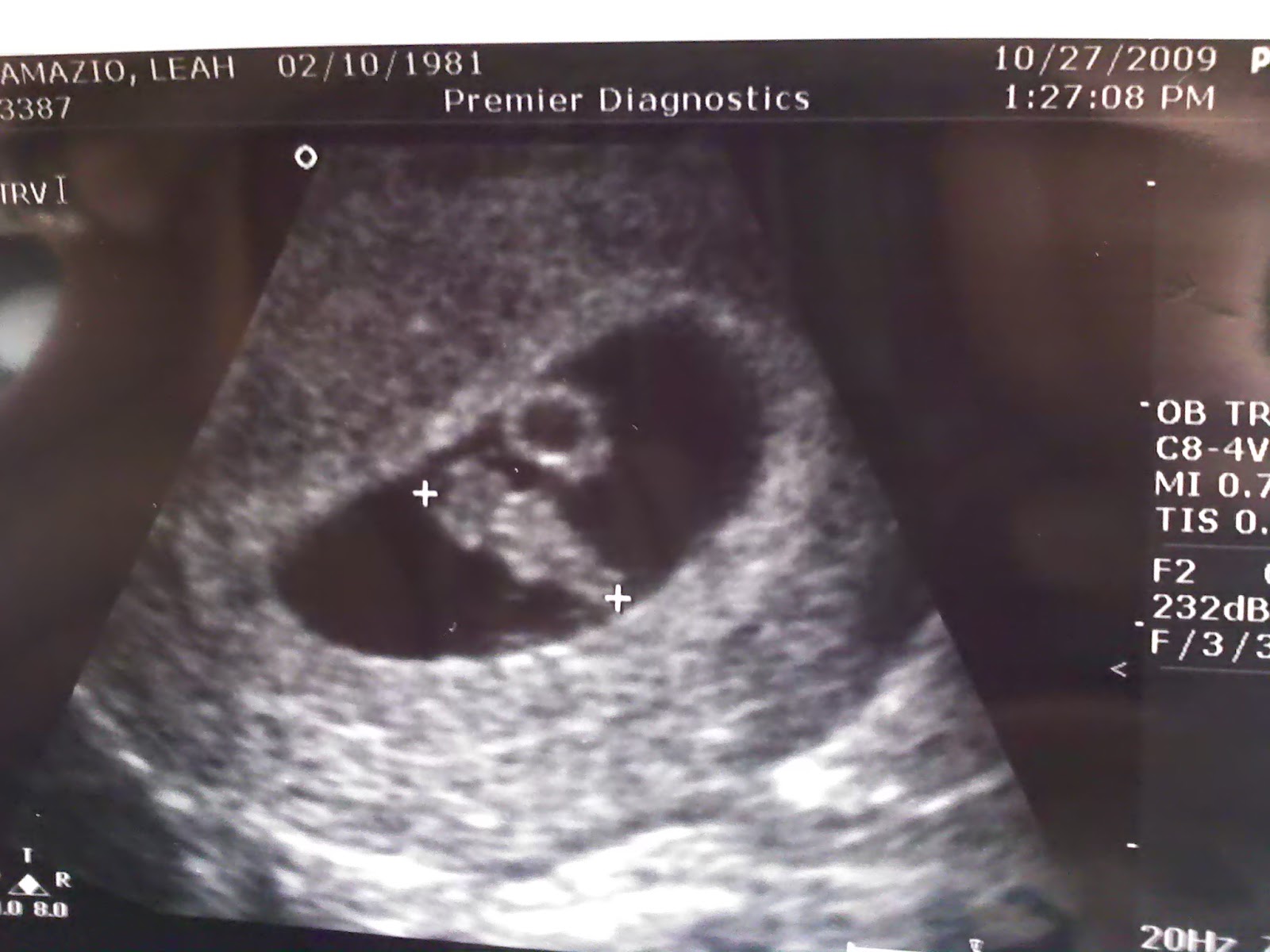
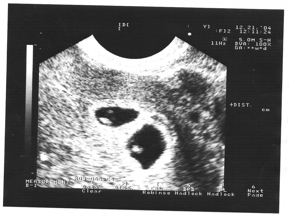
If twins are expected, the ultrasound image at this time will be approximately the same.
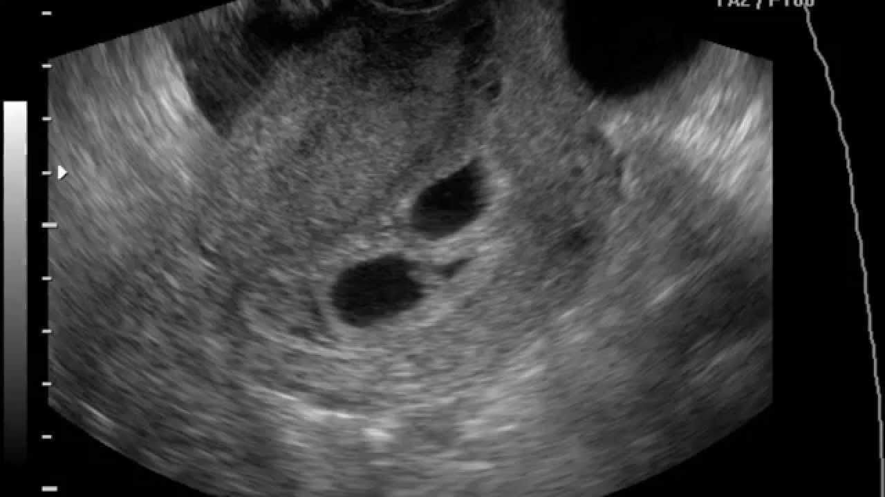
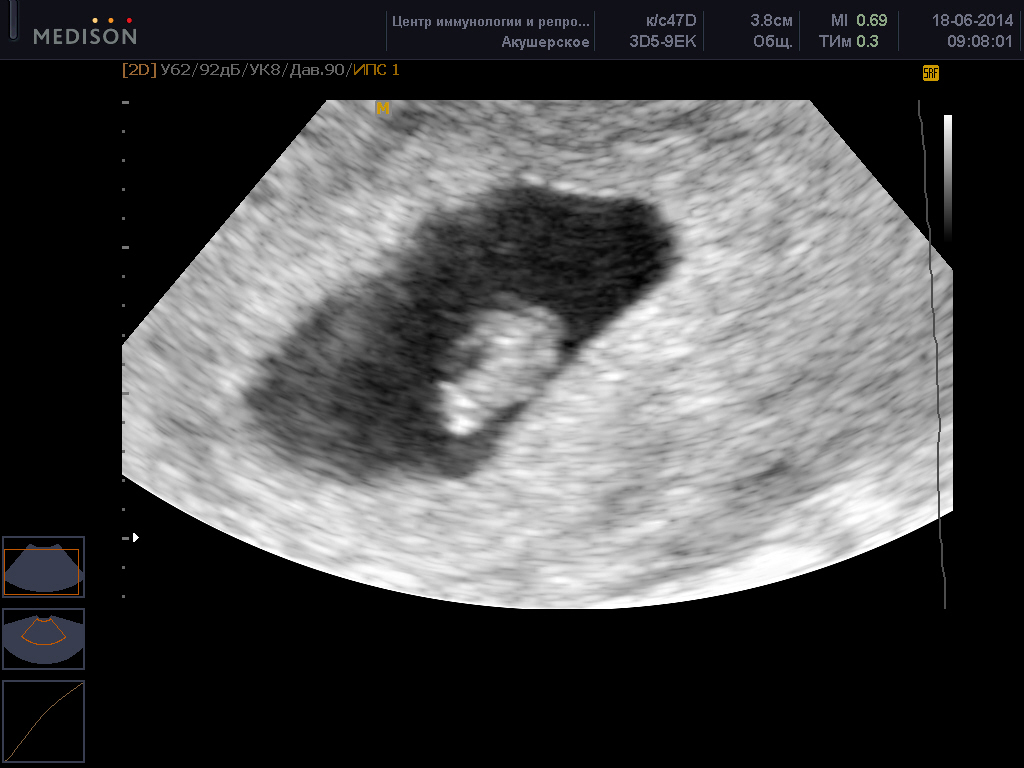
Identical twins at week 7 look like this.