Ultrasound in the 18th week of pregnancy: fetal size and other features
18 week of pregnancy - a time of relative calm. The painful attacks of toxemia retreated, the sensations of gravity have not yet appeared. The mood of the future mother is excellent. It is during this period that she can receive referrals for an ultrasound scan. What diagnostics will show at this time and what to prepare for, we will tell in this article.
Objectives of the survey
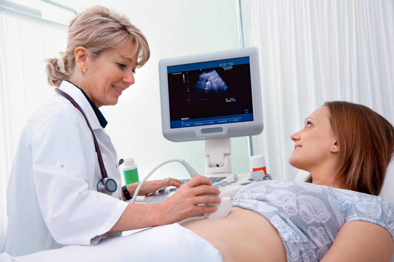
The concept of “screening” is already familiar to the expectant mother, because in the first trimester she had already undergone an ultrasound scan and donated blood for laboratory biochemical research. At 18 weeks another screening awaits her, second on account. The Ministry of Health recommends that it be held from 18 to 21 weeks., so now is the right time to re-look at the long-awaited baby.
Screening ultrasound is supported by data obtained from the laboratory. According to the results of both studies, it is possible to judge the possible pathological conditions, some malformations of the fetus.

Ultrasound examination at week 18 has an important goal - identify problems in the development of the baby. But this is not the only reason why scanning is done. On this period, the ultrasound scan helps to orient in the timing of pregnancy, if they are not fully elucidated, and also to assign an estimated date of delivery.
In this period already You can try to see the sex of the child, because the external genitalia are fully formed.
If the baby is not shy and shows her sex to the doctor, the woman will be able to find out who she expects is her son or daughter.
The issues that are solved during the screening do not imply sex determination, therefore this service is paid, while all other diagnostic measures within the framework of prenatal diagnosis are completely free for all women in an “interesting position”.

An unscheduled ultrasound scan at week 18 can be done according to indications - this is a sudden pain, discharge that cannot be considered normal for pregnant women, the discrepancy between the height of the uterus standing and the obstetric period.
Preparation for the procedure and features of
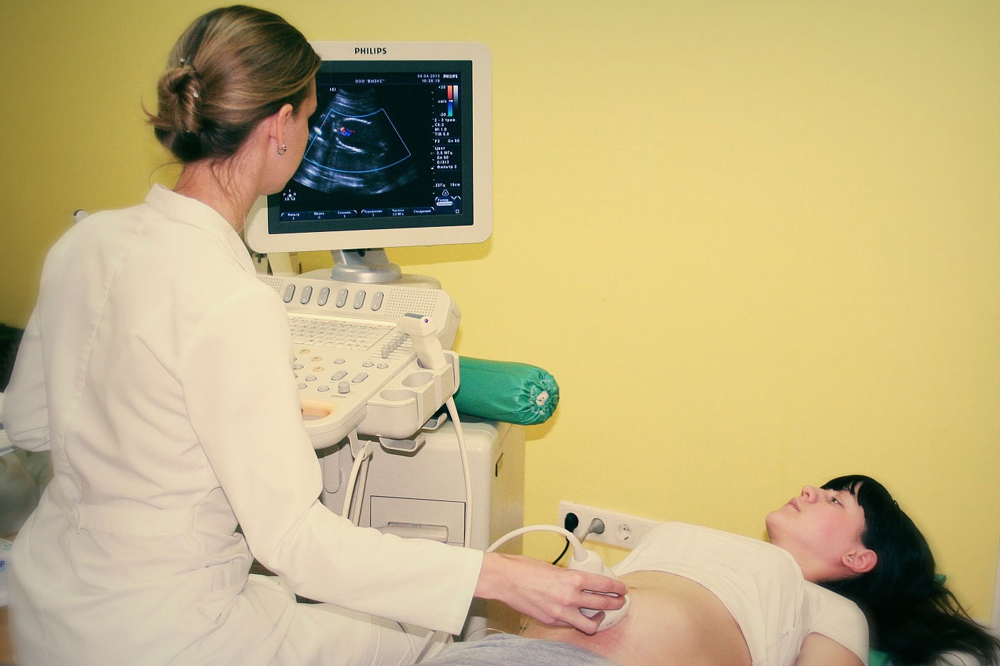
An ultrasound scan at week 18 can be performed in two ways - internal or external. Internal, called transvaginal, gives a good view through the vaginal wall, if it is difficult through the peritoneum. It can be difficult for a doctor to examine a child if the expectant mother is the owner of extra pounds and fat folds on the stomach.
The vaginal sensor is also indispensable for threatened abortion, as it allows you to see the cervix and cervical canal more clearly at week 18.
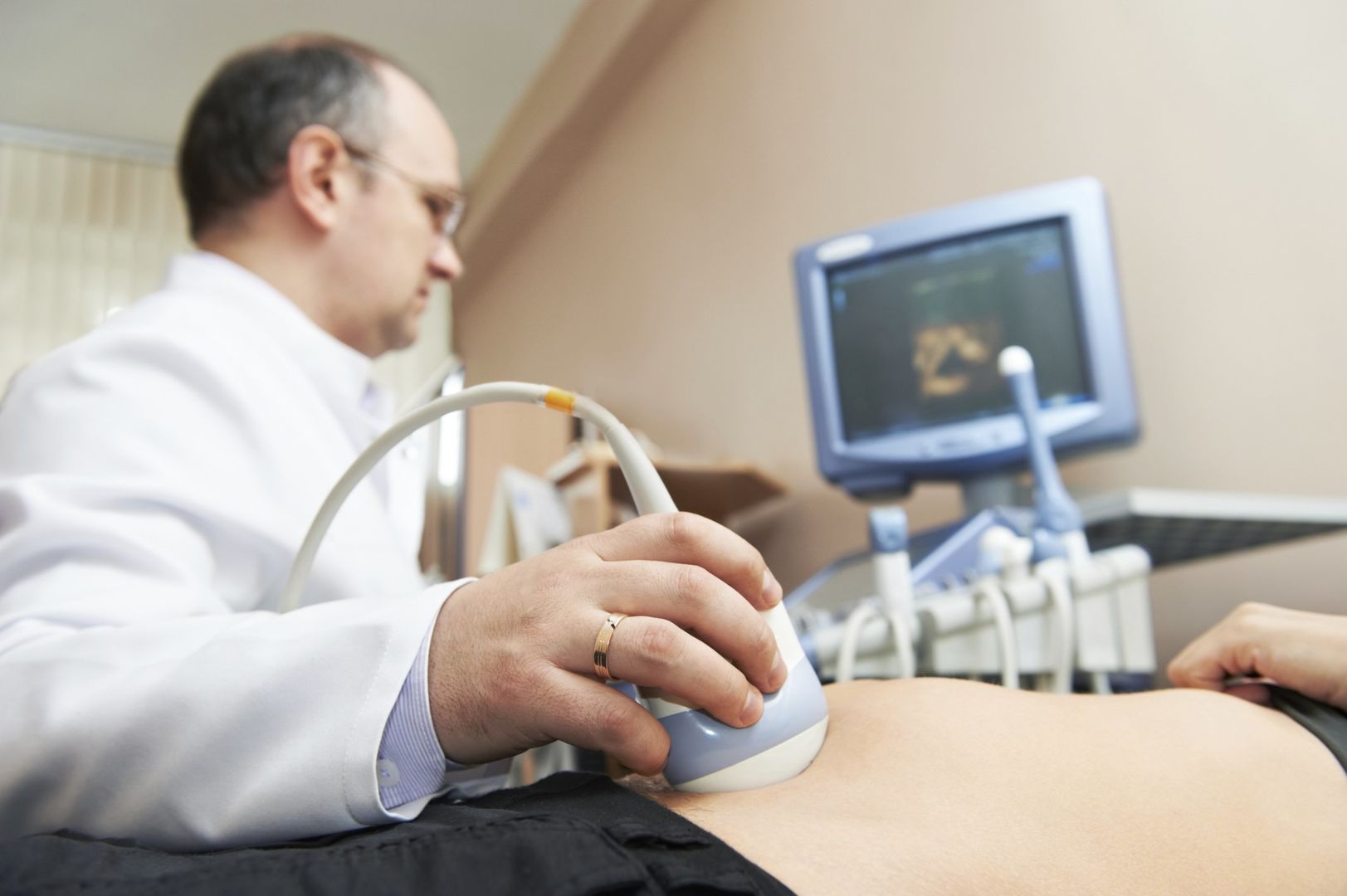
If a woman has a normal or thin physique, this week of pregnancy, you can conduct a study in a transabdominal way - through the stomach. Preparation for ultrasound is not required, no matter how it was carried out. No need to drink water or tea to fill the bladder, there is no need to adhere to a diet that reduces flatulence in the intestines.
What can be seen on the ultrasound?
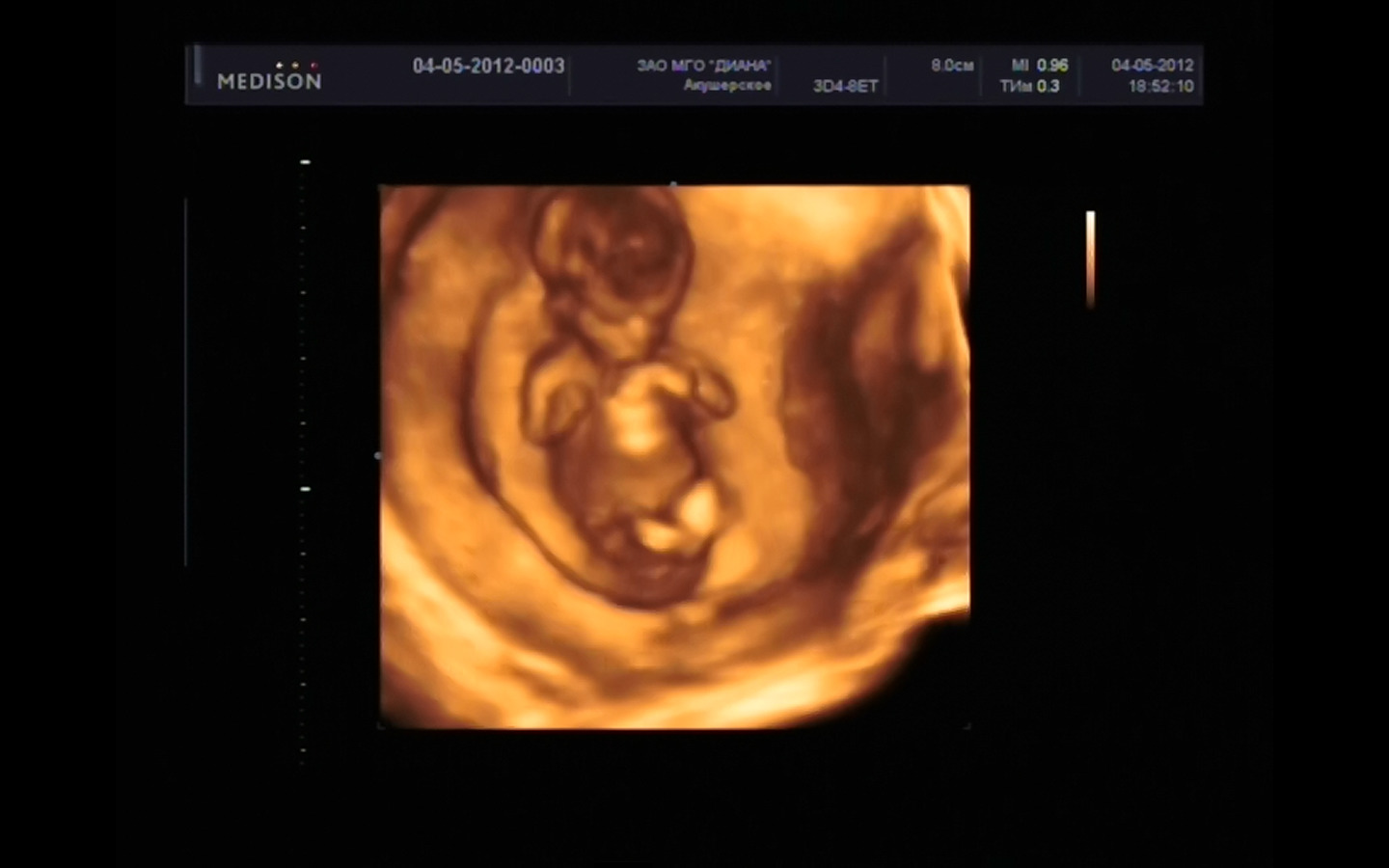
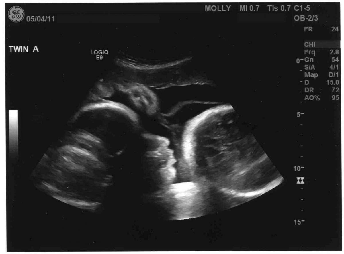
At week 18, the baby looks like a little man, but his head still remains disproportionately large. The height of the crumbs from heels to the crown is almost 20 centimeters, and its weight approaches 200-240 grams. Such dimensions allow him to freely tumble, swim, push off from the walls of the uterus, make a variety of movements with his hands and feet. It is during this period that some pregnant women begin to feel the first movements. These are mostly women awaiting appearance. twins or triplets, as well as multiparous.
Even if a woman still does not feel perturbations, you can enjoy the movements of your baby during the ultrasound procedure. The skin of the child is still very thin, blood vessels appear through it. The brain is developing rapidly, the endocrine system begins to work this week. This ultrasound scan, of course, does not show.
But mom will be able to see a small heart, listen to how it knocks, if the level of equipment permits, then you can see how the baby squeezes and unclenches his fists, spreads his fingers.

The crumb already distinguishes sounds. He already got used to the beat of his mother's heart, the noise of blood in her vessels, but everything new, that which comes from outside, interests and scares him at the same time. Therefore, the baby can liven up to the unfamiliar voice of the health worker who is conducting the study, and, conversely, may get scared and calm down.
The genitals are clearly visible at this time if the baby is conveniently located for inspection. It is possible that the cord will be clamped between the legs. And then the girl can be confused with the boy. Shy little boys can hide their “dignity” between the legs and they can easily be mistaken for a girl. The percentage of error, however, on this period is small - no more than 2-4%.
Norms and decoding
The description is compiled in three main areas - fetal size (fetometric indicators), fetal anatomy (the presence and proper formation of internal organs), as well as a description of auxiliary organs and media - placenta, umbilical cord surrounding the amniotic fluid. In addition, the doctor assesses the state of health of the woman herself - whether everything is in order with her reproductive organs, whether there is a threat of abortion.
Fetal photometry
It is possible to draw conclusions about how the head of the child is developed, by two parameters, their diagnosticians are measured in the first place. This is the longitudinal (LZR) and transverse size (BPR). Head circumference completes the picture. Considered important and such size as abdominal circumference of the baby. At this time, measurements are also carried out on paired bones - femur, shin, shoulder and forearm. At week 18, the bones of the nose are also measured.
Child head sizes:
Deadline, week | BPR, mm | LZR, mm | Head circumference, mm |
17-18 week | 38-42 | 50-54 | 135-146 |
Sizes of paired bones and bones of the nose:
Deadline, week | Thigh length, mm | Shin length, mm | Shoulder length, mm | Forearm length, mm | Nose bone length |
17-18 week | 22-25 | 21-24 | 21-24 | 18-20 | 5,4 -6,0 |
Anatomical features
A child in the womb is already big enough for the doctor to examine his internal organs. During the study, examine the heart, excluding the possible defects of this organ and large vessels. The doctor is also interested in the kidneys, bladder of the child, intestines, stomach, lungs, gall bladder, brain structures and cerebellum.
In the absence of defects and visible problems, the doctor indicates that the internal organs of the toddler do not have any features.
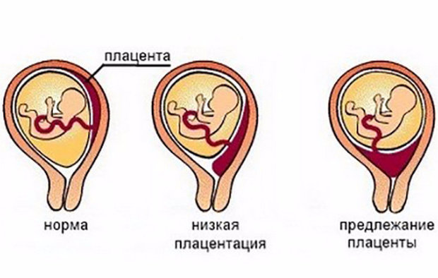
Placenta, umbilical cord, amniotic fluid
Standards and deviations are compared with this table:
Deadline, week | Placenta - norms | Amniotic Fluid Amount | Umbilical cord |
17-18 | Maturity - 0, thickness - 19.4 - 20.2 mm | 127-133 mm | Has 3 vessels |
The diagnostician must mark where the placenta is. For most pregnant women, this temporary body is responsible for providing the child with everything necessary for 9 months, is located on the back of the uterine wall, however, and the front location should not be surprising.This is a variant of the norm, only requiring more careful management of pregnancy by the attending physician.
Possible problems
In the process of ultrasound scanning at week 18, some pathological conditions may become apparent. On the most common tell more:
The size of the child is behind or ahead of the norm.
Any discrepancies with the standards that are peculiar to the fruit for a certain period, require careful study by doctors. Small lags or advances - can be caused by completely harmless reasons - heredity, features of the baby’s body constitution. Thin parents usually have the same kind of children.
Additional surveys deserve a situation where the dimensions deviate in one direction or another for 2 weeks or more.
In this case, you should pay attention to exactly which dimensions exceed the norm, whether the process is symmetrical (when all sizes are increased), or there is asymmetry (only one part of the body is increased or one size is increased).
An increase and a decrease in the head of a child can be a sign of genetic malformations, hydrocephalus or microcephaly. If the dimensions are increased symmetrically, then there is a chance that the woman is carrying the fetus with a tendency to heavy weight (more than 4 kilograms) or to a giant weight (more than 5 kilograms).
Symmetric reduction can talk about trouble which the baby experiences, up to intrauterine growth retardation.
However, there is no need to hurry with conclusions - the children grow up in the womb “jumps”, and it is possible that in a couple of weeks on the control ultrasound the baby “aligns” in the parameters.
The bones of the nose are behind the norm.
The length of the bones of the spout is a marker of chromosomal abnormalities. In children with Down syndrome, Patau, Turner and other incurable diseases, the profile of the face is flattened, the nose is much smaller than in healthy peers. This fact is taken as a basis in the measurement of this parameter. A slight lag in the length of the nasal bones should also not greatly disturb.
Rather, this is the basis to carefully look in the mirror to the mother herself and examine the appearance of her relatives. Maybe the family has small noses - a hereditary feature.
From this point of view, moderate increases in the size of the spout should not cause panic.

When measuring this parameter, very often errors and errors occur that are due to the fact that the equipment on which the diagnostics is carried out is outdated and has a low resolution.
If the diagnostician is still inclined to consider the deviation to be significant, and blood tests confirm the imbalance of hormones and proteins in the body, then this is reason to go to the reception to genetics and, possibly, to undergo an invasive procedure - an amniocentesis or a chorion biopsy in order to know with an accuracy of more than 99% whether the child is healthy.

The placenta is low
The verdict of the diagnostician on the low location of the placenta should not introduce a pregnant woman into a stupor. Pregnancy has not yet reached even half, the most intensive growth of the uterus is still ahead. The “baby seat” has every chance to rise higher with the growing uterine wall. In most cases, this is what happens.
However, the installed low placentation imposes several restrictions on women - she should not lift weights, bend down sharply and squat, should take vitamin complexes, eat well, avoid fast walking, fatigue, more often lie in a horizontal position. In some cases, doctors offer hospitalization in the department of gynecology or pathology of pregnant women, and you should not refuse it.

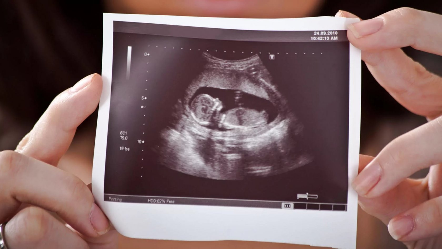
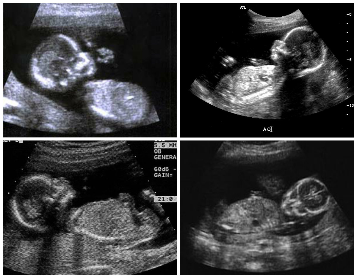
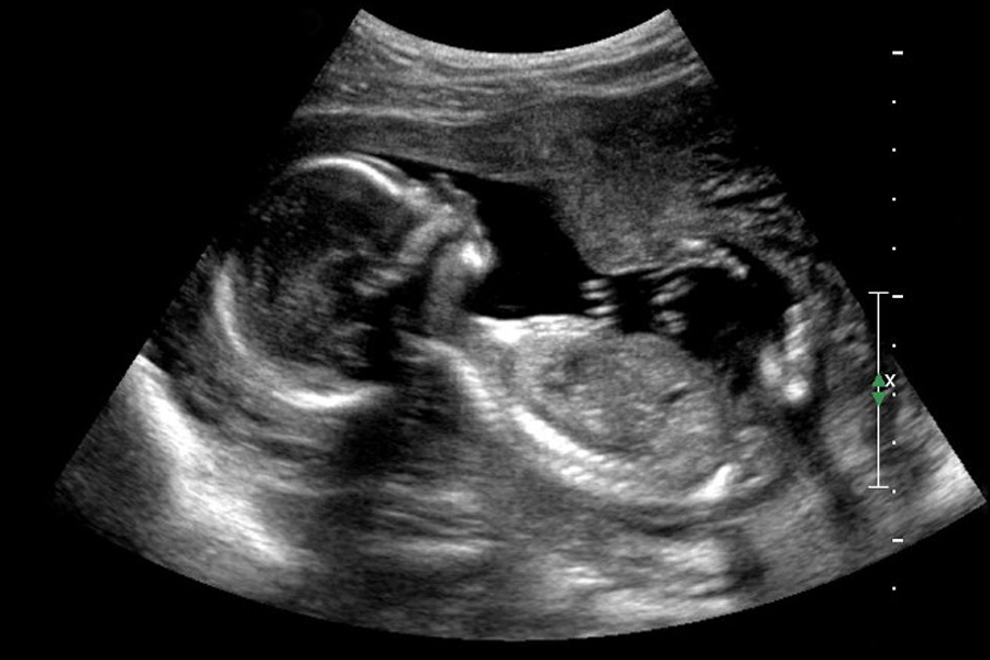
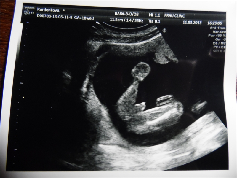
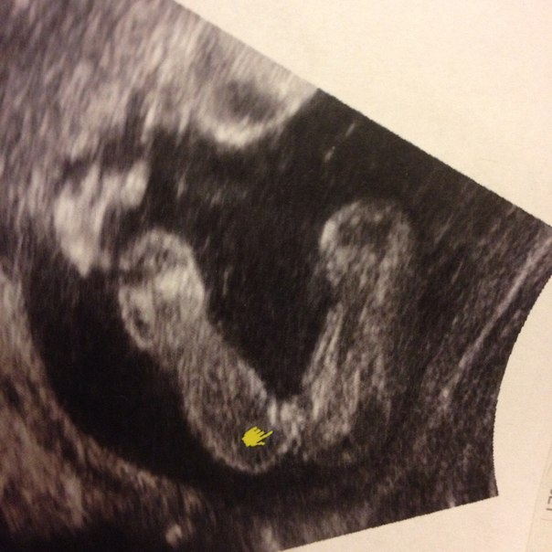
Ultrasound images
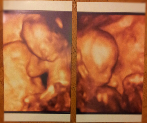
Even on a conventional two-dimensional ultrasound at week 18, you can consider not only the baby’s profile, but also the spine, ribs, fingers. Three-dimensional ultrasound can be performed at this time, but so far nothing particularly interesting can be detected in the 3D images. Scarce still does not grimace, does not "pose." For bright and emotional "photo" better to visit a three-dimensional ultrasound after 21 weeks.
If the doctor succeeds in “getting close” to the sensor from the bottom, then in such a projection one can clearly see the sex signs - the boy and the girl from this angle can be distinguished from each other with a naked non-professional eye.
If you want to save a photo from this ultrasound for history, you should ask the doctor to give a digital copy, because the ultrasound images tend to fade quickly and lose the contrast of the image.