What is doppler ultrasound during pregnancy, why and how to do it?

Modern methods of ultrasound during pregnancy also include the use of Doppler. This study allows to identify various pathologies of intrauterine development in the fetus at the earliest stages of its growth in the womb. This article will tell future moms about what a Doppler study is and when it is needed.
What it is?
The use of dopplerography (USDG) is quite often used in both obstetrics and gynecology. This is due to the fact that these methods do not cause any dangerous consequences either in the body of the future mother or in the baby in her womb.
At its core, Doppler research is based on the study of the speed of the modified wavelength. The principle of operation of the equipment used to conduct such surveys is quite simple. It generates waves of a certain frequency, which penetrate well through the skin and reach certain moving objects. Reflecting from them, the waves fall back into the sensor. This process is further processed by the device itself.

This physical feature of the apparatus explains that with the help of Doppler the work is evaluated only of "mobile" organs. In the composition of the blood there is a large number of various cells - red blood cells, platelets, leukocytes. They are in the bloodstream in constant motion. Such activity determines the possibility of using Doppler devices for blood testing.
The main use of this test - assessment of uteroplacental blood flow. It should be noted that doctors use dopplerometry to establish various disorders related to the blood supply to the internal organs. Using this research method during pregnancy allows you to identify various pathologies that occur both in the placenta and in the uterus blood vessels feeding the fetus.


These types of examinations are carried out by physicians who have certificates for both ultrasound diagnostics and the implementation of USDG. It is very important that only experienced and qualified specialists work with future moms. This is especially necessary in cases where a pregnant woman has any pathology during the course of the pregnancy.
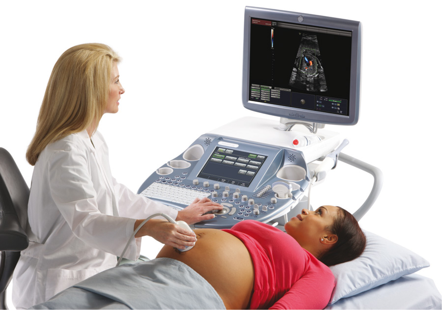
On the part of doplerometry very similar to conventional ultrasound. The survey is also carried out using a special apparatus. During pregnancy, several such studies may be conducted. Frequency and the need for their conduct is determined by the attending gynecologist.

The first studies were carried out strictly in M-mode. This method did not allow doctors to get all the necessary information about the presence in the body of the future mother of various pathologies.
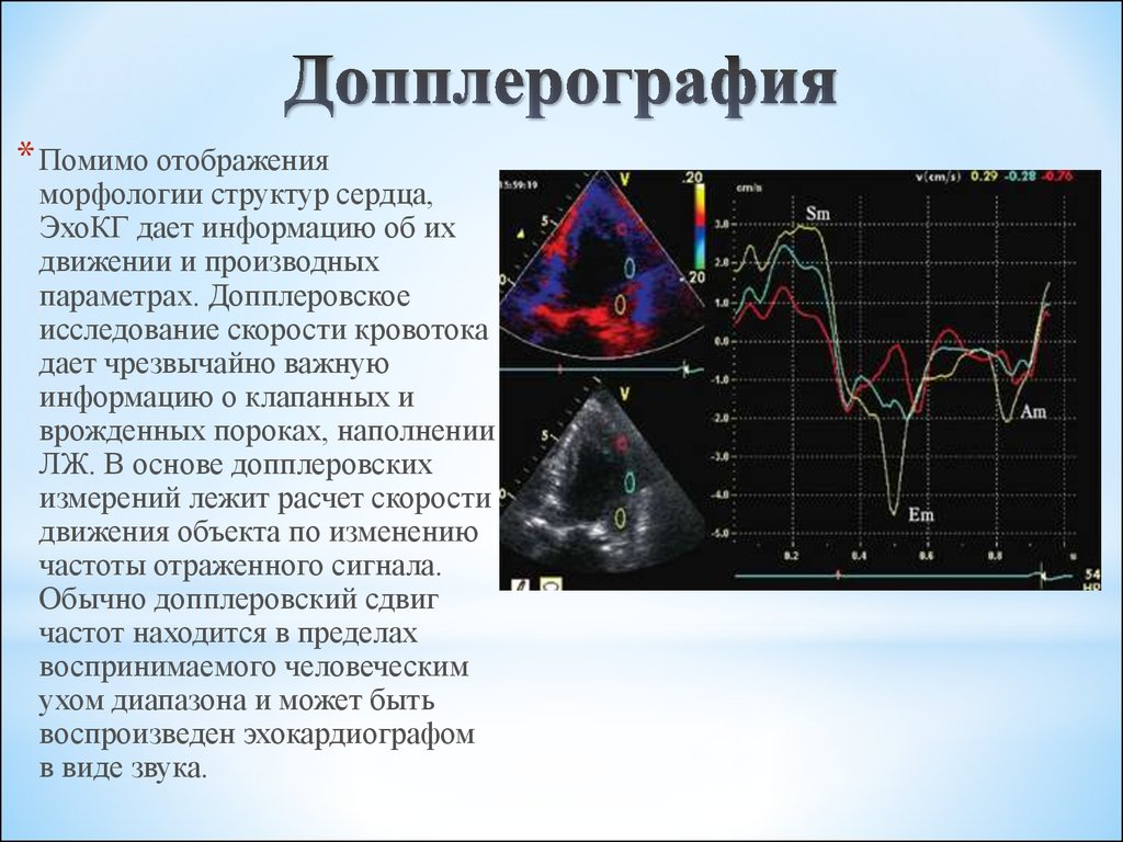
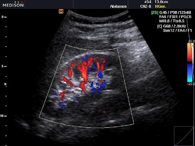
Technical progress has contributed to the fact that such tests have been replaced by more modern methods. These include echocardiography (ECHO - CG), as well as color mapping. These studies are most successfully used to identify various heart defects in the fetus and are usually performed in the 3rd trimester of pregnancy.
When is it held?
Many researchers believe that often dopplerography of uterine blood flow is very harmful for the future fetus.Therefore, in women in whom the pregnancy proceeds without any significant deviations, this study, as a rule, is not carried out.
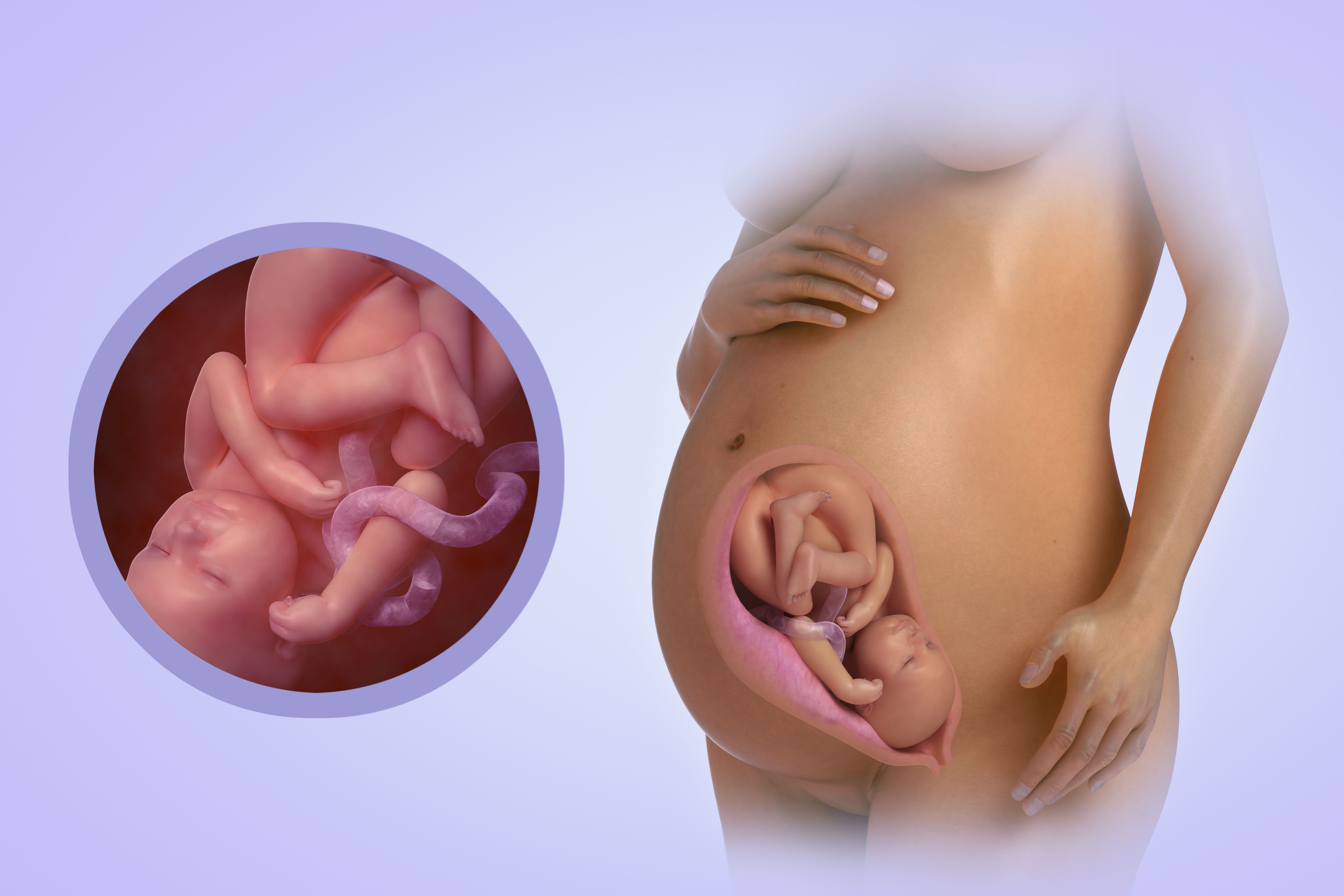
Doppler sonography may be prescribed from the 20th week of pregnancy. This is due to the fact that by this time the fetus already has signs of active blood supply and the heart is working. There are strict medical indications for Doppler ultrasound at this stage of pregnancy. The need for such a study is determined by an obstetrician-gynecologist who observes the expectant mother.

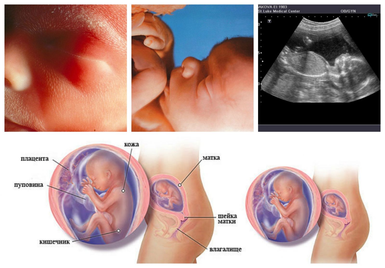
In most cases, USDG methods are used much later. As a rule, they are applied at 30-34 weeks of pregnancy. Such a study is not conducted by all expectant mothers, but only if they have strict medical indications. Often, Doppler in the third trimester of pregnancy is combined with an ultrasound. This combination of methods allows you to track the current state of the fetus at a certain stage of its intrauterine development.

Doppler echography - This is one of the latest research, which has become actively used in obstetrics in recent years. It allows you to identify various disorders in the work of the cardiovascular system in an unborn child at the stage of its prenatal development. Competent specialists, conducting such a study, can identify a variety of irregularities in the work of the small heart of the fetus.
The quality of this test also allows you to identify future congenital heart defects.
Modern equipment used to conduct such research allows ultrasound diagnostics doctors to also receive truly unique information. So, doctors can determine blood flow in most vital fetal blood vessels. They assess the adequacy of the blood supply, assessing the movement of blood particles along the middle cerebral artery, umbilical cord and aorta of the future baby.
More experienced specialists with a long clinical record of Doppler imaging can also evaluate the blood flow to some vital blood vessels that feed the internal organs of the fetus. Such a study allows to identify various anatomical defects at the earliest possible stages of their formation. In some cases the development of some anomalies can be prevented even in the period of fetal development.

With the help of the UZDG very different indicators can be traced both arterial and venous blood flow. To do this, doctors use special tables that contain all the necessary values of the norm. In each term of pregnancy, these figures will be different. With the use of such medical documents, ultrasound doctors can draw the right conclusion about the result of the study.
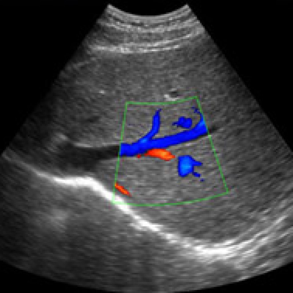
The use of various modes allows not only to assess the direct internal structure of blood vessels, but also to study the speed of movement of blood particles during blood flow.
Switching to another mode allows you to get even a color three-dimensional image. Such a three-dimensional image gives doctors a more complete picture of the violations of the fetus.
Mothers who carry several babies at once at the same time dopplerography is performed quite often. The use of this method allows to detect blood flow disorders in each of the twins. Also, using the Doppler sonography, one can also establish indirect signs of strong compression or clamping of umbilical cord blood vessels.
If the expectant mother suffers from diabetes or has a concomitant disease of the cardiovascular system, then a Doppler sonography is carried out for her for medical reasons.In case of kidney and urinary tract diseases, the conduct of this study is also an extremely necessary condition for diagnosis.

Pathological course of pregnancy occurring on the background of preeclampsia is the reason leading to the UZDG for a pregnant woman. Performing such a study in this state allows time to identify various deviationswhich can even lead to miscarriage or death of the fetus in the womb.
With the help of Doppler, you can assess a variety of disorders arising in the fetal membranes. Various abnormalities in the structure of the umbilical cord are also well identified using Doppler sonography. Fetal intrauterine growth retardation syndrome is another significant medical indication that carries USDG. Conducting this test can quite accurately identify this serious pathology.


If a mother and her future baby have a different Rh factor, then this can cause the development of many pathological disorders both in the woman’s body and in her future baby. This condition is quite dangerous. In some cases, rhesus conflict may cause spontaneous miscarriages. In order to identify various violations in this case, dopplerography is performed.
The entanglement of the umbilical cord of the fetus is an urgent indication for a Doppler study. In this case, the sooner a survey is conducted, the greater the chances for a favorable resolution of the situation. The emergence of sudden bloody discharge from the genital tract, especially in the late stages of carrying a baby, becomes a weighty medical indication for conducting this study.

In some cases, UZDG can be performed to expectant mothers who are carrying babies after 35 years of age. Doctors believe that by this age the number of possible pathologies during pregnancy increases significantly.
To identify the very first signs of various, even the most minimal pathologies of uteroplacental blood flow, dopplerometry is performed.
How to prepare for the study?
Many pregnant women begin to worry a few days before the study. Do not do this. Doppler sonography is absolutely painless examination, which does not bring any pain and discomfort.
If it will be held in the clinic, then the expectant mother should take bed sheet or towel. They will be needed in order to lay them on the couch before the procedure. Currently, pharmacies sell special disposable products. In some cases, using them is much more convenient than carrying a towel from home.

In private clinics, all disposable sheets and napkins are provided free of charge. All this is already included in the established cost of the procedure. If the study is conducted in an ordinary clinic, then you should also take a few more paper napkins with you. They may need it in order to remove the remnants of a special gel with which the stomach will be processed before the procedure.
Some kind of strict diet before the study is usually not required. However, on the eve of the analysis Do not abuse vegetables and fruits. Such products contain quite a lot of coarse fiber, which can enhance gas formation. A stomach swollen from gas will make it much more difficult for a doctor to visualize (inspect) internal organs.


Methodology
You should not eat a lot of water and drink plenty of water just before the test. This may lead to the fact that the doctor simply does not see anything, performing dopplerometry. Also, overeating and excessive fluid intake can lead to nausea or even vomiting in a horizontal position in a pregnant woman.
For the study should choose the most comfortable clothes.The doctor's office ultrasound diagnosis - this is not a fashion podium. Clothing should not hold down movements. You should choose pretty warm and comfortable things at the same time.
Also suitable clothes that can be easily removed, exposing the belly for the study.


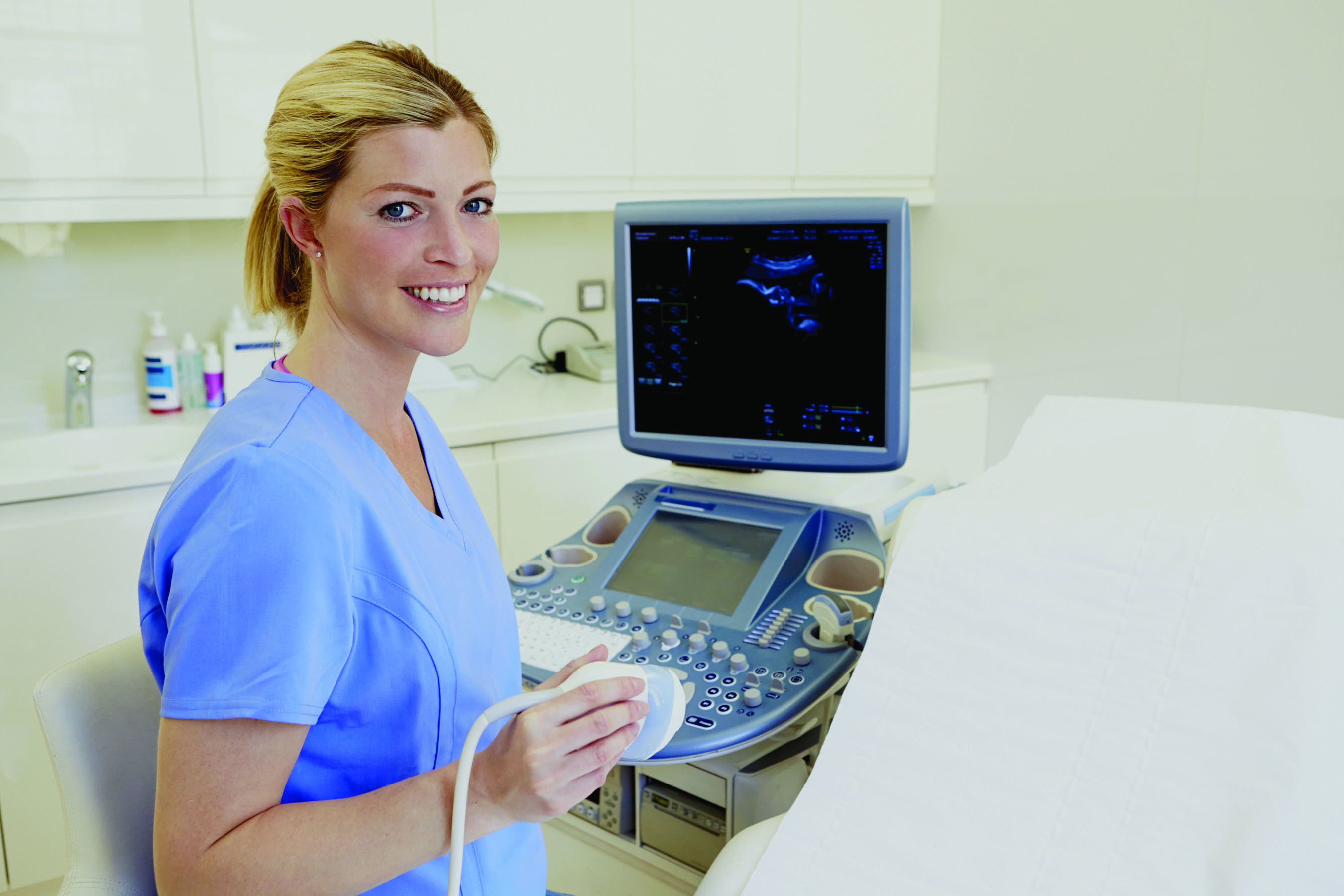
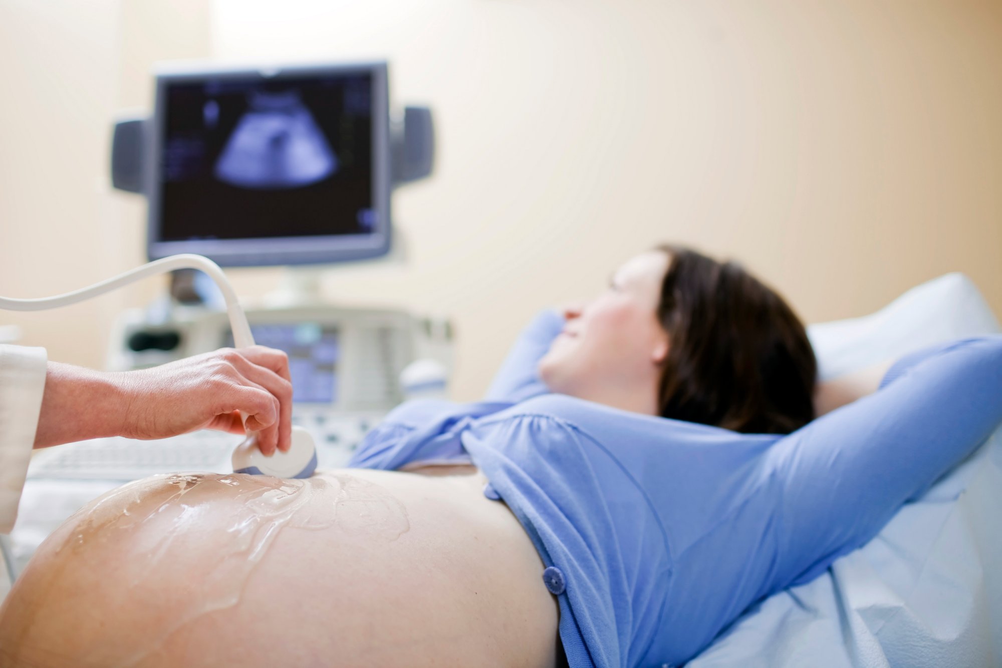
This study is conducted in the doctor's office ultrasound diagnosis. Coming to such an appointment, a pregnant woman knows the exact time at which it will be held. The duration of this procedure depends on many different factors. Experienced specialists dopplerography is usually done in 20-30 minutes. However, during the pathological course of pregnancy, the time of the study may be significantly longer.
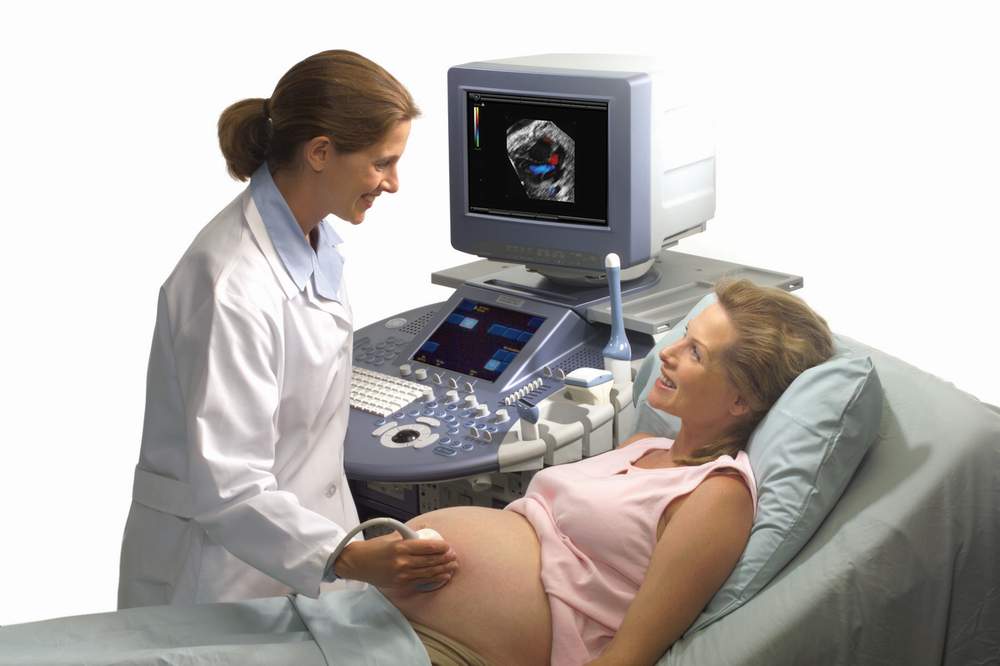
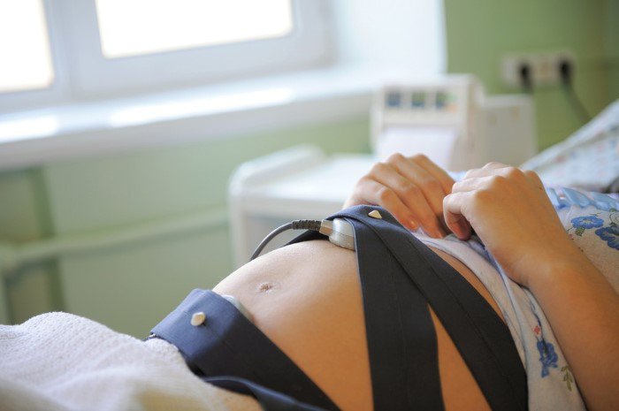
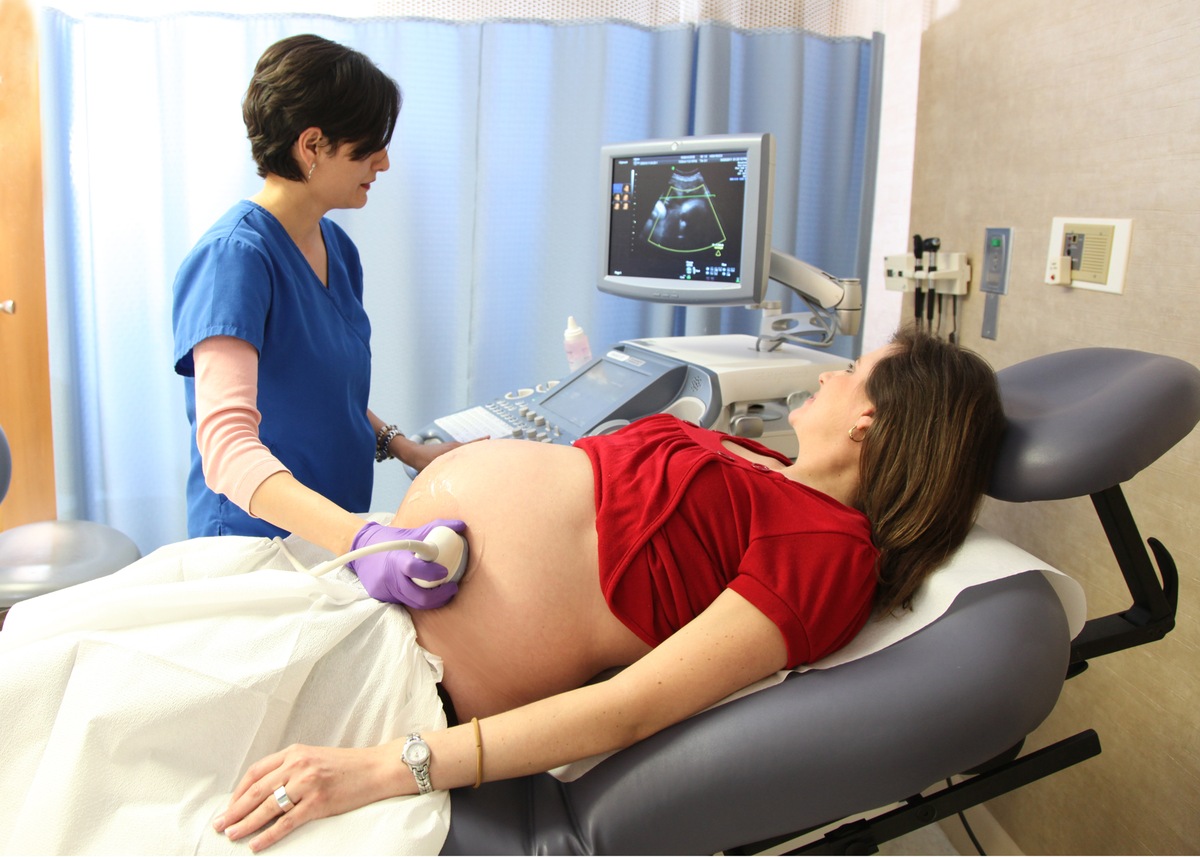
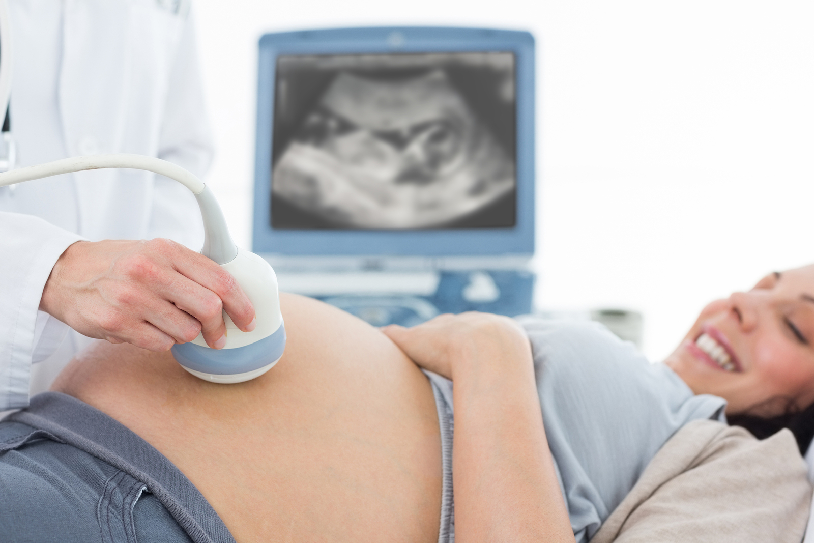
Future mother before the survey is placed on a special couch. The standard position is on the back. Only in some cases, the doctor may ask the pregnant woman to turn to the left. Usually this situation is possible in the third trimester of pregnancy. In the position on the left side of the body, the pressure of the uterus, in which the baby is already quite large in size, on the inferior vena cava is significantly reduced.
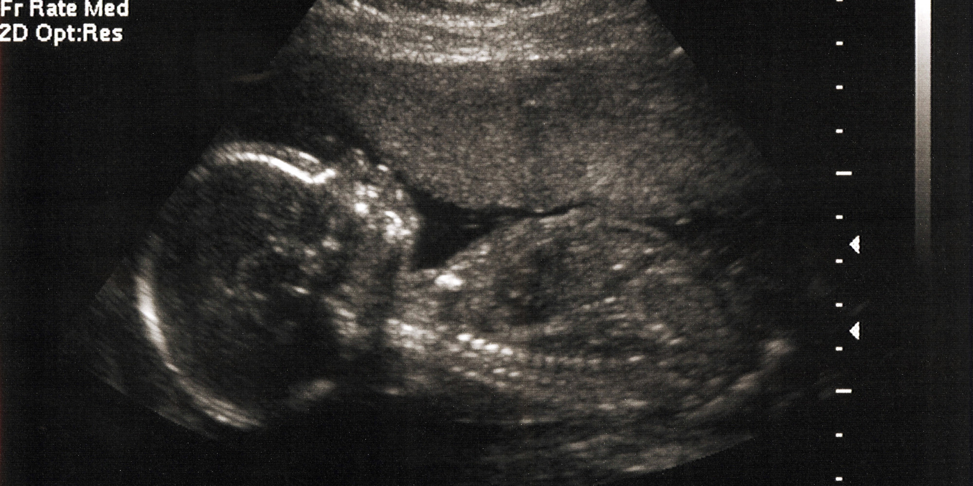
After preparation, the doctor takes a special sensor. With the help of him, he will conduct research. The sensor touches the skin, and various images appear on the screen of the device.

Before carrying out the main procedure of the study, the specialist should establish basic anatomical landmarks. These include: the main arteries of the uterus and their branches, as well as the blood vessels of the umbilical cord. They are very important for determining the main indicators of uteroplacental blood flow.
Then, for better visualization, the doctor applies a special gel on the woman's abdomen. The composition of this gel is the most safe and hypoallergenic. These drugs undergo multi-stage testing before being approved for use in pregnant women. The structure of this gel is usually sticky, the color and smell are completely absent.


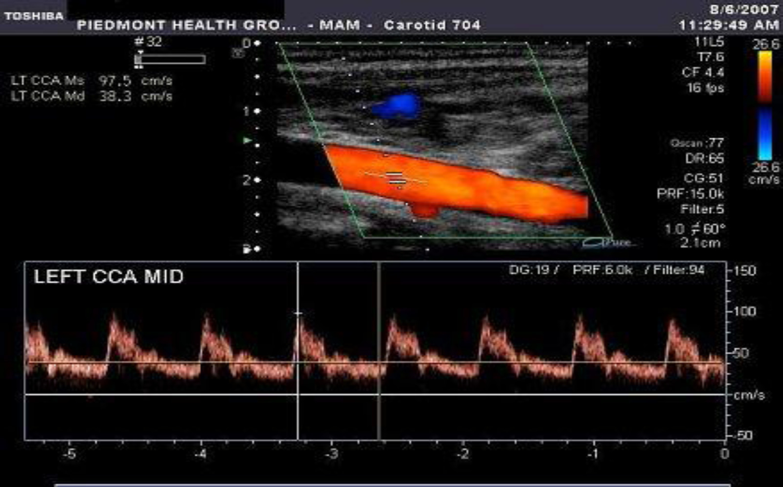
For the evaluation of Doppler, the most important are not the absolute values of the fetal uterine blood flow, but their quality indicators. Also, their ratios are also interpreted. To obtain such values, various indicators are measured in different periods of the cardiac cycle - during systolic and diastolic contractions.
As a result of measuring such indicators, doctors get systolodiastolic ratios, pulsating indices, resistance indices, as well as an average blood flow velocity. To assess the results of the doctor compares the values with normal valuesused in this period of pregnancy.

After the study, doctors determine which group the future mom can be assigned to. There are 2 groups - one of them includes women who do not have any violations of the uteroplacental blood flow, the second includes future moms who have reduced blood flow rates in the main main vessels.
Decoding results
Doppler sonography shows a variety of abnormalities that occur even in the early stages of fetal development. Doctors divide all pathological disorders according to several degrees (classes) of severity. It is important to note that the main criteria are not absolute values or any correlation, but the presence of localization of pathology.
The table below presents the main studied parameters that are used by doctors for issuing a USDG-conclusion:
Gestation time (weeks) | Systo-diastolic ratio | Resistance index |
12-13 | 2-3,5 | 0,52-0,71 |
14-16 | 1,9-3,1 | 0,48-0,68 |
17-19 | 1,7-2,6 | 0,44-0,62 |
20-24 | 1,6-2,5 | 0,41-0,61 |
25-31 | 1,7-2,4 | 0,4-0,59 |
32-37 | 1,6-2,3 | 0,35-0,58 |
38-40 | 1,4-2,1 | 0,32-0,55 |
If pathological abnormalities have arisen in the uteroplacental blood flow, then these values belong to class 1A. In this case, there are no serious abnormalities in the blood circulation in the fetus. Also in this situation there are no signs of intrauterine growth retardation and development of the future baby.
1B violations are characterized by a reverse pattern.In this case, no pathological disorders in the uteroplacental blood flow does not occur. The decrease in the estimated parameters occurs in the blood vessels connecting the fetus with the mother. In this clinical situation, signs of intrauterine growth retardation and growth of the future baby may also appear.


The second degree of disorders is characterized by the development of the most diverse disorders affecting many blood vessels. In this case, changes occur in both the uteroplacental blood flow and in the placental and fetal arteries. This condition is already more unfavorable. However, such indicators are quite adequate for the fetus to live and not die.
Extreme or third degree of violation is already a rather dangerous sign, indicating the presence of a strong disruption of the blood supply to the fetus. In this case, the blood flow in the supplying blood vessels between the future baby and the mother is significantly disturbed. If at this time it is not urgent for doctors to intervene and not correct this situation, then the fetus may die.
Any disturbances in the blood flow in a pregnant woman are the reason for giving her special treatment. In this case, repeated Doppler sonography may also be required to establish the dynamics of the development of pathological changes.