Ultrasound in the 10th week of pregnancy: fetal size and other features
If a woman has not yet been registered at the antenatal clinic, then the 10th week of pregnancy is the most suitable time for this, because after a couple of weeks the first prenatal screening is to take place. Ultrasound at 10 weeks is not considered mandatory, but they actually do it quite often. Many are worried about what to expect from such a procedure at this time.
The purpose of the diagnosis
It is still too early for the first ultrasound screening, because it is still quite difficult for the little man to stop being considered an embryo just a couple of weeks ago to become an embryo and become a fruit at the official level. The tenth week is only about 2 months from conception. However, such a method as an ultrasound scan may still be needed for other reasons. At this time for an ultrasound, a future mother can be sent for a number of indications:
- Clarification of the term. This problem is usually faced by women with an unstable and irregular menstrual cycle. When registering, they sometimes can not exactly name the first day of their last periods, and because of this confusion arises.
- Pathology of pregnancy. In the tenth week, a woman can be tormented by toxemia, and she may experience pain that is uncharacteristic of an “interesting situation”. Also, if not recorded, during a manual examination, the gynecologist may reveal a discrepancy between the size of the uterus and the expected date.
- Multiple pregnancy. If a woman has already done an ultrasound scan to confirm that she is pregnant, and there she was made happy with information about the upcoming birth of twins, then an examination at week 10 can be recommended by a doctor to clarify the viability of both babies and the secondary confirmation of multiple fertility.
Features and preparation
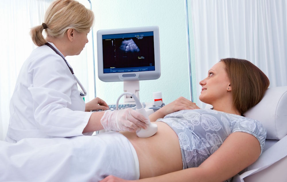
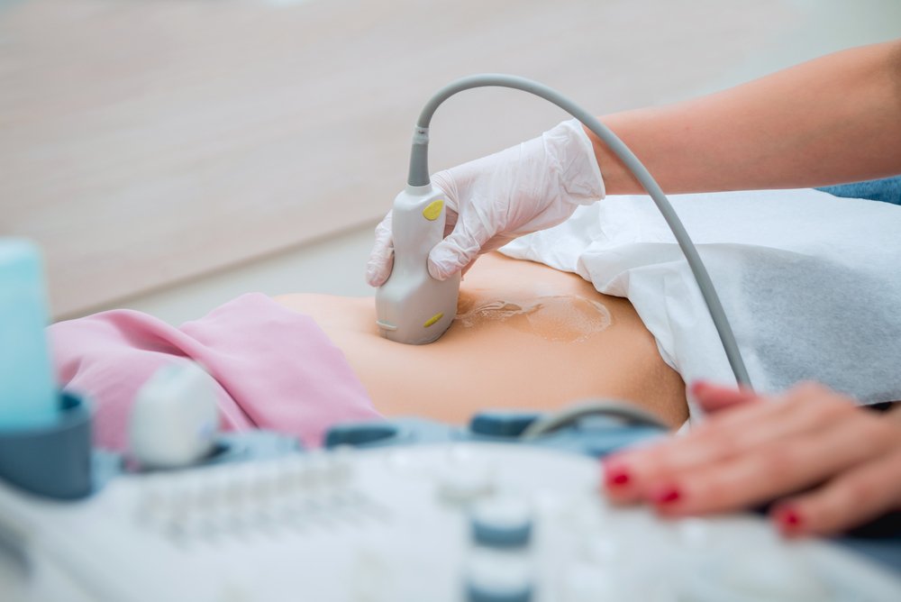
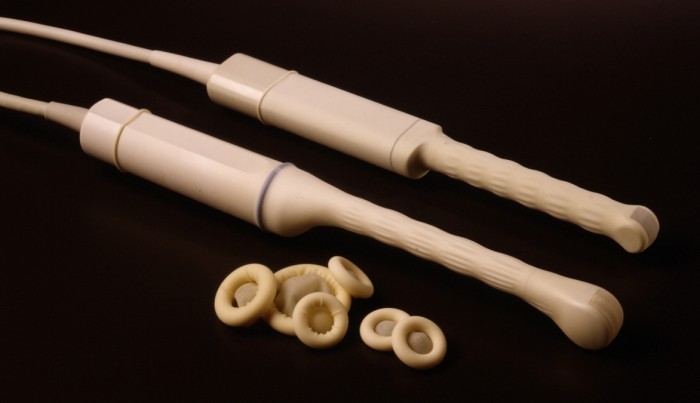
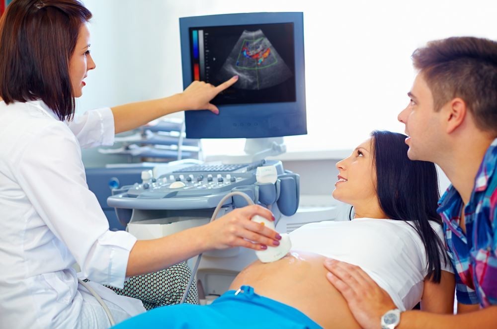
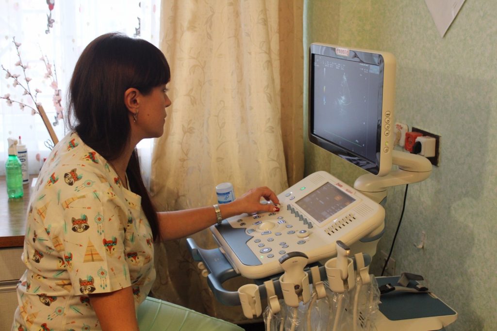
The child is still very small, little amniotic fluid, and because of this, looking at the baby through the anterior abdominal wall is rather difficult. At this time, doctors use ultrasound for ultrasound diagnostics. vaginal sensor. This method is called transvaginal. The diagnostician puts a condom on the scanner's sensor, and it is carried out while lying horizontally on a couch or on a special examination chair, which is available in the office of each gynecologist.
At such a short time, visualization through the vaginal wall is clearer and better than through the peritoneum.
It is a common misconception among women that before an ultrasound scan you should drink plenty of water to enter the diagnostic room with a full bladder. Previously, when ultrasound was performed only by the external method (through the abdomen), such preparation was justified; Now for scanning through the wall of the vagina to fill the bubble is not necessary. It is better to even appear on the examination with an empty bladder and intestines. The problem can create intestinal gases, which in the early stages deliver many unpleasant sensations to pregnant women.
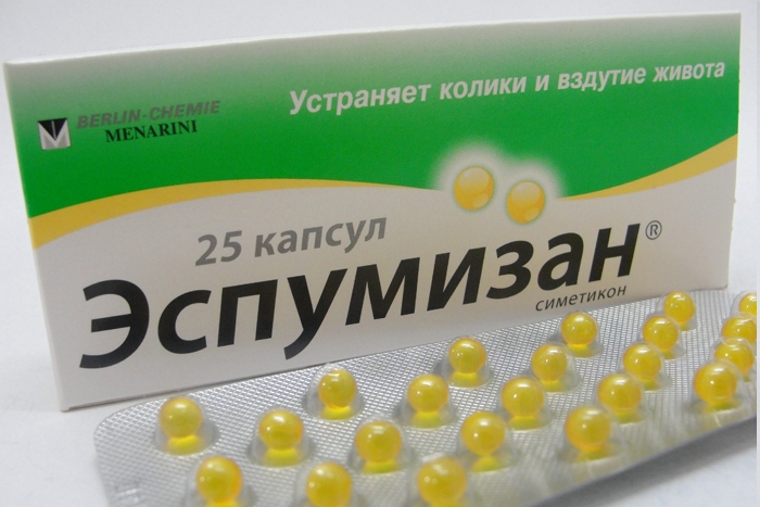
If the intestine swells with increased gas formation, its loops begin to squeeze the organs of the small pelvis. This may affect the results of the study.Therefore, a few days before the ultrasound scan you should not eat foods that promote flatulence (legumes, fatty dairy products, carbonated drinks), and a few hours before going to the doctor worth taking "Espumizan», to get rid of intestinal gas accumulations.
On the ultrasound at week 10, they take a second shoe or shoe cover, a small towel or cloth, a diaper to put it on a couch or a viewing chair.


What will the ultrasound show?
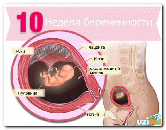
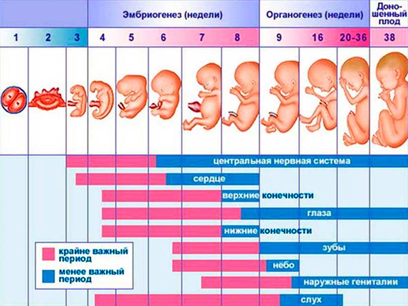
The child is completing the laying of organs and systems this week. Its height is about 35-40 mm, and its weight is only about 5-7 grams. However, this crumb is very much like a man. He now has no fetal tail, but there are hands and legs, which can be viewed on the monitor of the ultrasound scanner.
The baby’s skin is still transparent, the hair does not grow on it, but small ears and mouth are already formed. The baby from time to time opens and closes the mouth, brings the handles to the face. There is a formation of the brain, the nervous system is “tuned”. At the tenth week of the ultrasound, it will not be possible to see all of this, but mom will be able to listen to how her crumbs beat fast and often, many future mothers admit that this is an incomparable sensation.
It’s better not to pester the doctor with the question about the sex of the child at this time, because external genitals are not yet formed. Both girls 'ovaries and boys' testicles are located in the abdominal cavity, but the production of sex hormones has begun, which will support the process of the formation of visual sex. In the meantime, the so-called genital tubercle is in place of the genitalia, almost the same in boys and girls.
Definition of sex on this term is difficult.

Results and norm indicators
On ultrasound this week is well determined by the number of children. Each of the fruits is determined heartbeat and motor activity. If the heart knocks and there are signs of movement, the doctor indicates that the fetus is alive and the pregnancy is developing. About the duration of pregnancy can tell the size of the ovum, in which the baby develops. It is determined by the interval between the inner walls, and therefore this parameter is called average internal diameter of the ovum or simply - SVD.
In the tenth week, its figures are as follows:
Exact term | SVD, mm |
9 weeks + 1 day | 36-37 |
9 weeks + 2 days | 38 |
9 weeks + 3 days | 39 |
9 weeks + 4 days | 40-41 |
9 weeks + 5 days | 42 |
9 weeks + 6 days | 43 |
10 weeks exactly | 44 |
The appearance of the ovum is also considered important. With a normal developing pregnancy, it has clear and even contours, does not look deformed, crushed. The yolk bag, which, for the time being, performs the function of a food storehouse for a child, is already ready to hand over the functions of feeding the crumbs of the placenta, which has almost completed its formation. The size of the yolk sac on the tenth week is from 5.0 to 5.1 mm.
The most important indicator that can tell a doctor a lot about the pace of development of a little man, and also helps in correcting the duration of pregnancy, is the coccyx parietal size. KTR should not be confused with the growth of the child. Measure from the top of the tailbone, and not to the heels. KTR this week has on average the following meanings:
Exact term (weeks and days) | KTR of norm, mm | KTP range of permissible values, mm |
9 +1 | 23,7 | 18,6 — 28,8 |
9 +2 | 25 | 19,9 — 30,2 |
9 +3 | 26,4 | 21,2 – 31,6 |
9+4 | 27,8 | 22,5 — 33,1 |
9+5 | 29,2 | 23,8 — 34,6 |
9+6 | 30,7 | 25,2 — 36,1 |
10 weeks exactly | 32,1 | 26,6 — 37,6 |

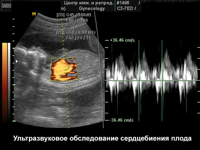
Another important dimension concerns the work of the heart of the baby. The doctor registers the frequency of contractions of the small heart. In conclusion, this is abbreviated as heart rate. In the tenth week, the average is 170 beats per minute. Variations from 161 to 179 beats per minute are possible. You should not look in the heart rate of signs of gender of the child.
Many pregnant women in Internet forums ask who is more like a heartbeat at 155 beats per minute - a boy or a girl? The sex of the child in the early stages does not affect the heart activity. In late periods, experienced obstetricians predict gender in the frequency and tone of the heartbeat, although such a diagnosis from the point of view of medicine has no evidence.
The tenth week is still a high risk of miscarriage, and therefore the doctor will definitely examine the ovaries and fallopian tubes, the uterus, the condition of the endometrium, and the cervix.
If signs of thickening of the uterine walls, changes in the cervical canal are detected, the threat of abortion may be put.

Possible problems
During screening at week 10, the following concerns may arise.
Fetal egg does not meet the deadline
This is the most common and disturbing future moms situation. If the tenth week of the SVD below the norm, then you should not despair. It is best to undergo a follow-up ultrasound scan in a couple of weeks, when the study starts in the first screening. If there is an opportunity, you should ask the attending physician for an expert class ultrasound, such equipment is available in the medical genetic centers.
In itself, the discrepancy between the ovum and the real term can also have harmless causes - for example, ovulation was late, the fetus was fixed later, and therefore development occurs later than the woman herself thinks. Pathological causes of inconsistencies may lie in developmental delay. The kid just will not start to "lag".
There are always preconditions for this - poor nutrition of the mother, lack of vitamins and microelements in her blood, bad habits (smoking, alcohol, narcotic substances), infectious diseases transferred by the mother in early pregnancy, as well as medication. Growing up slowly, the baby may also because of its genetic pathologies, gross malformations, which are not visible on ultrasound.
The first scheduled prenatal screening in the next 1-2 weeks will help clarify this issue.
If the fertilized egg is not deformed, the child gives signs of life, then there is no need to panic, it is quite possible that the baby will “pick up” its own and catch up with the norms in the near future. If there are additional signs of trouble (tone of the uterus, deformation of the ovum, lack of heartbeat), doctors can talk about missed abortion or the incidence of miscarriage. In any case, attentive observation of the situation is required.

KTR does not meet the deadline
Reduction of the coccyx-parietal size without signs of other pathologies in a child (there is a heartbeat and physical activity is noted) is a reason to revise the terms of pregnancy, perhaps they are not calculated correctly. However, the decrease in CTE and the absence of signs of vital activity - basis for immediate hospitalization and surgical curettage procedure uterine cavity with subsequent biopsy of fetal tissue, to understand why the baby died in utero.
The result, in which the KTR is less than the norm, and there are signs of life, requires re-checking. At the upcoming first screening, doctors will try to find the true cause, because chromosomal abnormalities can accompany the growth of the embryo, and then the fetus.
It may be normal to decrease CTE in twins or triplets, in the case of multiple pregnancies such deviations are not considered pathological.
Risk of miscarriage
If the walls of the uterus are thickened, the diagnostician indicates the risk of losing the child, the woman should always listen to the opinion of the attending physician. If he insists on hospitalization, do not argue. If you are allowed to be treated at home, you should take good care of your situation, take the prescribed medications, observe a calm and measured regimen.
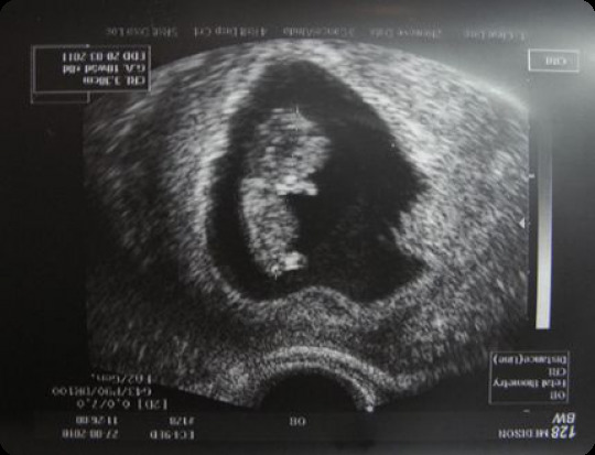
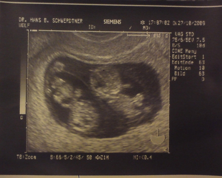
Snapshots
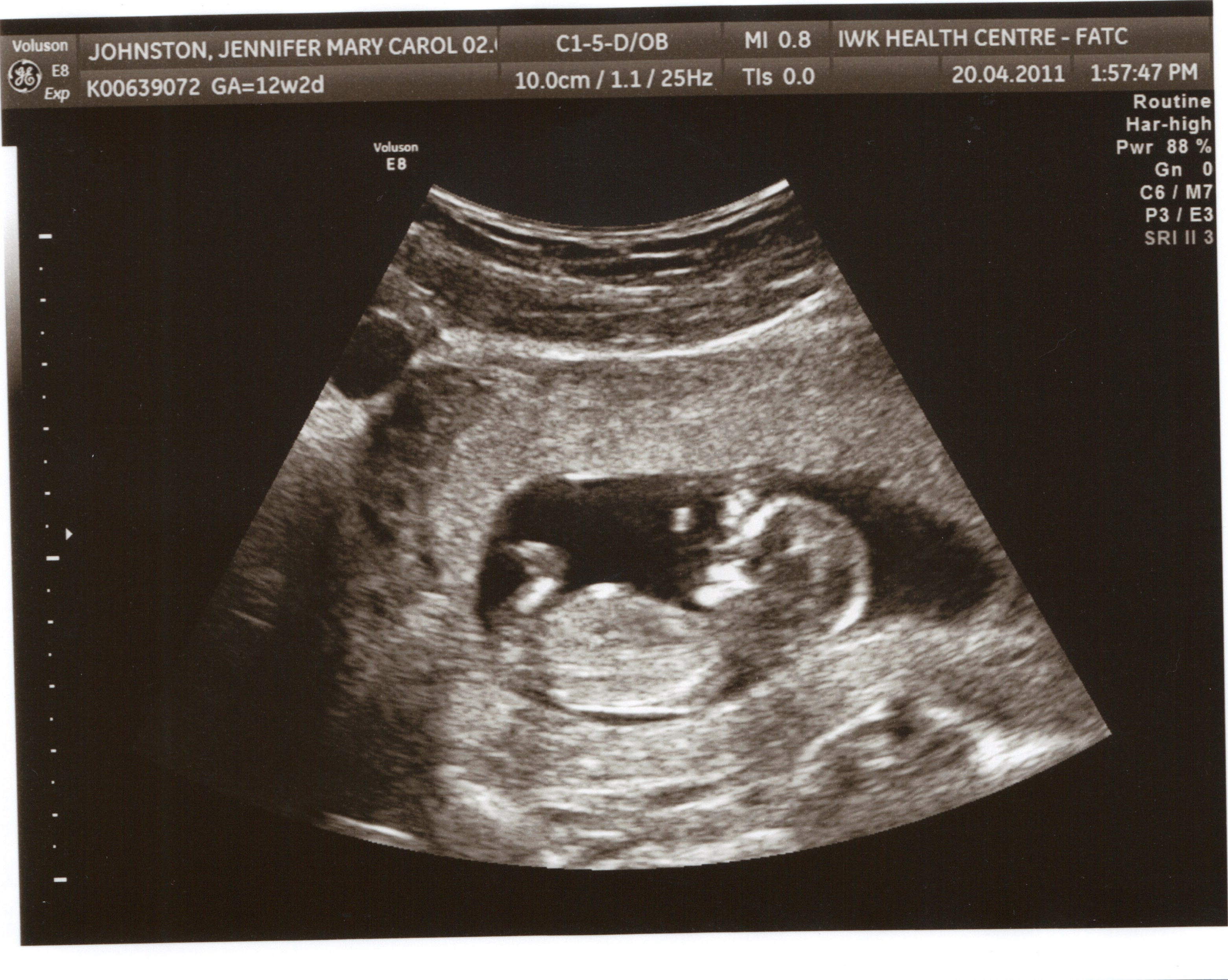
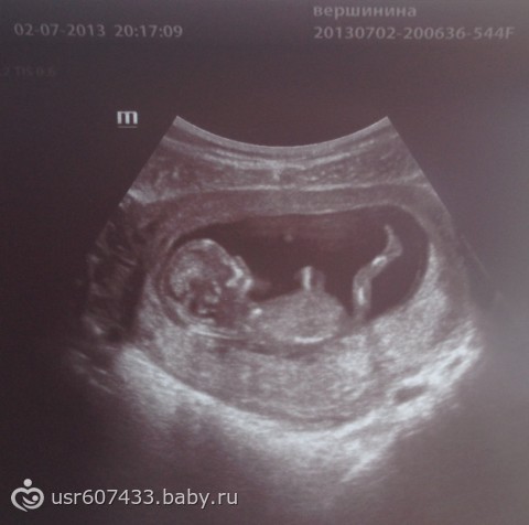
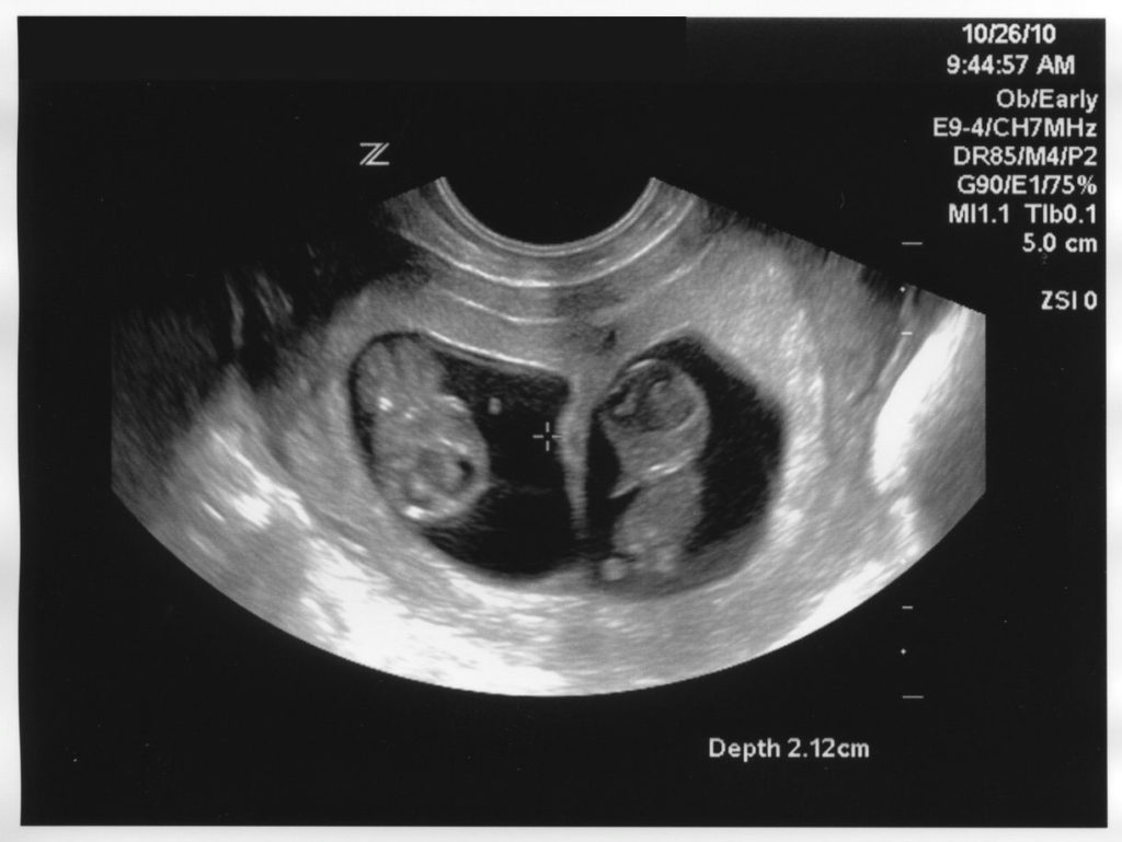
In the pictures taken on ultrasound at 9-10 weeks, the baby most likely will not show anything special, except for a disproportionately large head and handles (if lucky). In the pictures twins looks like that.There is no reason to make fashionable 3D-ultrasound today on this date, for now the three-dimensional image is not capable of giving a clear picture in which mom and dad could see the features of their unborn child.
However, the baby does not at all resemble a grain or pea, as it were month earlier, but because such a picture may well be the first in the family photo album.
All about the development of the baby in the 10th week of pregnancy, see the following video.


